*e-mail: [email protected]
1. Introduction
The NiTi alloy is extensively used in the biomedical ield due to its notable mechanical properties of superelasticity and shape memory1. Comparative studies of biocompatibility
between alloys2 show that the solubilized nickel may
present several adverse reactions. Despite the reports of some authors3,4 of the lack of cytotoxicity of alloys, a
study conducted by Fili5 found that Ni-rich precipitates
in NiTi osseous implants chemically passivized with HF, HNO3 and H2O 300 days after implantation. A number of methodologies were proposed with the objective of creating a barrier that would block the release of nickel, resulting in an increased biocompatibility of the alloy and corrosion resistance: anodization6,7, heat treatments for supericial oxidation8,9, sputtering10,11, plasma electrolytic
oxidation12, plasma immersion ion implantation13,
hydroxiapatite/zirconia composite coating14, TiN coating15,
sol-gel technology16 metal vapor vaccum arc plasma
source17. The correlation of the results from these studies
in regards to the corrosion resistance is complex due to the different simulated body luids that were used as electrolytes. The American Society for Testing and Materials (ASTM)
standard to determine the corrosion susceptibility of small implant devices does not specify the electrolyte formulation – only an ion concentration range is mentioned18. However,
the different electrolyte compositions may result in distinct aggressiveness and corrosion mechanisms. Some of the most used solutions to simulate body luids include: Ringer’s solution19, Hanks solution20,21, Hanks balanced salt solution
(HBSS)22, saline body luid solution (SBF)23.
A study of Qiu et al.14 used mechanically polished and
hydroxiapatite/zirconia (HAP/ZrO2) electrodeposited NiTi. The electrochemical corrosion test was performed in an SBF solution. These authors observed a corrosion current density (icorr) of 3.98 × 10⁻⁷ A/cm2 for the NiTi uncoated
sample, and of 7.00 × 10⁻⁹ A/cm2 for the HAP/ZrO 2 coated
sample, which is almost 60 times lower14. They also found
that the breakdown potential (Ebr) of the bare NiTi sample appeared at nearly 600 mV, while coated samples did not break at studied potentials14. Zhao et al.17, on the other
hand, used a commercial NiTi polished alloy samples, as well as samples polished by metal vapor vaccum arc plasma (MEVVA 100) with different parameters. The electrochemical tests were conducted in Hank’s solution.
The Electrochemical Behavior of the NiTi Alloy
in Different Simulated Body Fluids
Alana Witt Hansena*, Luciane Taís Führb, Leonardo Marasca Antoninia,
Denis Jardim Villarinhob, Cláudia Eliana Bruno Marinoc, Célia de Fraga Malfattia
aCorrosion Research Laboratory – LAPEC, Federal University of Rio Grande do Sul – UFRGS,
9500 Bento Gonçalves avenue, Porto Alegre, RS, Brazil
bExact and Technology Sciences Institute – ICET, Feevale University,
2755 ERS-239, Novo Hamburgo, RS, Brazil
cMechanical Engineering Department, Federal University of Paraná – UFPR,
1299 XV de Novembro street, Curitiba, PR, Brazil
Received: June 26, 2014; Revised: February 3, 2015
In order to improve the NiTi alloy biocompatibility, surface treatments become very important. Nevertheless, researchers use different solutions to simulate the body luids in electrochemical assays, and the correlation between the obtained results is dificult and might not even be possible. The present paper evaluated the electrochemical behavior of polished NiTi surfaces exposed to different simulated body luid solutions: Hanks solution, Hanks’ balanced salt (HBSS) solution, saline body luid (SBF) solution, and Ringer solution. The electrochemical behavior of NiTi was evaluated by open circuit potential (OCP) and cyclic voltammetry tests. The surfaces of the samples were also characterized by scanning electron microscopy, which was performed after the electrochemical tests. The results demonstrated that the NiTi alloy shows the same corrosion mechanism (pitting) in all simulated body luids that were studied. However, the corrosion potential changes for each electrolyte, being HBSS, SBF and Ringer the most corrosive solutions. Furthermore, the Hanks and HBSS solutions demonstrated good reproducibility of the electochemical results. Considering that the HBSS represents an extreme environment, this solution seems to be the most indicated to study the corrosion behavior of NiTi treated surfaces.
Nb-NiTi samples exhibit much higher Ecorr (-396 mV) and Ebr (1094 mV) than the untreated NiTi samples (Ecorr = - 478 mV, Ebr = 420 mV). The authors state that Ebr is an indication to evaluate the susceptibility to pitting corrosion, thus, higher Ebr values means higher resistance to pitting corrosion17. They also found that the i
corr values
of the Nb-NiTi samples (5.89 × 10⁻¹⁰ A/cm2) were much
lower than that of the untreated NiTi (1.07 × 10⁻⁸ A/cm2),
suggesting a much slower eroding rate. In both studies, the developed surface treatment improved the corrosion resistance of the NiTi. Even though the Nb alloy treatments developed lower current densities, it is important to notice that the corrosion densities found by Zhao et al.17 were
also lower than the values obtained by Qiu et al.14 for
the NiTi without surface treatment. Therefore, when the same substrate (NiTi) is analyzed in different electrolytes that simulate body luids, there may be problems in interpreting the results obtained by different authors.
Shahrabi et al.11 studied bulk NiTi, polished to 1500 grit, in both Ringer’s and Hank’s solutions. The authors observed a higher Ebr in Ringer’s solution (747 mV). Furthermore, the passive region of the bulk in Ringer’s solution has shifted to the right (higher current density) in comparison with Hank’s solution11. Another
work, by Liang and Mou24, studied the effects of different electrolytes (Ringer’s, PBS and Hank’s solution) on anticorrosion biometallic materials (SUS316L stainless steel, Co-Cr alloy and Ti-6Al-4V). The results indicate that the corrosion caused by the Ringer’s solution is the strongest, followed by the PBS and Hank’s solutions. In addition, the decrease of the pH of the solution significantly increased the corrosion rate and the susceptibility to localizated corrosion of SUS316L and Co-Cr. On the other hand, Ti-6Al-4V alloys exhibited stability and only a slight increase of corrosion rate with decreasing pH.
I n g e n e r a l , a s m e n t i o n e d b e f o r e , s t u d i e s envolving biomedical alloys already showed different electrochemical results when distincts electrolytes were used to simulated the body luids, which indicates the important influence of the electrolyte composition. Besides, different alloys can present distint behavior in the same electrolyte. In this context, the aim of this work is to evaluate the inluence of the different electrolytes used to simulate the body luids on assessing the NiTi corrosion resistance. For this purpose, the Ringer, Hanks, HBSS and SBF solutions were chosen. Additionally a 0.9% NaCl solution was used following some authors suggestions that its corrosion potential is similar to the body fluid due to its high chloride concentration1,25,26.
2. Experimental
2.1. Sample preparation
The near-equiatomic superelastic NiTi (Ni 55.8 wt%) alloy was used for this study. NiTi samples were cut using the Electric Discharge Machining (EDM). The test specimen dimensions were 15 mm x 15 mm x 1 mm. All samples were polished with silicon carbide sandpaper up to the 600 grade
and subsequently isolated with Araldite, deining an area of 0.91 cm² for electrochemical tests.
2.2. Preparation of electrolytes
The following electrolytes were prepared according to published articles: Ringer27, Hanks28, HBSS22 and SBF29.
A 9 g.L–1 (153. mmol/L) NaCl (0.9% NaCl) distilled water
solution was prepared. The ion concentration of each prepared solution is described on Table 1. For comparison purposes, the human blood plasma ion concentrations is also demonstrated on Table 128.
The pH measurements were perfomed in a Sanxin PHS-3D pHmeter at 37 ºC for Hanks, HBSS, SBF and Ringer solutions, and 25 °C for NaCl 0.9% (Table 2).
2.3. Electrochemical characterization
Open circuit potential (OCP) measurements and potentiodynamic polarization curves were performed threefold, under an inert atmosphere, in an Autolab PGSTAT302N potenciostat/galvanostat, according to the ASTM F2129-08 standard18. The test temperature was of 25°C (0.9% NaCl), with a maximum variation of 1°C, and of 37 °C – for the other solutions, with the same variation. The 0.9% NaCl solution was prepared at room temperature to reproduce the methodology described by several authors1,25,26. The OCP and potentiodynamic polarization
were performed in a conventional three-electrode electrochemical cell. The working electrode was the NiTi samples, the counter electrode was a platinum wire, and the reference electrode was a satured calomel electrode (SCE). The OCP was monitored for 1 hour, the period that was necessary to stabilize the electrochemical system. The cyclic voltammetry was performed from the OCP potentials until 2 V in reference to the SCE with a scan rate of 1 mV.s–1.
2.4. Supericial characterization
The superficial characterization was performed in polished NiTi after potenciodynamic polarization in 0.9% NaCl, Hanks, HBSS, Ringer and SBF solution using an scanning electron microscopy (SEM), JEOL 6060. Characterization was also made in polished NiTi without the electrochemical test for comparison purposes.
3. Results and Discussions
3.1. Electrochemical characterization
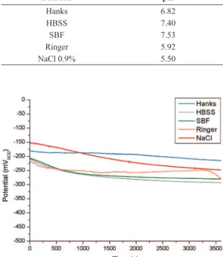
The OCP values that were obtained are shown in Figure 1. The lowest OCP value, measured at 3600 seconds (- 291 mV), which is the most active potential, was developed in HBSS solution. Considering the electrolytes studied at 37 °C, HBSS, SBF and Ringer demonstrated the highest corrosion potential, which may be associated to a higher Cl– concentration in these electrolytes (Table 1). NiTi alloy
is susceptible to pitting corrosion in chloride containing solutions30. Attack by Cl– in NiTi result in Ni being released
into the solution and decrease in the local Ni concentration at the pitting sites31. The remaining Ti reacts with dissolved
oxygen from the solution to form titanium oxides in the corroded area31. The corrosion product layer expands over
TiO with depleted Ni31. The change in the Ni release rate
is related to the corrosion defects such as pitting pores on the NiTi specimens31. In the early immersion period,
nickel ions area released gradually into the surrounding solution31. As the immersion continues, corrosion results in
the formation of pitting pores that promote nickel release, leading to a high Ni release rate31. However, if the immersion
time is long enough, the corrosion-induced titanium oxide layer seals the pitting pores on the NiTi surface, thereby reducing the Ni release rate31. Early pitting corrosion
takes place on many sites on the surface. Early corrosion process take place on surface defects initially and involves the breakdown of original thin oxide layer and Ni-Ti bond after attack by Cl–, and this process releases Ni ions31. As
time elapses, the pitting sites propagate and noticeable pores form on the surface31. Ni released from the bulk materials
into the solution reduces the local concentration of Ni in the materials31. Meanwhile, the remaining Ti reacts with
dissolved oxygen in the solution to form titanium oxides around the pitting sites and pores31. As the corrosion process
proceeds, the titanium oxide layer grows and propagates over the entire surface of the NiTi specimen31. The pores formed
during corrosion are possibly blocked by the titanium oxides formed inside the pores. As a result, a uniform and dense oxide layer is formed31. After the titanium oxide layer has
formed on the entire surface of the NiTi specimen, it will serve as a passivation ilm to retard the corrosion process31.
This susceptibility increases when the chloride concentration is higher30. In the Hanks solution, the NiTi alloy developed
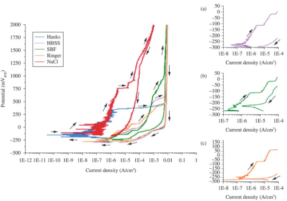
a less active potential (- 214 mV). Figure 2 presents the voltammograms of NiTi in different studied solutions. A slight increase in the current density occurred in the anodic potential scan, indicating a supericial oxide disruption allowing the current low. This phenomenon of anodic dissolution in the surface indicates the beginning of the oxide layer breakdown. When the applied potential reaches 2 V, it is reversed, and for all systems the current density of the inverse scan is higher (Figure 2). This behavior is associated to the pitting corrosion of the NiTi alloy in the studied solutions27.
In the 0.9% NaCl solution, the NiTi samples developed current densities that were one order of magnitude lower than the others studied solutions (Figure 2) indicating that a lower temperature (25 °C) and the absence of other ions, such as HCO3-1, can make the medium less aggressive to this alloy. However the difference in temperature, 25 °C for the test in 0.9% NaCl solution compared to 37 ºC for the other electrolytes can contribute for the differences observed between NaCl solution and the other solutions. Few studies have investigated the effects of temperature on the corrosion resistance of nitinol, but a study in Hank’s physiological solution at temperatures ranging from 10 to 80 °C show that the ability of NiTi to repassivate is signiicantly reduced by an increase in temperature32. Studies
using other alloys proved that a bicarbonate ion can induce pitting corrosion33. Furthermore, in a 0.9% NaCl solution
it is possible to observe the disruption of the passive layer at 757 mV (Figure 2). Considering the solutions that were analyzed at 37 °C, the Hanks solution demonstrated the lowest current densities in the passive zone of the polarization curve (Figure 2). This is probably associated to HCO3– ion concentration in the Hanks solution (Table 1). The oxide layer was irstly ruptured, in Hanks solution, when the applied potential reached 99 mV (current density increased from 1.55 × 10⁻⁷ A/cm2 to 8.72 × 10⁻⁷ A/cm²)
while this value was – 78 mV (current density increased
Table 1. Simulated body luids and human blood plasma ion concentrations.
Solution Ion concentrations (mmol/L)
Na+ K+ Mg+2 Ca+2 Cl- HCO
3
- HPO
4
-2 SO
4 -2
Hanks 142.00 5.00 1.50 2.50 103.00 27.00 1.00 0.50
HBSS 141.60 5.81 0.81 1.26 144.80 4.09 0.78 0.81
SBF 142.00 5.00 1.50 2.50 147.80 4.20 1.00 0.50
Ringer 113.60 1.88 - 1.08 115.30 2.38 -
-NaCl 0.9% 153.00 - - - 153.00 - -
-Plasma 142.00 5.00 1.50 2.50 103.00 27.00 1.00 0.50
Table 2. pH measurements of studied solutions at 37 °C
(Hanks, HBSS, SBF and Ringer) and 25 °C (NaCl 0.9%).
Solution pH
Hanks 6.82
HBSS 7.40
SBF 7.53
Ringer 5.92
NaCl 0.9% 5.50
Figure 1. Open circuit potentials in different simulated body luids
from 3.20 × 10⁻⁶ A/cm² to 1.17 × 10⁻⁵ A/cm2) for Ringer, - 119 mV (current
density increased from 2.80 × 10⁻⁶ A/cm² to 1.17 × 10⁻⁵ A/cm2)
for HBSS and -148 mV (current density increased from 2.80 × 10⁻⁶ A/cm2 to 1.39 × 10⁻⁵ A/cm²) for SBF. In the Hanks
solution the NiTi shows a lower tendency to corrosion due to a minor passive current and a higher pitting potential, when compared to other solutions studied at 37°C. This results demonstrated a lower corrosive tendency of NiTi in Hanks solution in comparison with other studied solutions. The variation in the passive zone of the polarization curve of different alloys due to the concentration of bicarbonate in the electrolyte was described by other authors34. Ions
bicarbonate accelerated the cathodic reactions, promoting an oxide growth on the surface, and resulting in lower anodic current densities. Despite the curves in the other solutions having similar behavior (Figure 2), the potentials in which sudden increases of current density occurred varied. This fact suggests that it is not possible to compare the results of studies performed in different electrolytes, even if they have all been performed in simulated body luids. A standardization of all of the parameters for corrosion tests is important to compare the different studies about the protection of NiTi surfaces. It should be mentioned that, according to some authors, it is dificult to choose an ideal solution to simulate the body luids due to the complexity of the human physiological enviroment24. Other authors
suggest that organic acids found in the blood should also be considered in the preparation of simulated body luids27.
In the present paper, the Hanks and the HBSS solutions demonstrated a good reproducibility of the results.
Considering that all samples were made of the same alloy and were submitted to the same supericial treatment, and the electrochemical tests were performed under the same parameters (temperature, sealing and scan rate), the corrosion potential variance is probably due to the different aggressiveness levels of the studied electrolytes, despite the slight difference in the pH among the solutions, which can inluence the cathodic curves, thus affecting the corrosion potential. Nevertheless, this influence is probably not relevant, since all of the solutions are in neutral pH range. A study developed in Hank’s physiological solution with pH varying from 1 to 9 showed that corrosion resistance of NiTi is not signiicantly affected by pH values32. The
Hanks solution showed the lowest aggressiveness in comparison to the other simulated body luid solutions, which is evidenced by the less active corrosion potential and by the evolution of minor current densities. Other authors also demonstrated that the Ringer solution is stronger than the Hanks. This variation was associated to the presence of Na2PO4 and KH2PO4, which beneits the biomaterials against corrosion, in Hanks solution11,24. It is important to
notice that the Cl– ions concentration is lower in the Hanks
solution, in comparison to the others studied solutions. Studies indicate that the NiTi alloy is suscetible to pitting corrosion in chloride solutions, and in increasing chloride concentration, the pitting potential decreases30. In the HBSS
solution the NiTi alloy developed a higher current density and more active potentials (Figure 2), which was expected due to the results obtained by the OCP (Figure 1).
Figure 2. Potentiodynamic polarization in different simulated body luids at 37 °C and in 0.9% NaCl at 25 °C. Voltammogram detail
3.2. Supericial characterization
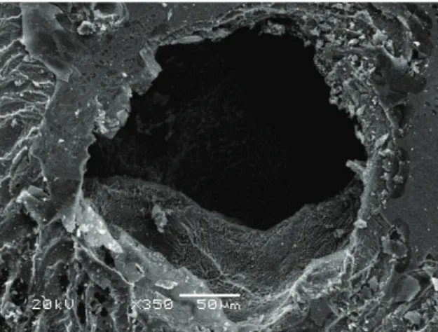
After the electrochemical tests, the NiTi samples were analyzed by scanning electron microscopy in order to determine the relation to the corrosion mechanism found in the electrochemical tests (Figures 3-8). Although the electrochemical results demonstrated that the NiTi alloy shows the same corrosion mechanism (pitting) in all studied
simulated body luids, only with the HBSS solution it was possible to notice the presence of oxidation products in the surface (Figure 6), resulting from more active potentials (Figure 2) and a higher current density; such products have been described by other authors35,36 as titanium hydroxides,
which are more soluble, less protective than the oxides, and are known by an increase in the current between ~1,0 and
Figure 3. Mechanically polished NiTi images obtained by scanning electron microscope.
Figure 4. NiTi samples after electrochemical tests in Hanks solution, by scanning electron microscope.
Figure 5. NiTi samples after electrochemical tests in SBF solution, by scanning electron microscope.
Figure 6. NiTi samples after electrochemical tests in HBSS solution, by scanning electron microscop.
Figure 7. NiTi samples after electrochemical tests in Ringer solution, by scanning electron microscope.
1,5 V. Other studies37 have showing pitting corrosion on
NiTi, in physiological solution, at potentials between +190 and +280 mV versus SCE. Oshida et al.38, evidenced
NiTi low pitting potential values, and presence of numerous oscillations of anodic passive current for potentials lower than 500 mV.
4. Conclusions
Evaluating coatings through an appropriated method is as important as developing surface treatments for NiTi alloys. Considering that the corrosion resistance is a major issue in these alloys, it is relevant to standardize the electrochemical procedure parameters, as the electrolytes, allowing a comparison between the various coatings developed by several researchers.
This study compared the most commonly used electrolytes to simulated body luids: Hanks, HBSS, SBF, Ringer and 0.9% NaCl. Electrochemical and morphological results, show that the NiTi presents the same corrosion mechanism (pitting induced by Cl- and HCO
3
- ions) in all studied simulated body luids. However, the Hanks solution demonstrated less aggressiveness in comparison to other simulated body luids, which is evidenced by its less active corrosion potential and by the development of lowest current densitie values, probably due to a lower chloride concentration. In the HBSS, SBF and Ringer solutions, the NiTi showed higher current densities and a more active potential. The Hanks and HBSS solutions presented the best reproducibility of results. Considering that the HBSS represents an extreme environment, this solution seems to be the most indicated to study the corrosion behavior of NiTi treated surfaces.
References
1. Shabalovskaya SA, Rondelli GC, Undisz AL, Anderegg JW, Burleigh TD and Rettenmayr ME. The electrochemical
characteristics of native Nitinol surfaces. Biomaterials. 2009; 30(22):3662-3671. http://dx.doi.org/10.1016/j. biomaterials.2009.03.034. PMid:19345407
2. McMahon RE, Ma J, Verkhoturov SV, Munoz-Pinto D, Karaman I, Rubitschek F, et al. A comparative study
of the cytotoxicity and corrosion resistance of nickel-titanium and nickel-titanium-niobium shape memory alloys. Acta Biomaterialia. 2012; 8(7):2863-2870. http://dx.doi. org/10.1016/j.actbio.2012.03.034. PMid:22465573
3. Dinca VC, Soare S, Barbalat A, Dinu CZ, Moldovan A, Stoica I, et al. Nickel–titanium alloy: Cytotoxicity evaluation on microorganism culture. Applied Surface Science. 2006; 252(13):4619-4624. http://dx.doi.org/10.1016/j. apsusc.2005.07.093.
4. Wever DJ, Veldhuizen AG, Sanders MM, Schakenraad JM and van Horn JR. Cytotoxic, allergic and genotoxic activity
of a nickel-titanium alloy. Biomaterials. 1997; 18(16):1115-1120. http://dx.doi.org/10.1016/S0142-9612(97)00041-0. PMid:9247349
5. Fili P, Lausmaa J, Musialek J and Mazanec K. Structure
and surface of TiNi human implants. Biomaterials. 2001; 22(15):2131-2138. http://dx.doi.org/10.1016/S0142-9612(00)00404-X. PMid:11432593
6. Bernard SA, Balla VK, Davies NM, Bose S and Bandyopadhyay A. Bone cell-materials interactions and Ni ion release of anodized equiatomic NiTi alloy. Acta Biomaterialia. 2011; 7(4):1902-1912. http://dx.doi.org/10.1016/j. actbio.2011.01.004. PMid:21232641
7. Horiuchi Y, Horiuchi M, Hanawa T and Soma K. Effect of surface modification on the photocatalysis of Ti-Ni alloy in orthodontics. Dental Materials Journal. 2007; 26(6):924-929. http://dx.doi.org/10.4012/dmj.26.924. PMid:18203500
8. Dhage SR, Choube VD, Samuel V and Ravi V. Synthesis of nanocrystalline TiO2 at 100 °C. Materials Letters. 2004; 58(17–18):2310-2313. http://dx.doi.org/10.1016/j. matlet.2004.02.021.
9. Vojtěch D, Novák P, Novák M, Joska L, Fabián T, Maixner J, et al. Cyclic and isothermal oxidations of nitinol wire at
moderate temperatures. Intermetallics. 2008; 16(3):424-431. http://dx.doi.org/10.1016/j.intermet.2007.12.006.
10. Shen Y, Wang G, Chen L, Li H, Yu P, Bai M, et al. Investigation of surface endothelialization on biomedical nitinol (NiTi) alloy: Effects of surface micropatterning combined with plasma nanocoatings. Acta Biomaterialia. 2009; 5(9):3593-3604. http://dx.doi.org/10.1016/j.actbio.2009.05.021. PMid:19477302
11. Shahrabi T, Sanjabi S, Saebnoori E and Barber ZH. Extremely high pitting resistance of NiTi shape memory alloy thin film in simulated body fluids. Materials Letters. 2008; 62(17– 18):2791-2794. http://dx.doi.org/10.1016/j.matlet.2008.01.052.
12. Huan Z, Fratila-Apachitei LE, Apachitei I and Duszczyk J.
Porous NiTi surfaces for biomedical applications. Applied Surface Science. 2012; 258(13):5244-5249. http://dx.doi. org/10.1016/j.apsusc.2012.02.002.
13. Liu XM, Wu SL, Chu PK, Chung CY, Chu CL, Yeung KWK, et al. Effects of water plasma immersion ion implantation on surface electrochemical behavior of NiTi shape memory alloys in simulated body fluids. Applied Surface Science. 2007; 253(6):3154-3159. http://dx.doi.org/10.1016/j. apsusc.2006.07.008.
14. Qiu D, Wang A and Yin Y. Characterization and corrosion behavior of hydroxyapatite/zirconia composite coating on NiTi fabricated by electrochemical deposition. Applied Surface Science. 2010; 257(5):1774-1778. http://dx.doi.org/10.1016/j. apsusc.2010.09.014.
15. Lifeng Z, Yan H, Dayun Y, Xiaoying L, Tingfei X, Deyuan Z, et al. The underlying biological mechanisms of biocompatibility differences between bare and TiN-coated NiTi alloys. Biomedical Materials (Bristol, England). 2011; 6(2):025012. http://dx.doi.org/10.1088/1748-6041/6/2/025012. PMid:21441653
16. Gupta R and Kumar A. Bioactive materials for biomedical
applications using sol-gel technology. Biomedical Materials (Bristol, England). 2008; 3(3):034005. http://dx.doi. org/10.1088/1748-6041/3/3/034005. PMid:18689920
18. American Society for Testing and Materials – ASTM. F2129-08: Standard Test method for conducting cyclic potentiodynamic polarization measurements to determine the corrosion susceptibility of small implant devices. West Conshohocken; 2008. http://dx.doi.org/10.1520/F2129-08. 19. Liu XM, Wu SL, Chu PK, Chung CY, Chu CL, Chan YL, et al.
In vitro corrosion behavior of TiN layer produced on orthopedic nickel–titanium shape memory alloy by nitrogen plasma immersion ion implantation using different frequencies. Surface and Coatings Technology. 2008; 202(11):2463-2466. http://dx.doi.org/10.1016/j.surfcoat.2007.08.017.
20. Bayat N, Sanjabi S and Barber ZH. Improvement of corrosion resistance of NiTi sputtered thin films by anodization. Applied Surface Science. 2011; 257(20):8493-8499. http://dx.doi. org/10.1016/j.apsusc.2011.05.001.
21. Shi P, Cheng FT and Man HC. Improvement in corrosion resistance of NiTi by anodization in acetic acid. Materials Letters. 2007; 61(11-12):2385-2388. http://dx.doi. org/10.1016/j.matlet.2006.09.020.
22. Chrzanowski W, Neel EAA, Armitage DA and Knowles JC.
Effect of surface treatment on the bioactivity of nickel-titanium. Acta Biomaterialia. 2008; 4(6):1969-1984. http://dx.doi. org/10.1016/j.actbio.2008.05.010. PMid:18565807
23. Park HH, Park IS, Kim KS, Jeon WY, Park BK, Kim HS, et al.
Bioactive and electrochemical characterization of TiO2 nanotubes on titanium via anodic oxidation. Electrochimica Acta. 2010; 55(20):6109-6114. http://dx.doi.org/10.1016/j. electacta.2010.05.082.
24. Liang C-h, Mou Z. Effects of different simulated fluids on anticorrosion biometallic materials. Transactions of Nonferrous Metals Society of China. 2001;11(4):579-582.
25. Flamini DO, Saugo M and Saidman SB. Electrodeposition of polypyrrole on Nitinol alloy in the presence of inhibitor ions for corrosion protection. Corrosion Science. 2014; 81:36-44. http://dx.doi.org/10.1016/j.corsci.2013.11.063.
26. Maho A, Delhalle J and Mekhalif Z. Study of the
formation process and the characteristics of tantalum layers electrodeposited on Nitinol plates in the 1-butyl-1-methylpyrrolidinium bis(trifluoromethylsulfonyl)imide ionic liquid. Electrochimica Acta. 2013; 89:346-358. http://dx.doi. org/10.1016/j.electacta.2012.11.026.
27. Khalil-Allafi J, Amin-Ahmadi B and Zare M. Biocompatibility
and corrosion behavior of the shape memory NiTi alloy in the physiological environments simulated with body fluids for medical applications. Materials Science and Engineering C. 2010; 30(8):1112-1117. http://dx.doi.org/10.1016/j. msec.2010.06.007.
28. Siu HT and Man HC. Fabrication of bioactive titania coating on nitinol by plasma electrolytic oxidation. Applied Surface Science. 2013; 274:181-187. http://dx.doi.org/10.1016/j. apsusc.2013.03.008.
29. Kokubo T and Takadama H. How useful is SBF in predicting in vivo bone bioactivity? Biomaterials. 2006; 27(15):2907-2915. http://dx.doi.org/10.1016/j.biomaterials.2006.01.017. PMid:16448693
30. Li X, Wang J, Han EH and Ke W. Influence of fluoride and
chloride on corrosion behavior of NiTi orthodontic wires. Acta Biomaterialia. 2007; 3(5):807-815. http://dx.doi.org/10.1016/j. actbio.2007.02.002. PMid:17467350
31. Hu T, Chu C, Xin Y, Wu S, Yeung KWK and Chu PK. Corrosion products and mechanism on NiTi shape memory alloy in physiological environment. Journal of Materials Research. 2010; 25(02):350-358. http://dx.doi.org/10.1557/
JMR.2010.0051.
32. Trépanier C and Pelton AR. Effect of temperature and pH on the
corrosion resistance of Nitinol. In: Medical Device Materials II: Proceedings from the Materials & Processes for Medical Devices Conference 2004; 2004. St. Paul Minnesota: ASM International; 2005. p. 392.
33. Han J, Zhang J and Carey JW. Effect of bicarbonate on
corrosion of carbon steel in CO2 saturated brines. International Journal of Greenhouse Gas Control. 2011; 5(6):1680-1683. http://dx.doi.org/10.1016/j.ijggc.2011.08.003.
34. Torres-Islas A, Gonzalez-Rodriguez JG, Uruchurtu J and
Serna S. Stress corrosion cracking study of microalloyed pipeline steels in dilute NaHCO3 solutions. Corrosion Science. 2008; 50(10):2831-2839. http://dx.doi.org/10.1016/j. corsci.2008.07.007.
35. Milošv I, Strehblow H-H, Navinšek B and Metikoš-Huković
M. Electrochemical and thermal oxidation of TiN coatings studied by XPS. Surface and Interface Analysis. 1995; 23(7-8):529-539. http://dx.doi.org/10.1002/sia.740230713.
36. Tomashov ND, Chukalovskaya TV, Myedova LL and Yegorov FF. Corrosion and anodic behaviour of carbide, nitride and boride of titanium in sulphuric and phosphoric acids. Zaschyta Myetallov. 1985;5:682-688.
37. Rondelli G. Corrosion resistance tests on NiTi shape memory
alloy. Biomaterials. 1996; 17(20):2003-2008. http://dx.doi. org/10.1016/0142-9612(95)00352-5. PMid:8894095
38. Oshida Y, Sachdeva R, Miyazaki S and Fukuyo S. Biological