w w w . r b o . o r g . b r
Original
article
The
effect
of
platelet-rich
plasma
on
the
repair
of
muscle
injuries
in
rats
夽
Marcelo
Luiz
Quarteiro
∗,
João
Ricardo
Filgueiras
Tognini,
Everton
Lucas
Flores
de
Oliveira,
Izabelli
Silveira
UniversidadeFederaldeMatoGrossodoSul,CampoGrande,MS,Brazil
a
r
t
i
c
l
e
i
n
f
o
Articlehistory: Received2June2014 Accepted12September2014 Availableonline29August2015
Keywords: Muscles/injuries Platelet-richplasma Rats
a
b
s
t
r
a
c
t
Objective:Theneedfortherapeuticoptionsformuscleinjuries,whichareincreasingly fre-quentamongsportspractitioners,wasthemotivationforthisexperimentalstudy,which hadtheaimofevaluatingthehistologicaleffectsofplatelet-richplasma(PRP)onrepairsto muscletissuesofrats.
Methods:PRPwasobtainedbymeansofdoublecentrifugationofbloodfromfiveanimals.In 30rats,aninjurywasproducedinthemiddlethirdofthebellyofthegastrocnemiusmuscle ofeachhindlimb.Theseinjuriesdidnotreceiveanytreatmentinsixrats(12legs).In24rats, 0.9%physiologicalserumwasinjectedintotheinjuryintheleftlegandPRPintotheinjury intherightleg.Samplesfromthetreatedanduntreatedtissuewereevaluatedhistologically 7and21daysaftertheprocedures.
Results:ThequantityofcollagenintheinjuriestreatedwithPRPwassignificantlylower thanthatintheotherinjuries,intheevaluationmade7daysaftertheprocedure,butit becameequaltotheothergroups intheevaluationdoneonthe21st day.Therewasa significantincrease(p<0.001)inthequantityofcollagenfromthe7thtothe21stdayinthe injuriestreatedwithPRP,butthiswasnotseenintheinjuriestreatedusingothermethods. TheinflammatoryprocesswasshowntobemoreintenseintheinjuriestreatedwithPRP thanintheinjuriesoftheothertreatmentgroups,intheevaluationdone7daysafterthe procedure.However,themorphologicalaspectsoftheseinjurieswereseentobesimilarto thoseoftheuntreatedinjuries,21daysaftertheprocedure.
Conclusion:PRPpromotedcompletetissuerestitutionbetweenthe 7thand21st daysin experimentalmuscleinjuries.
©2014SociedadeBrasileiradeOrtopediaeTraumatologia.PublishedbyElsevierEditora Ltda.Allrightsreserved.
夽
WorkperformedintheLaboratóriodeFisiologiaAnimal,UniversidadeFederaldeMatoGrossodoSul,CampoGrande,MS,Brazil.
∗ Correspondingauthor.
E-mail:[email protected](M.L.Quarteiro).
http://dx.doi.org/10.1016/j.rboe.2015.08.009
O
efeito
do
plasma
rico
em
plaquetas
no
reparo
de
lesões
musculares
em
ratos
Palavras-chave: Músculos/lesões
Plasmaricoemplaquetas Ratos
r
e
s
u
m
o
Objetivo: A necessidadedeopc¸õesterapêuticasparalesõesmusculares,cadavezmais frequentesentreosesportistas,fundamentouesteestudoexperimental,cujoobjetivofoi avaliarosefeitoshistológicosdoplasmaricoemplaquetas(PRP)noreparodotecido mus-cularderatos.
Métodos: OPRPfoiobtidoporduplacentrifugac¸ãodosanguedecincoanimais.Em30ratos, foiproduzidoumtraumanoterc¸omédiodoventredomúsculogastrocnêmiodecada mem-brotraseiro.Essaslesõesnãoreceberamtratamentoemseisratos(12patas).Em24ratos, injec¸õesintralesionaisdesorofisiológicoa0,9%edePRPforamaplicadasnaspatas esquer-dasedireitas,respectivamente.Amostrasdotecidotratadoenãotratadoforamavaliadas histologicamentesetee21diasapósosprocedimentos.
Resultados: AquantidadedecolágenonaslesõestratadascomPRPfoisignificativamente menordoqueadasdemaislesõesnaavaliac¸ãofeitasetediasapósoprocedimento,masse equiparouàdosdemaisgruposnaavaliac¸ãofeitano21◦dia.Houveaumentosignificativo
(p<0,001)naquantidadedecolágenodosétimoparao21◦dianaslesõestratadascomPRP,
oquenãoocorreunaslesõestratadasdeoutraforma.Oprocessoinflamatóriosemostrou maisintensonaslesõestratadascomPRPemcomparac¸ãocomaslesõesdosoutrosgrupos detratamentonaavaliac¸ãofeitasetediasapósoprocedimento;todavia,osaspectos mor-fológicosdessaslesõessemostraramsimilaresaodaslesõesnãotratadas21diasapóso procedimento.
Conclusão: OPRPpromoveucompletarestituic¸ãotecidualentreosétimoeo21◦diaem
lesõesmuscularesexperimentais.
©2014SociedadeBrasileiradeOrtopediaeTraumatologia.PublicadoporElsevier EditoraLtda.Todososdireitosreservados.
Introduction
Muscleinjuriesaredefinedasmorphologicalorhistochemical alterationsthatcausedysfunctionofthelocomotorsystem.1 Theycanbecausedbytwomechanisms:directtraumasuchas bruisesandlacerations,andindirecttraumasuchasischemia, denervationandstrain.2
Approximately30% ofinjuriesdiagnosed bydoctorsare relatedtothemusclesystem,3and muscleinjuryisoneof themostcommonformsoftraumathatoccur duringsport practice,causing10–55%ofallinjuries.2Sportinjuriesappear tobearesultofexercisesperformedinastrenuous, inadver-tentorinappropriatemanner.Theprevalenceandincidence oftheseepisodesareunderestimatedbecauseoftheabsence ofnotificationswithintheworldofsports.4Thereported inci-denceofinjuriestohamstringmusclesisoftheorderof12% insoccerplayers,550.9%insprintathletes6and42%in breast-strokeswimmers.7
Depending on the severity and location of the injury, differentformsoftherapeuticmanagementare used, from conservative and drug treatment to surgical treatment.8 Exceptfor casesof complete muscle tearing, avulsionand myositisossificans, the standard treatment used foracute muscleinjuriesconsistsofresting,protection,ice, compres-sionandelevation.Beyondtheseprinciples,thereisnoclear consensusabouttreatmentsforacutemuscleinjuries.9Thus, questions stillremain, especially regardingthe effects and
resultsofvariouscommonlyusedtreatmentsforstimulating theprocessofmusclerepair.
Platelet-rich plasma (PRP) isa product from autologous blood that, since 1990, has been proposed for treatments becauseitpromotesstrongstimulationtotissuerepair.10Itis obtainedthroughcentrifugationofperipheralbloodandthe platelet concentrationshould ideally be higherthan 338%, incomparisonwiththatoftheperipheralblood.11 PRPhas healingpropertiesthathavebeenattributedtotheincreased concentrationsofautologoustissuegrowthfactorsand pro-teins at cellular level. These factors, when introduced to theareaoftheinjury,areexpectedtoincreaserecruitment, proliferation and differentiation of cells involved in tissue repair, andtopromoteacceleratedrepairwithbettertissue differentiation.12
Various clinical uses ofPRP have been studied, includ-ing the repair of chondral13 and tendon injuries,14 repair ofinjuriesandboneregeneration,15 andtreatmentof plan-tar fasciitis16 and severe diabetic foot ulcers.17 The repair ofchronicAchillestendinopathywithintralesionalinjection of PRP has shown promising results from histological and morphological evaluation of the neoformed tissue.18 Both experimentalandclinicalstudieshaverevealedtheeffectsof intralesionalinjectionofPRPinmuscleinjuriesand, gener-ally,thesestudieshavereportedbettermuscleregeneration, increasedneovascularizationandreducedfibrosis.19–25
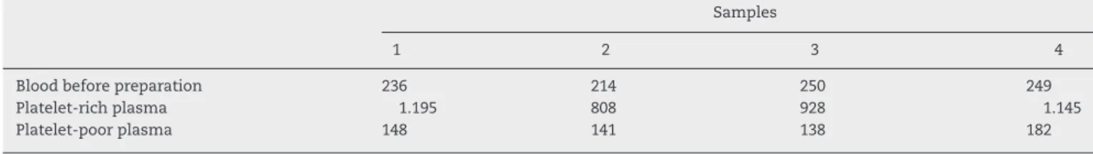
Table1–Plateletquantification(103/l)observedinthefourbloodsamplesextractedfromfiveratsforpreparationofPRP.
Samples
1 2 3 4
Bloodbeforepreparation 236 214 250 249
Platelet-richplasma 1.195 808 928 1.145
Platelet-poorplasma 148 141 138 182
effectivemuscleregeneration,thepresentexperimentalstudy wasperformedinordertoascertaintheeffectofhomologous PRP onrepairstomusclelesionsthat were inducedinrats throughapplyingan impact.Thisstudy evaluated collagen depositionandmadeaqualitativemorphologicalanalysison thetissuerepairprocess,underamicroscope,7and21days aftertreatment.
Material
and
methods
ThisstudywasapprovedbytheEthicsCommitteeunderthe protocol334/2011.Theexperimentwasconductedina labo-ratoryfromApriltoJuly2012.Thirty-fiveisogenicmaleWistar rats(Rattusnorvegicusalbinus)ofEPM-1lineagefromthe cen-tralvivariumoftheFederalUniversityofMatoGrossodoSul wereused.Theratswere12weeksoldandtheirmeanweight was320±20g.
Initially, overa 30-dayperiod,the animals underwent a periodofadaptationandweightgain,duringwhichtheywere keptin standard boxes forfive animals, madeof polypro-pyleneandwithagalvanizedmetalliclid.Theenvironment wasclimate-controlled,withatemperatureof22±3◦C, arti-ficial lighting with12-h light/dark cycles and airhumidity of56±13%.The animalswere fedwithNuvilab® CR1feed
(NuvitalAlimentoseProdutosVeterináriosLtda®,Curitiba,PR,
Brazil)andfilteredwater,adlibitum.
Fourstudygroupswereformedrandomly:
Group 1: five rats underwent blood sampling in order to preparePRP;
Group2:theleftlegsof24ratswhosemuscleinjurieswere treatedwith0.9%physiologicalsalinesolution.
Group3:therightlegsof24ratswhosemuscleinjurieswere treatedwithPRP.
Group4:therightandleft legs(12legs)ofsixratswhose muscleinjurieswerenottreated.
Protocolforproducingtheexperimentalinjury
Thedeviceandthetechniqueusedforproducingthe exper-imentalinjurieswerethesame asdescribed byNogueira.26 For creatingmuscle injuries,the device developedbySene wasused,27whichconsistsoftwoadjustabletelescopicmetal rods,throughwhichispossibletomarkoutaheightof30cm, andaplasticbaseofarea 272.5cm2.Arectangularmetallic surfaceofareaof12.25cm2 wasattachedtothisbase.This surfaceservedasasupportfordroppingtheweightandfor attachingtheanimal’shindleginapredeterminedplace,thus concentratingtheweightinthecentralareaoftheleg.Ametal structurewasattachedtotheupperendofthemetalrods,in ordertoprovidestabilityand tohold apulleywheelacross
whichaguidewireheldtheweightthatwastobereleased. Transparentacrylicchannelingwassetupbetweentherods toguidetheweightduringthe30cmfreefall,inordertoavoid deviationandoscillationoftheweight.
Thedeviceforproducinginjuriesthroughimpactwasfixed withclipstothesurgicaltableinordertostabilizeitinsuch awayastoavoidany oscillationastheweightdropped.In ordertoensurethattheinjurywouldoccurinthesamearea, theloadreleasedfrom30cmhighwaschanneledbytheacrylic guideandbyawireattacheddirectlytotheweight,whichwas releasedcentrallybymeansofapulleywheelthatwasplaced ontherodsofthedevice.
Theanimalswerepreviouslyanesthetizedwithketamine (60mg/kg)andxylazine(15mg/kg)andthenunderwenta sin-gletraumaticeventineachlimb,inthemiddlethirdofthe bellyofthegastrocnemiusmuscleandwereseparated accord-ingtotheexperimentalgrouptowhichtheywouldbelong.The 24animalswhoselegscomprisedgroups2and3underwent contusioninjuriesintheirhindlegs.Inthecentralposterior areaoftheleftlegs,0.1mlof0.9%salinesolutionwas admin-istratedand,intherightlegs,0.1mlofPRP.
Preparationandapplicationoftheplatelet-richplasma
CardiacpuncturewasperformedusingaBDneedle(22g×1′′; 0.70mm×25mm)attachedtoa20mldisposablesyringe(Viet Jet®;LaborImportComércio,Importac¸ãoeExportac¸ãoLtda., Osasco,SP,Brazil)with1mlof10%sodiumcitrate(Bioclin®; QuibasaLtda.;batch0067/2011).Foursamplesof8mlofblood withanticoagulantwereobtainedfromthefiveratsthat com-prised group 1. Theblood with anticoagulant immediately underwent cell counting in an automated device (Sysmex XE-2100D).Afterthefirstcentrifugation,theplasmawas sep-aratedfromtheredbloodcellconcentrate.Duringthesecond centrifugation,thesupernatantportionwaseliminated,such thatonlyapproximately1mloftheheaviercentrifuged mate-rial remained.This fractionwas called the PRP or platelet concentrate. The homogenized PRP and the platelet-poor plasmaunderwentautomatedcellcountingagain,asshown inTable1.
Sacrifice
Afterdissectionandmusclesamplecollectionforanalysis,the animalswerediscardedthroughincinerationinan appropri-ateenvironment.
Formsofevaluation
Thegastrocnemiusofeachanimalwasremovedthrough pos-terior incision in the hind legs of the animals in ventral decubitusposition,withbluntdissectionofskinandsoft tis-sues.Muscle integritywaspreserved, withmaintenanceof theoriginandtheinsertion(femur–muscle–calcaneus).The pieceswereattachedtoasolidsurfaceusingpinsandwere storedin10%formaldehyde.Theyweresentforhistological analysis,inwhichtheyreceived routinetreatment consist-ingofprogressivedehydrationinalcohol,inclusioninparaffin blocks and cutting of sagittal and longitudinalsections of thickness5musingamicrotome(inthecentralthirdofthe musclebelly). Theslides thus produced were stained with picrosiriusredandhematoxylineosin (HE).Examinationof thehistologicalcharacteristicsandquantityofcollagenwas performedintheToxicologyandMedicinalPlantLaboratory ofAnhangueraUniversity(UNIDERP).
Usinganopticalmicroscope coupledtoacomputer,the imagesoftheslidesweredigitizedandcapturedbyanimage processingand analysissystem (ImageLabTM). Thissystem wasdevelopedformorphometricanalysisandimage subtrac-tionandcanbeusedforspecimensatbothmacroscopicand microscopicscale.Therearemanyunitconversionsystems, imagecorrectionfilters, exportation formatsand means of communicationwithothersoftware.Onthecomputerscreen, thesystempresentstheoriginalimagedigitizedfromthe his-tologicalslideand,alongsidethis,threefrequencyhistograms showingtheimageintensitiesR(red),G(green)andB(blue). Fromtherepresentationofthesehistograms,thesystem cal-culatesthe desiredquantifications. All thedata relating to thesecalculationsarepresentedinaspreadsheetwithinthis software,whichcanbeconvertedintoaspreadsheetofthe MicrosoftOfficeExcel4.0software.
Themaininstruments and procedures of the study are showninFig.1.
Statisticalanalysis
The data were tabulated in spreadsheets in the Microsoft OfficeExcel(2010)softwareandthenormalityofthesamples wasevaluatedusingtheBioestat5.0software.Calculationsfor comparingthedataandproducinggraphsweremadethrough the GraphPad Prism 4.0 software. The measurements of thenumericalvariableswereexpressedasmeans±standard deviations.Intragroupcomparisonswereperformedusingthe Studentttestonthesampleswithnormaldistributionand theWilcoxontestonthoseofnon-normaldistributions. Inter-group analyseswere performed using analysis ofvariance (ANOVA)andtheposthocTukeytestonsampleswithnormal distributionandtheKruskal–WallistestandposthocDunntest onsampleswithnon-normaldistribution.Thenormalityof thegroupswasevaluatedthroughtheShapiro–Wilktest.The valueofp≤0.05wasadoptedfordeterminingthesignificance levelofthedifferencesfound.
Fig.1–(A)Deviceusedforproducingmuscleinjuriesin animals.(B)Positionoftheloadreleasedoneachlimbof theanimals.(C)Administrationofsalinesolutioninthe posteriorareaoftheleftlegoftheanimals.(D)
Table2–Means(andstandarddeviations)ofthe quantificationofcollagenfibersobservedinthelegsof thecontrolratsandtheratsthatreceivedPRPandsaline solution,7and21daysaftermuscleinjury.
Controlgroup
n=12
PRP
n=24
Salinesolution
n=24
7days 34.44±6.65 30.69±4.99 35.35±5.19
21days 38.29±6.58 38.52±6.47 35.02±6.73
Results
Atotalof60legswereevaluatedandthemeanvalues(and standarddeviations)ofthequantityofcollagenfibersineach groupateachevaluationtime(7and21days)areshownin
Table2.Intheintragroupanalysis,comparingthecountsthat weremadeonthe7thand21stdayafterinjury,therewere nodifferencesinthemeanquantitiesofcollagenfibersinthe controlgroup(p=0.094)orinthegroupofratsthatreceived salinesolution(p=0.817),asshowninFig.2AandC.Inturn, thequantityofcollagenobserved21daysaftertheinjurywas significantlygreater(p=0.00021)thanthequantityobserved 7daysaftertheinjuryinthegroupofratsthatreceivedPRP (Fig.2B).Intheintergroupanalysis,inthecountperformed 7daysaftertheinjury,themeanquantityofcollagenfibers wassignificantlysmaller(p=0.014)intheratstreatedwithPRP thaninthosetreatedwithsalinesolution,butnotin compar-isonwiththecontrolgroup.Ontheotherhand,inthecount performed21daysaftertheinjury, therewasnodifference betweenthegroupsregardingthemeanquantityofcollagen fibers(Fig.3).
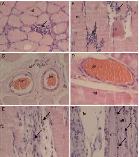
Regarding the morphological findings, circumstantial macrophageswereobservedintheinterstitialtissueofthe skeletalmuscleinthecontrolgrouponthe7thday.The mus-clefibershadnormalappearanceatthistime.Also,myoblasts appearedamongthemusclefibers(Fig.4).Atthesametime(7 days),inthegroupofratstreatedwithPRP,anevident inflam-matoryprocessduetomononuclearcells,redbloodcellsof normalappearanceandmyotubeswereobservedamongthe musclefibers(Fig.5).Intheratstreatedwithsalinesolution, myotubes,macrophagesandredbloodcellsofnormal appear-ancewereobserved(Fig.6).At21days,themusclefibersand bloodvesselsalreadypresentedthenormalappearanceofthe tissuein the controlrats and in the rats treatedwith PRP (Figs.5and6).Intherats treatedwithsaline solution,foci ofmacrophagesandsomemyotubeformationwereobserved (Fig.7).
Discussion
Thereisevidencethatgrowthfactorsplayanessentialrolein thehealingprocessoftissues.28However,inadditiontothe factthatuseofmanygrowthfactorsseparatelyisstill impos-sibleinclinicalpractice,themechanismsofactionofallthe differentfactorsinvolvedinthisprocessarenotcompletely clear. From knowledgethat the alpha-granules ofplatelets concentratelargequantitiesofspecificgrowthfactorssuchas PDGFandTGF-beta,atechniqueforobtaininghigh concentra-tionsofgrowthfactorsthroughpreparationofautologousPRP
A
Control group7 days 21 days
20 25 30 35 40 45 50 55 Collagen fibers
Student’st test; p=0.0949 t
B
PRP group7 days 21 days
20 25 30 35 40 45 50 55 60 Collagen fibers
Wilcoxon’s test; p=0.00021
C
Saline solution group7 days 21 days
20 25 30 35 40 45 50 Collagen fibers
Student’st test; p=0.8179
Fig.2–Graphicrepresentationofthemeansandstandard deviationsofthequantitiesofcollagenfibers7and21days aftertheinjuriescausedintheratsofthecontrolgroup(A), thosetreatedwithPRP(B)andthosetreatedwithsaline solution(C).
wasproposed.11Thistechniquebasicallyconsistsof seques-trating and concentratingplatelets from the blood plasma, whichresultsinaproductthatcanbeappliedtotheinjury healingarea.29
07 days
Control PRP Saline
20 25 30 35 40 45 50 55
*
Collagen fibers
Kruskal-wallis; p=0.0145; *PRP versus SF: p<0.05
21 days
Control PRP Saline
20 25 30 35 40 45 50 55 60
Collagen fibers
ANOVA; p=0.1806
Fig.3–Graphicrepresentationofthemeansandstandard deviationsofthequantitiesofcollagenfibers7and21days aftertheinjuriescausedintheratsofthethreedifferent studygroups.
canbediscussedfromtheresultsofthisstudy.Nonetheless, in an experimental model of bruising that was also con-ductedonthegastrocnemiusmuscleofrats,whoseinjuries were injected withautologousplatelet-rich serum, Wright-Carpenteretal.19observedacceleratedactivationofsatellite cells, 30–48h after injury, and an increase in the diame-terofthemusclefibersundergoingregeneration,duringthe firstweekafterinjury.Theseauthorsalsoobservedincreased concentrationsofFGF-2(460%)andTGF-beta1(82%)through ELISAandsuggestedthatthesecould,atleastpartially,have beenresponsiblefortheacceleratedregeneration,duetotheir proliferativeandchemotacticcharacteristics.
Inmajorsprainsintheanteriortibialmuscleofratstreated withPRPorplacebo,Hammondetal.20observedthatthetime taken formuscle regenerationwas significantly shorterin thegrouptreatedwithPRPandsuggestedthatacceleration ofmyogenesiswasprobablythemechanismresponsiblefor thiseffectfromPRP,becauseofthehigherconcentrationsof differentgrowthfactorsintheproduct.
Harrisetal.22injectedPRPorsalinesolutionintovarious muscletissuesofhealthyrabbits(withoutinjuries),and sam-pleswerehistologicallyevaluated2,6and12weeksafterthe procedure.After6and12weeks,theyobservedpersistentbut decreasing amountsofinflammatoryinfiltrate atsites that receivedPRP,but notatthosethatreceivedsalinesolution. Likewise,theyonlyobservedfibroblasts,collagenformation andneovascularizationinthefocalareasofthescartissueof thelegsthatreceivedPRP.Thiswasnotobservedatanysitein whichsalinesolutionwasinjected.Theseauthorsconcluded thatPRPpromotedaninflammatoryresponseinnormalsoft tissues of rabbits. Their assertion supportsthe hypothesis defendedbythepresentstudy,i.e.thatPRPinitiallypromotes intensificationoftheinflammatoryprocessinmuscleinjuries. Incontrast,Giganteetal.23didnotobserveanydifference regardingtheinflammatoryprocesswhencomparinginjuries
Fig.5–PhotomicrographofratstreatedwithPRPafter7days.Aninflammatoryprocessduetomononuclearcells (continuousarrows),musclefiberintransversesection(mf),musclefiberinlongitudinalsection(mfl),myotubes(dashed arrows),bloodvessels(bv),fatcells(fc)andconnectivetissue(ct)canbeseen.HE/200×.
producedinthelongusmuscleofrats,betweenthosetreated withPRP and those thatwere not treated.However, better muscleregeneration,increasedneovascularizationandslight fibrosisreductionwereobservedamongthetreatedinjuries.
Itneedstobeemphasizedthattheconcentrationsof differ-entgrowthfactorsinPRPobtainedfromdifferentspecies(rats, rabbits,sheepandhumans)presentsignificantvariation.This hasadirectinfluencebothonexperimentalstudiesandon clinicaltrials.30Hence,thereisaneedforstandardized proto-colsinordertoachieverealexpansionofknowledgeregarding theeffectsofPRPintreatingmuscleinjuries.
The use of PRP for regenerating bones and soft tis-sues has been a focus ofattention among clinical doctors andresearchers.Itsuse indifferentsurgicalspecialtieshas alsobeenreported.Sincethetimeoftheinitiallyproposed techniqueforproducingautologousPRP,inwhichthe discon-tinuousmethodofcellseparationdemandedagreatamount of blood, several other protocols have been proposed and havecontributedtowardthe evolutionoftheoriginal tech-nique.Theadvanceshaveincludedlowerbloodvolumeneeds, useofbenchtopcentrifuges,lowercosts,shorterproduction time,easierapplicationinoutpatientsettingsandlowerstress onthepatient’scardiovascularsystem.Thesafestandmost
effectiveprotocolsseemtorelatetothedoublecentrifugation techniquesthatwereusedforperformingthepresentstudy.29 Some studies have shown that platelet concentrations, 338% higherthan normallyfoundinthe blood,boostbone andsofttissuehealinginhumanbeings.11Otherauthorshave maintained thattheexpectedeffects ofPRP would onlybe reachedwhenthe plateletconcentrations wereeighttimes higherthantheconcentrationsinthecirculatingblood.Inour experimentalstudy,inthefourbloodsamplescollectedfrom fiveratsforproducingPRP,weobtainedameanplatelet con-centrationthatwasapproximatelyfourtofivetimeshigher thanwhatwasobservedintheblood.
Fig.6–Photomicrographofratstreatedwithsalinesolutionafter7days.Musclefiberinlongitudinal(mfl)andtransverse (mf)sections,myotubes(continuousarrows),macrophages(dashedarrows)andbloodvessels(bv)canbeseen.HE/200×.
treatedwithPRP,which,inthislastevaluation,presenteda meanquantityofcollagenthatwassimilartothatoftheother groups.
Degradationofcollagenisknowntobeginearlyandthere isintenseactivity in theinflammatory process.Thisevent occursduringthe first stageofhealing. Infact, apartfrom theinjuriestreatedwithsalinesolution,alowermean quan-tityofcollagenwasobservedintheevaluationperformedon the7th day afterinjurythan inthe oneperformed onthe 21stday.However,inthefirstevaluation(7days),themean quantityofcollagenwassignificantlylowerininjuriestreated withPRP than inthe controlinjuries and inthose treated with saline solution. This finding seems toratify the the-oryaccordingtowhichtheinflammatoryprocessisprobably altered in the presence of PRP, thereby sometimes reduc-ingtheperiodofinflammationoftheinjuryandsometimes alteringthereleaseofcytokines.20Thus,theresultsmakeit possibletoputtogetheranewhypothesisthatcanbetested, therebysuggestingthattheinflammatoryphaseisextended or has higher intensity. This would lead to more intense
degradationofcollageninthefirstphaseofmusclehealing. However,nostudieshaveyettestedthesehypothesesmore rigorously.
On the other hand, during the repair and remodeling phases, depositionofcollageninanorganizedand gradual manneristhemostimportantcharacteristicforassuring bal-ance betweenlysis ofthe old cell matrix and synthesisof the newmatrix.Thisisanessentialconditionfor success-fulregenerationoftheinjuredmuscletissue.Inaddition,the initiallyproducedcollagenisthinnerthanthecollagenfrom thehealthytissue;thisinitialcollagenisthenreabsorbedand thickercollagenisproducedalongthetensionlines,andthisis positivelycorrelatedwithincreasesintensilestrength.Inthis study,themeanquantityofcollagensignificantlyincreased from the7th tothe21st dayaftertheinjectionofPRP,but thisdidnotoccurintheuntreatedinjuriesorinthosetreated withsalinesolution. Theincreaseofcollagenfromthe 7th to the21st dayin theanimals that receivedPRP seems to havehelpedinthecollagendegradation–depositionbalance, throughmechanismsthatstillneedtobeelucidated.
IninjuriestreatedwithPRP,theinflammatoryprocesswas moreevidentthanintheothergroupsofthestudy,inthe eval-uationperformed7daysaftertheprocedure.Thisobservation seemstoconfirmthatPRPcanintensifytheinflammatory pro-cess.
Inthefinalevaluationat21days,themorphological find-ingsfromthecontrolinjuriesandtheinjuriestreatedwithPRP weresimilar,whichindicatesthatgoodregenerationofthe injuredmuscleoccurredinbothgroups.Theadministration ofPRPdoesnotseemtohaveshowndifferencesin morpho-logicalfeaturesafter21days,attheendofthemusclerepair process,incomparisonwiththecontrolgroup,sincethesame characteristicswereobservedintheuntreatedinjuries.Onthe otherhand,intheinjuriestreatedwithsalinesolution,the tis-suestilldidnotshowfullregeneration,withmusclefibersstill undergoingformationandwithoutvascularization.
Thismorphologicaldescriptive analysiscorroboratesthe quantitativefindings regardingcollagen fiber deposition in musclescarsbecauseitshowsthatinthegroupthatreceived salinesolutionalone,therewasstillaninflammatory reac-tionafter21days,whileintheothertwogroups,thetissue hadalreadybeencompletelyregenerated.Thisperhaps sug-geststhattherewasbetterorganizationofthemusclerepair processinthegrouptreatedwithPRP.
Thecontribution ofthe present study is limited to the observationthatPRPsignificantlyactivatedtheinflammatory process7daysafterbeinginjectedintobluntinjuriesthathad beenproducedinthegastrocnemiusmuscleofrats.The rea-sonforthisseemstohavebeengreaterdegradationofcollagen overthisperiod.Ontheotherhand,thisresultedina signif-icantincreaseincollagenbetweenthe7thandthe21stday aftertheprocedure,whichsuggeststhatfulltissuerecovery wasachieved.
Conflicts
of
interest
Theauthorsdeclarenoconflictsofinterest.
r
e
f
e
r
e
n
c
e
s
1. ClebisNK,NataliMRM.Lesõesmuscularesprovocadaspor exercíciosexcêntricos.RevBrasCiênMov.2001;9(4):47–53.
2. JärvinenTA,JärvinenTL,KääriäinenM,KalimoH,JärvinenM. Muscleinjuries:biologyandtreatment.AmJSportsMed. 2005;33(5):745–64.
3. RizziCF,MaurizJL,FreitasCorrêaDS,MoreiraAJ,ZettlerCG, FilippinLI,etal.Effectsoflow-levellasertherapy(LLLT)on thenuclearfactor(NF)-kappaBsignalingpathwayin traumatizedmuscle.LasersSurgMed.2006;38(7):704–13.
4. KettunenJA,KujalaUM,KaprioJ,KoskenvuoM,SarnaS. Lower-limbfunctionamongformerelitemaleathletes.AmJ SportsMed.2001;29(1):2–8.
5. RollsA,GeorgeK.Therelationshipbetweenhamstring muscleinjuriesandhamstringmusclelengthinyoungelite footballers.PhysTherSport.2004;5(4):179–87.
6. PastreCM,CarvalhoFilhoG,MonteiroHL,NettoJuniorJ, PadovaniCR.Lesõesdesportivasnaelitedoatletismo brasileiro:estudoapartirdemorbidadereferida.RevBras MedEsporte.2005;11(1):43–7.
7.GroteK,LincolnTL,GambleJG.Hipadductorinjuryin competitiveswimmers.AmJSportsMed.2008;32(1):104–8.
8.DeCarliA,VolpiP,PelosiniI,FerrettiA,MelegatiG,MossaL, etal.Newtherapeuticapproachesformanagementof sport-inducedmusclestrains.AdvTher.2009;26(12):1072–83.
9.ChanYS,LiY,FosterW,FuFH,HuardJ.Theuseofsuramin, anantifibroticagent,toimprovemusclerecoveryafterstrain injury.AmJSportsMed.2005;33(1):43–51.
10.HaynesworthSE,KadiyalaS,LiangL,ThomasT,BruderSP. Chemotacticandmitogenicstimulationofhuman mesenchymalstemcellsbyplateletrichplasmasuggestsa mechanismforenhancementofbonerepair.In:48thAnnual MeetingoftheOrthopaedicResearchSociety;2002.
11.MarxRE,CarlsonER,EichstaedtRM,SchimmeleSR,Strauss JE,GeorgeffKR.Platelet-richplasma:growthfactor
enhancementforbonegrafts.OralSurgOralMedOralPathol OralRadiolEndod.1998;85(6):638–46.
12.FosterTE,PuskasBL,MandelbaumBR,GerhardtMB,Rodeo SA.Platelet-richplasmafrombasicsciencetoclinical applications.AmJSportsMed.2009;37(11):2259–71.
13.SunY,FengY,ZhangCQ,ChenSB,ChengXG.The
regenerativeeffectofplatelet-richplasmaonhealinginlarge osteochondraldefects.IntOrthop.2010;34(4):589–97.
14.SánchezM,AnituaE,AzofraJ,PradoR,MuruzabalF,AndiaI. Ligamentizationoftendongraftstreatedwithanendogenous preparationrichingrowthfactors:grossmorphologyand histology.Arthroscopy.2010;26(4):470–80.
15.TorresJ,TamimiF,TresguerresIF,AlkhraisatMH,KhraisatA, BlancoL,etal.Effectofcombiningplatelet-richplasmawith anorganicbovineboneonverticalboneregeneration:early healingassessmentinrabbitcalvariae.IntJOralMaxillofac Implants.2010;25(1):123–9.
16.PeerboomsJC,VanLaarW,FaberF,SchullerHM,Vander HoevenH,GosensT.Useofplateletrichplasmatotreat plantarfasciitis:designofamulticentrerandomized controlledtrial.BMCMusculoskeletDisord.2010;11:69.
17.VillelaDL,SantosVL.Evidenceontheuseofplatelet-rich plasmafordiabeticulcer:asystematicreview.Growth Factors.2010;28(2):111–6.
18.DeVosRJ,WeirA,VanSchieHT,Bierma-ZeinstraSM,Verhaar JA,WeinansH,etal.Platelet-richplasmainjectionforchronic Achillestendinopathy:arandomizedcontrolledtrial.JAMA. 2010;303(2):144–9.
19.Wright-CarpenterT,OpolonP,AppellHJ,MeijerH,WehlingP, MirLM.Treatmentofmuscleinjuriesbylocaladministration ofautologousconditionedserum:animalexperimentsusing amusclecontusionmodel.IntJSportsMed.2004;25(8):582–7.
20.HammondJW,HintonRY,CurlLA,MurielJM,LoveringRM. Useofautologousplatelet-richplasmatotreatmusclestrain injuries.AmJSportsMed.2010;37(6):1135–42.
21.HamidMS,MohamedAliMR,YusofA,GeorgeJ.Platelet-rich plasma(PRP):anadjuvanttohastenhamstringmuscle recovery.Arandomizedcontrolledtrialprotocol
(ISCRTN66528592BMC).MusculoskeletDisord.2012;13:138.
22.HarrisNL,HufferWE,VonStadeE,LarsonAI,PhinneyS, PurnellML.Theeffectofplatelet-richplasmaonnormalsoft tissuesintherabbit.JBoneJointSurgAm.2012;94(9):786–93.
23.GiganteA,DelTortoM,ManzottiS,CianforliniM,Busilacchi A,DavidsonPA,etal.Plateletrichfibrinmatrixeffectson skeletalmusclelesions:anexperimentalstudy.JBiolRegul HomeostAgents.2012;26(3):475–84.
24.BubnovR,YevseenkoV,SemenivI.Ultrasoundguided injectionsofplateletsrichplasmaformuscleinjuryin professionalathletes:comparativestudy.MedUltrason. 2013;15(2):101–5.
26.NogueiraGB[dissertac¸ão]Efeitodomeloxicanelaserde baixaintensidadenaterapêuticadelesõesmusculares induzidasemratos.CampoGrande:UniversidadeFederaldo MatoGrossodoSul;2012.
27.SeneGL[dissertac¸ão]Influênciadolaserdebaixaintensidade naspropriedadesmecânicasdomúsculogastrocnêmio submetidoàlesãomuscularpormecanismodeimpacto.São Carlos:EscoladeEngenhariadaUniversidadedeSãoPaulo; 2005.
28.LynchSE,BuserD,HernandezRA,WeberHP,StichH,FoxCH, etal.Effectsoftheplatelet-derivedgrowthfactor/insulin-like growthfactor-Icombinationonboneregenerationaround
titaniumdentalimplants.Resultsofapilotstudyinbeagle dogs.JPeriodontol.1991;62(11):710–6.
29.MessoraMR,NagataMJH,FurlanetoFAC,DeliberadorTM, MeloLGN,GarciaVG.Análisedaeficiênciadoprotocolode duplacentrifugac¸ãoparaopreparodoplasmaricoem plaquetas(PRP):estudoexperimentalemcoelhos.RSBO. 2009;6(3):291–6.