MicroRNAs, cancer and ionizing radiation: Where are we?
GUSTAVO NADER MARTA1*, BERNARDO GARICOCHEA2, ANDRÉ LOPES CARVALHO3, JULIANA M. REAL4, LUIZ PAULO KOWALSKI5
1M.D. – Physician at the Radiotherapy Service of the Hospital Sírio-Libanês, Physician of the Radiotherapy Service at the Instituto do Câncer do Estado de São Paulo (Icesp), São Paulo, SP, Brazil 2PhD – Physician at the Clinical Oncology Service, Hospital Sírio-Libanês, São Paulo, SP, Brazil
3PhD – Physician at the Head and Neck Surgery Division, Hospital de Câncer de Barretos – Fundação Piu XII, Barretos, SP, Brazil 4PhD – Researcher at the Instituto Sírio-Libanês de Ensino e Pesquisa, São Paulo, SP, Brazil
5PhD – Head of the Head and Neck Surgery, and Otorhinolaryngology Division, A.C. Camargo Cancer Center, São Paulo, SP, Brazil
S
UMMARYStudy conducted at the Hospital Sírio-Libanês, Radiotherapy Service,
São Paulo, SP, Brazil
Article received: 8/18/2014
Accepted for publication: 8/24/2014
*Correspondence:
Address: Rua Dona Adma Jafet, 91 São Paulo, SP – Brazil
Postal code: 01308-050 gmarta@uol.com.br
http://dx.doi.org/10.1590/1806-9282.61.03.275
Conflict of interest: none
The aim of this study is to describe the biogenesis of microRNA, its relations with carcinogenesis, and the correlation between microRNA and ionizing radiation (IR), focusing on radioresponsiveness. It is known that microRNA biogenesis is well es-tablished and involves different enzymatic cleavages, resulting in the production of mature microRNA. MicroRNAs are involved in carcinogenesis. Their interac-tion is related to the genetic and epigenetic changes associated with activainterac-tion of proto-oncogenes or inactivation of tumor suppressor genes. Several studies have shown that the levels of expression of some microRNAs vary signiicantly after ir-radiation. There are evidences that microRNAs can inluence cellular response af-ter IR. In addition, microRNAs are related to modulation of the expression of sev-eral post-transcriptional targets in DNA damage response pathways, and to the DNA damage repair regulation mechanism. Future studies can clarify a possible clinical use of microRNAs as a new class of radiosensitive agents.
Keywords: microRNAs, radiotherapy, neoplasms.
I
NTRODUCTIONMicroRNAs (miRNA, miR) are a class of endogenous small (16 to 22 nucleotides) non-coding RNAs that con-trol the degradation and translation of target mRNAs
and regulate almost 30% of human genes.1 MicroRNA
genes have been normally found in cancer-related
genom-ic areas.2 They can have diverse roles, such as acting as
tu-mor suppressors or oncogenes depending on tissue type
and the speciic targets.3
The capacity of microRNAs to perform as regulators for gene expression permits them to inluence signaling pathways that might modify several cellular processes, in-volved in irradiation response.
Ionizing radiations (IR) exert their effects through both direct and indirect actions. Direct action means direct injury to the DNA, while indirect action is um-pired through free radicals created through ionization
of H2O. Therefore, DNA damage might appear in
dif-ferent forms such as single-strand breaks (repaired via base excision repair) and double-strand breaks (DSBs), repaired via homologous recombination repair, which is accurate, or non homologous end-joining, which is error-prone. Whereas several cells suffer apoptosis after
IR, cell death also can be related to IR-induced injury that triggers cell inactivation and becomes lethal after
some cell divisions.4
The DNA injury response pathways have an impor-tant role in deining cell survival, radiosensitivity and cell cycle checkpoints. Cellular sensitivity to IR is com-monly determined by DNA DSB repair. However, it is important to know other issues that could be associat-ed with IR, and their effects on tissues: microRNAs as inherent gene regulators may offer new likelihoods in this context.
The aim of this study was to review the microRNA biogenesis, carcinogenesis actions, and the most recent evidences of microRNA and IR regarding radiosensitivi-ty response.
R
ESULTSMicroRNA: biogenesis
MicroRNAs are found in most eukaryotes and their bio-genesis involves several enzymatic cleavages that result in
the production of mature microRNA.5
RNA polymerase II transcribes microRNA genes and
Af-ter enzymatic action and the removal of introns, the result-ing RNA shows the changes in the usual 5’(cap) and 3’ (in-sertion of adenine tail) sequences forming a molecule with thousands of bases (pri-microRNAs). A pri-microRNA is cleaved by complex formed by the RNase III Drosha and its cofactor DGCR8 (DiGeorge syndrome critical region in gene 8) to create a hairpin structured forerunner, or pre-microRNA with 70-100 nucleotides in the nucleus. Pre-mi-croRNAs are brought to the cytoplasm via nuclear
recep-tor-dependent exportin-5 RanGTP.7,8
In the cytoplasm, pre-microRNA is cleaved by Dicer (RNase) to create a microRNA duplex intermediate 22 base pairs. In this process, the Dicer interacts with the protein TRBP (trans-activation response RNA-binding protein) kinase and PRKRA/PACT (interferon-inducible
double stranded RNA-dependent activator).9,10 An
Argo-naute (Ago) protein binds the duplex and integrates the mature, single-stranded microRNA into the Ago:RNA complex to produce mature RNA-induced silencing
com-plex (RISC), whilst the other strand is often rejected.11-13
MicroRNA and carcinogenesis
There are two major mechanisms used in gene expression regulation by repression and degradation of the mRNA translation target (RISC). This interaction is related to the complementary bases intensity between mRNA and
microRNA.14,15
MicroRNAs are involved in the carcinogenesis pro-cess. Their interaction is related to genetic changes asso-ciated with the activation of proto-oncogenes or inacti-vation of tumor suppressor genes. Thus, microRNAs can be grouped into two categories: oncomicroRNAs (nega-tively regulate tumor suppressor genes), and anti-microRNA (act in the negative regulation of
onco-genes).16,17 However, a single microRNA can exert both
actions, depending on their performance in the target tis-sue.18
The relationship of microRNAs in tumorigenesis was irst described in chronic lymphocytic leukemia
(microR-NA-15 and microRNA-16).19 Subsequently, microRNAs
modiication was shown in several neoplasms, such as breast cancer, lymphoma, lung, head and neck,
colorec-tal, prostate, liver, pancreas and thyroid.20 As an example,
the family of microRNAs let-7 regulates some oncogenes (HmgA2, Myc, and Ras) and they are poorly expressed in lung, prostate, glioblastoma. In addition, its low expres-sion correlates with a worse prognosis in patients with
lung cancer.21-23 Conversely, the microRNA 17-92 clusters
can also act as oncogene and they are overexpressed in
some types of lung and kidney cancers.24,25
MicroRNAs are normally located in genome areas
re-lated to alterations in cancer.26,27 Mutations could be
ob-served in the primary transcripts of microRNA-15a and microRNA-6-1, involved in reducing expression of the
two microRNAs in leukemia.28
Moreover, deregulated microRNA expression may be associated with epigenetic alterations, such as changed DNA methylation. It was suggested that microRNA-34b/c and B-cell translocation gene 4 (BTG4) are novel tumor suppressors in colorectal cancer and that the microRNA-34b/c CpG island, which bidirectionally regulates mi-croRNA-34b/c and BTG4, is a common target of epigen-etic silencing in colorectal cancer.29
Likewise, another epigenetic system can disturb mi-croRNAs expression: histone acetylation. Histone deacet-ylase inhibition is followed by the vast and rapid
modii-cation of microRNA levels.30 For example, in breast
carcinoma cell lines, Rhodes et al.31 using histone
deacet-ylase inhibitor (HDACi) trichostatin A (TSA)
demonstrat-ed the suppression of in vitro clonogenicity in the
previ-ously described apoptosis resistance. Significant upregulation of 22 microRNAs and downregulation of 10 microRNAs in response to TSA treatment was observed. These results demonstrate that HDACi TSA exerts anti-cancer activity in apoptosis resistance. This activity is cor-related with TSA alteration of microRNA expression pro-iles indicative of a less aggressive phenotype.
Another important point is that microRNA expres-sion may be modulated as a result of deregulations in the
microRNA biogenesis.32 It was demonstrated that Dicer
silencing promotes cellular transformation and
tumori-genesis in vivo. Kumar et al.33 analyzed the consequences
of conditional Dicer1 mutation (Dicer1 “floxed” or Dicer1(l)) on several mouse models of cancer. Deletion of a single copy of Dicer1 in tumors from Dicer1(l) ani-mals led to reduced survival compared with controls. Tu-mors from Dicer1(l/l) animals always maintained one functional Dicer1 allele. Consistent with selection against full loss of Dicer1 expression, enforced Dicer1 deletion caused inhibition of tumorigenesis. These indings sug-gest that Dicer1 may be an important haploinsuficient tumor-suppressor gene. Moreover, there are some evi-dences suggesting that loss of Dicer and/or Drosha has been inversely related with outcome in different types of tumors.34-37
lev-observe the biological consequences. Changes in microRNA expression were observed for 17 microRNA species, follow-ing exposure to radiation, 23, after H2O2, and 45, after
et-oposide; seven microRNA species were commonly altered by all the agents. These results demonstrate a common microRNA expression signature in response to exogenous genotoxic agents. Furthermore, pre-treatment with cys-teine prevented radiation-induced alterations in microR-NA expression, which suggests that microRmicroR-NAs are re-sponsive to oxidative stress. It could be implied that microRNAs play a role in cellular defense against exoge-nous stress and are involved in the generalized cellular response to genotoxic oxidative stress.
Some microRNAs were associated with essential pro-tective mechanisms counteracting radiation cytotoxici-ty. MicroRNA-34 was found to be a straight target for p53, functioning downstream of the p53 pathway as a tumor suppressor. MicroRNA-34 targets Notch, HmgA2 and Bcl-2 genes involved in self-renewal and survival of
tumor cells.48 IR led to an important increase in the
ex-pression of microRNA-34a, equaled by a decline in the expression of its target oncogenes NOTCH1, MYC, E2F3, and cyclin D1.
A pivotal regulator of DNA methylation and ge-nome stability, lymphoid speciic helicase (LSH), was associated to microRNA-7. After IR, LSH was upregu-lated, while microRNA-7 was downregulated.
Kotur-bash et al.49 have analyzed the effects of X-ray
irradia-tion on microRNA expression in the hippocampus, frontal cortex, and cerebellum of male and female mice. It was shown that changes in the expression of the mi-croRNA-29 family could be related to altered
expres-sion of de novo methyltransferase DNMT3a and changed
global DNA methylation levels. Taken together, this seems to be a cellular protective effect against
radia-tion-induced hypomethylation.50
Additional authors have assessed the inluence of
mi-croRNA and cells reaction after irradiation. Kraemer et al.51
reported the functional role results of microRNAs in radi-ation response in immortalized and primary endotheli-al cells. Overendotheli-all suppression of microRNA expression was accomplished through downregulation of Dicer or Ago2. The decreases in those components led to improved cell death after irradiation, denoting a prosurvival role of mi-croRNAs. Moreover, whilst cell cycle checkpoint activa-tion and apoptosis were impaired, the low microRNAs levels did not disturb DNA DSB repair. The distinctive sensitivity of these pathways suggests the autonomous activation of the two response pathways rather than a
re-paired DNA damage response. Nevertheless, Surova et al.,52
els of microRNA-103/107 are associated with metastasis and poor outcome. Functionally, microRNA-103/107 award
migratory capacities in vitro and empower metastatic
dis-semination of otherwise non aggressive cells in vivo.
Inhibi-tion of microRNA-103/107 opposes migraInhibi-tion and metas-tasis of malignant cells. At cellular level, a key event fostered by microRNA-103/107 is induction of epithelial-to-mesen-chymal transition attained by downregulating microR-NA-200 levels.38
Finally, changes in microRNA expression can be a consequence of transcription variations due to an altered transcription factor action. MicroRNA can be either neg-atively or positively regulated by p53,39 MYC,40 or ZEB1.41
Thus, there are many uncertainties about the mech-anisms involved in microRNAs expression and tumors. Nonetheless, they are probably alike to those that affect the expression of genes, encoding proteins, such as DNA mutations (deletions, substitutions, insertions,
translo-cations and ampliitranslo-cations) and epigenetic alterations.42
Furthermore, there is evidence that microRNAs in can-cers may be deregulated and there are derangements and
transcriptional defects in their biogenesis.43
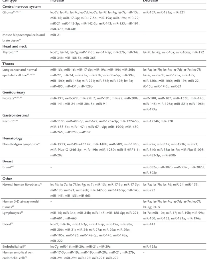
MicroRNA changes after IR
The roles of speciic microRNAs in IR response have only recently begun to be observed. Several studies have dem-onstrated that the expression levels of some microRNAs change notably after irradiation. This fact was conirmed in different cell types and at variable irradiation doses (X-ray or gamma (X-ray). Table 1 summarizes the levels of mi-croRNA expression after IR in some cell lines.
MicroRNAs can be directly involved in apoptosis regulation and cell cycle. It was shown that let-7 cluster of microRNA is associated with cellular differentiation and proliferation and likewise acts as a tumor
suppres-sor.44 Kras is one of the recognized targets of the let-7
family;45 RAS ampliied seems to protect tissues from
IR and sponsor growth,46 demonstrating that there may
be a positive relationship between the radiation response and 7. As it can be observed in Table 1, IR changed let-7 cluster expression in different cell lines: they are down-regulated or updown-regulated, showing discrepancies among the results. Additional studies are required to establish the main reason for the divergence between current re-porting papers and whether other mechanisms are in-volved.
Simone et al.47 have studied the alterations in
microR-NA expression in primary human ibroblasts following exposure to several stress-inducing anticancer agents
TABLE 1 MicroRNAs levels of expression after IR.
Cell type Increase Decrease
Central nervous system
Glioma31,32,33 let-7a; let-7b; let-7c; let-7d; let-7e; let-7f; let-7g; let-7i; miR-15a;
miR-16; miR-17-3p; miR-17-5p; miR-19a; miR-19b; miR-22; miR-21; miR-142-3p; miR-142-5p; miR-143; miR-155; miR-191; miR-379; miR-601
miR-107; miR-181a; miR-521
Mouse hippocampal cells and brain tissue34
miR-21 –
Head and neck
Thyroid35,36 let-7c; let-7d; let-7g; miR-17-3p; miR-17-5p; miR-27b; miR-34a;
miR-34b; miR-188-5p; miR-365
let-7f; let-7g; miR-10a; miR-106a; miR-152
Thorax
Lung cancer and normal epithelial cell line37,38,39
miR-15a; miR-16; miR-17-5p; miR-19a; miR-19b; miR-20b; miR-22; miR-24; miR-27a; miR-27b; miR-30a-5p; miR-99a; miR-106a; miR-148a; miR-221; miR-365; miR-126; let-7a; miR-495; miR-451; miR-128b
let-7a; let-7b; let-7c; let-7d; let-7e; let-7f; let-7i; miR-26b; miR-125a; miR-155; miR 130a, miR-106b, miR-19b, miR-22, iR-15b, miR-17-5p; miR-21
Genitourinary
Prostate40,41,42 miR-191; miR-379; miR-29b 7; miR-191; miR-22; miR-200c;
miR-141; miR-24 ; miR-30a-5p; miR-9-1
miR-100; miR-107; miR-133b; miR-143; miR-145; miR-196a; miR-521; miR-106b; miR-199a
Gastrointestinal
Rectum43,44 miR-1183; miR-483-5p; miR-622; miR-125a-3p; miR-1224-5p;
miR-188-5p; miR-1471; miR-671-5p; miR-1909; miR-630; miR-765; miR125b; miR137
miR-1274b; miR-720
Hematology
Non-Hodgkin lymphoma45 miR-1913; miR-Plus-F1147; miR-148b; miR-589; miR-106b;
miR-Plus-G1246-3p; miR-19b; miR-1280; miR-BHRF1-1; miR-20a
miR-29a; miR-335; miR-193b; miR-21; miR-340; miR-33a; let-7e; miR-Plus-E1098; miR-483-3p; miR-200b
Breast
Breast46 – miR-302a; miR-302b; miR-302c; miR-302d;
miR-302e
Other
Normal human fibroblasts47 let-7d; let-7e; let-7f; let-7g; let-7i; miR-15a; miR-17-3p; miR-17-5p;
miR-19b; miR-21; miR-26b; miR-142-3p; miR-142-5p; miR-143; miR-145; miR-155; miR-663
let-7a; let-7b; let-7d; miR-24; miR-155; miR-222
Human 3-D airway model tissues48
– let-7a; let-7b; let-7c; let-7d; let-7e; let-7f; let-7g; let-7i
Lymphocytes49 miR-16; miR-34a; miR-34b; miR-145; miR-188-5p; miR-221;
miR-601; miR-663
let-7e; miR-10a; miR-17; miR-19b; miR-99a; miR-100; miR-152; miR-181a; miR-196a Blood50 let-7f; miR-16; miR-17-3p; miR-17-5p; miR-19a; miR-20a;
miR-20b; miR-21; miR-24; miR-27a; miR-29a; miR-29c; miR-106a; miR-126; miR-142-5p; miR-145; miR-148a; miR-222
miR-143
Endothelial cell51 let-7g; miR-16; miR-20a; miR-21; miR-29c miR-125a
Human umbilical vein endothelial cells52
miR-17-5p; miR-19a; miR-19b; miR-20a; miR-21; miR-27b; miR-29a; miR-29c; miR-126; miR-221; miR-222
in non-small cell lung cancer cell lines, have found that Drosha and Dicer were expressed at higher levels in ra-dioresistance but not in sensitive cell lines. The downreg-ulation of either Dicer or Drosha had no effect on the sensitivity of cells to irradiation. In other words, knock-down of these proteins separately did not alter the num-ber of apoptotic cells in non-small cell lung cancer cell lines. This disparity might probably be associated with cell types, the degree of knockdown by small interfering RNAs, or the doses of IR used, which were different in the two studies.
In addition, Francia et al. have shown that reduction of Drosha or Dicer leads to decline in the number of cells
with foci related with DNA damage response (DDR) at
the injury site, but that reduction did not depend on mi-croRNAs (the process is related to damage-site-speciic sequence of RNAs managed by Dicer and Drosha).
MicroRNA has to do with modulating the expression of several post-transcriptional targets in the DDR path-way.54,55 Lal et al.56 have found that microRNA-24 is
up-regulated through post-mitotic differentiation of hema-topoietic cell lines and controls the histone variant H2AX, a protein that has a role in the DSBs response. MicroR-NA-24 downregulates the expression of H2AX and re-presses DNA repair. Thus, there is reduced cell capabili-ty to repair DNA DSBs. When DNA DSBs appear, microRNA-24 decreases its genomic stability and DNA damage repair capacity by regulating H2AX expression. Likewise, after DNA injury, microRNA-24-mediated down-regulation of H2AX increases cell death. DNA DSBs re-pair was promoted by suppressing microRNA-24
expres-sion and decreased cell sensitivity to DDR.57 Hu et al.58
have demonstrated that microRNA-421 controls the atax-ia-telangiectasia mutated (ATM) gene. ATM is a protein that plays a central role in the maintenance of genomic integrity by activating cell cycle checkpoints and support-ing repair of DNA DSBs. MicroRNA-421 suppresses ATM expression; ectopic expression of microRNA-421 result-ed in S-phase cell cycle checkpoint changes and an in-creased sensitivity to IR. Equally, the N-myc oncogene acts as a transcription factor on the microRNA-421 pro-moter region to upregulate microRNA-421 expression. As a result, inding a linear signaling pathway (microR-NA-421/N-Myc/ATM) could contribute to determine a role in regulating DNA synthesis in cell cycle and in pro-moting tumor radiosensitivity.
Other microRNAs were also related to regulatory mechanism DNA damage repair or radiosensitivity. Mi-croRNA-101 was associated with DNA-dependent pro-tein kinase catalytic subunit (DNA-PKcs) and ATM to
sensitize neoplasms to radioation;59,60 microRNA-210 and
miR-373 are overexpressed in hypoxic cells and regulate the expression of various aspects in DNA damage repair
pathways;61,62 microRNA-125b, microRNA-504, and
mi-croRNA-33 were correlated to p53 (one major factor in
DNA-damage checkpoint activation);63,64 microRNA-421
downregulation of ATM leading to clinical manifest tu-mor radiosensitivity in head and neck squamous cell
car-cinoma;65 microRNA-7 increases radiosensitivity of
hu-man tumor cells with activated epidermal growth factor
receptor (EGFR) associated signaling;66 microRNA-221
and microRNA-222 control radiation sensitivity by
con-trolling the PTEN/Akt pathway;67 microRNAs (-155, -20a,
-25, and -15a) are involved in the regulation of IR-induced
premature senescence;68 lin28-let7 modulates
radiosen-sitivity of human cancer cells with activation of K-Ras.69
In conclusion, it is not very clear if the global mech-anism of microRNA disturbs sensitivity to radiation, but it is evidence that cellular radiosensitivity could indeed be inluenced by microRNAs. The regulatory mechanisms of microRNA in the DNA injury repair process, if com-pletely clariied, could offer new understanding regard-ing cancer radiosensitivity.
C
ONCLUSIONThe observation that non-coding regions of the genome play an important role in regulating molecules that might control essential cellular functions is one of the most sig-niicant innovations in current oncology. This inding has supported insight into the mechanisms of cancer de-velopment, recognized possible biomarkers, and created possibilities for new therapies. The effort of understand-ing microRNA biology and convertunderstand-ing those indunderstand-ings to clinical practice is just beginning. There have been sever-al reports recording the sever-alteration in microRNA levels upon IR from diverse cell types and the particular role of many microRNAs on cell radiosensitivity. There is evi-dence that microRNAs are important players in the com-pound response to radiation. Hence, continued research on all fronts might be of equal importance to the even-tual clinical purpose of microRNA as a new class of ra-diosensitizing agents.
R
ESUMOMicroRNAs, câncer e radiação ionizante: em que ponto estamos?
do microRNA com a radiação ionizante (RI), com enfo-que na radiorresponsividade. Observou-se enfo-que a biogêne-se do microRNA está bem estabelecida e envolve diversas clivagens enzimáticas que resultam na produção do mi-croRNA maduro. Os mimi-croRNAs estão envolvidos na car-cinogênese. Sua interação está relacionada às alterações genéticas e epigenéticas, associadas à ativação de proto--oncogenes ou à inativação de genes supressores de tumor.
Vários estudos demonstraram que os níveis de expressão de alguns microRNAs variam signiicativamente após a ir-radiação. Há evidências de que os microRNAs podem in-luenciar a resposta celular após a RI. Além disso, os mi-croRNAs estão relacionados à modulação da expressão de vários alvos de pós-transcrição das vias de resposta aos danos no DNA e o do mecanismo de regulação de repa-ração de danos do DNA. Estudos futuros podem eluci-dar uma possível utilização clínica dos microRNAs como uma nova classe de agentes radiossensíveis.
Palavras-chave: microRNAs, neoplasias, radioterapia de alta energia.
R
EFERENCES1. Lewis BP, Burge CB, Bartel DP. Conserved seed pairing, often lanked by adenosines, indicates that thousands of human genes are microRNA targets. Cell. 2005; 120(1):15-20.
2. Calin GA, Sevignani C, Dumitru CD, Hyslop T, Noch E, Yendamuri S, et al. Human microRNA genes are frequently located at fragile sites and genomic regions involved in cancers. Proc Natl Acad Sci USA. 2004; 101(9):2999-3004. 3. Fabbri M, Ivan M, Cimmino A, Negrini M, Calin GA. Regulatory mechanisms of microRNAs involvement in cancer. Expert Opin Biol Ther. 2007; 7(7):1009-19. 4. Li L, Story M, Legerski RJ. Cellular responses to ionizing radiation damage.
Int J Radiat Oncol Biol Phys. 2001; 49(4):1157-62.
5. Perron MP, Provost P. Protein interactions and complexes in human microRNA biogenesis and function. Front Biosci. 2008; 13:2537-47. 6. 6. Cullen BR. Transcription and processing of human microRNA precursors.
Mol Cell. 2004; 16(6):861-5.
7. Han J, Lee Y, Yeom KH, Kim YK, Jin H, Kim VN. The Drosha-DGCR8 complex in primary microRNA processing. Genes Dev. 2004; 18(24):3016-27. 8. Lee Y, Ahn C, Han J, Choi H, Kim J, Yim J, et al. The nuclear RNase III Drosha
initiates microRNA processing. Nature. 2003; 425(6956):415-9.
9. Chendrimada TP, Gregory RI, Kumaraswamy E, Norman J, Cooch N, Nishikura K, et al. TRBP recruits the Dicer complex to Ago2 for microRNA processing and gene silencing. Nature. 2005; 436(7051):740-4.
10. Lee Y, Hur I, Park SY, Kim YK, Suh MR, Kim VN. The role of PACT in the RNA silencing pathway. EMBO J. 2006; 25(3):522-32.
11. Kwak PB, Iwasaki S, Tomari Y. The microRNA pathway and cancer. Cancer Sci. 2010; 101(11):2309-15.
12. Kim VN. MicroRNA biogenesis: coordinated cropping and dicing. Nat Rev Mol Cell Biol. 2005; 6(5):376-85.
13. Graves P, Zeng Y. Biogenesis of mammalian microRNAs: a global view. Genomics Proteomics Bioinformatics. 2012; 10(5):239-45.
14. Sun BK, Tsao H. Small RNAs in development and disease. J Am Acad Dermatol. 2008; 59(5):725-37.
15. Zhang W, Dahlberg JE, Tam W. MicroRNAs in tumorigenesis: a primer. Am J Pathol. 2007; 171(3):728-38.
16. Fabbri M, Ivan M, Cimmino A, Negrini M, Calin GA. Regulatory mechanisms of microRNAs involvement in cancer. Expert Opin Biol Ther. 2007; 7(7):1009-19. 17. Johnson SM, Grosshans H, Shingara J, Byrom M, Jarvis R, Cheng A, et al.
RAS is regulated by the let-7 microRNA family. Cell. 2005; 120(5):635-47.
18. Garzon R, Calin GA, Croce CM. MicroRNAs in cancer. Annu Rev Med. 2009; 60:167-79.
19. Calin GA, Dumitru CD, Shimizu M, Bichi R, Zupo S, Noch E, et al. Frequent deletions and down-regulation of micro-RNA genes miR15 and miR16 at 13q14 in chronic lymphocytic leukemia. Proc Natl Acad Sci USA. 2002; 99(24):15524-9.
20. Abba M, Mudduluru G, Allgayer H. MicroRNAs in cancer: small molecules, big chances. Anticancer Agents Med Chem. 2012; 12(7):733-43.
21. Lee ST, Chu K, Oh HJ, Im WS, Lim JY, Kim SK, et al. Let-7 microRNA inhibits the proliferation of human glioblastoma cells. J Neurooncol. 2011; 102(1):19-24. 22. Dong Q, Meng P, Wang T, Qin W, Qin W, Wang F, et al. MicroRNA let-7a inhibits proliferation of human prostate cancer cells in vitro and in vivo by targeting E2F2 and CCND2. PLoS One. 2010; 5(4):e10147.
23. Kumar MS, Erkeland SJ, Pester RE, Chen CY, Ebert MS, Sharp PA, et al. Suppression of non-small cell lung tumor development by the let-7 microRNA family. Proc Natl Acad Sci U S A. 2008; 105(10):3903-8.
24. Chow TF, Mankaruos M, Scorilas A, Youssef Y, Girgis A, Mossad S, et al. The miR-17-92 cluster is over expressed in and has an oncogenic effect on renal cell carcinoma. J Urol. 2010; 183(2):743-51.
25. Hayashita Y, Osada H, Tatematsu Y, Yamada H, Yanagisawa K, Tomida S, et al. A polycistronic microRNA cluster, miR-17-92, is overexpressed in human lung cancers and enhances cell proliferation. Cancer Res. 2005; 65(21):9628-32. 26. Calin GA, Sevignani C, Dumitru CD, Hyslop T, Noch E, Yendamuri S, et al.
Human microRNA genes are frequently located at fragile sites and genomic regions involved in cancers. Proc Natl Acad Sci U S A. 2004; 101(9):2999-3004. 27. Zhang L, Huang J, Yang N, Greshock J, Megraw MS, Giannakakis A, et al.
MicroRNAs exhibit high frequency genomic alterations in human cancer. Proc Natl Acad Sci U S A. 2006; 103(24):9136-41.
28. Raveche ES, Salerno E, Scaglione BJ, Manohar V, Abbasi F, Lin YC, et al. Abnormal microRNA-16 locus with synteny to human 13q14 linked to CLL in NZB mice. Blood. 2007; 109(12):5079-86.
29. Toyota M, Suzuki H, Sasaki Y, Maruyama R, Imai K, Shinomura Y, et al. Epigenetic silencing of microRNA-34b/c and B-cell translocation gene 4 is associated with CpG island methylation in colorectal cancer. Cancer Res. 2008; 68(11):4123-32.
30. Scott GK, Mattie MD, Berger CE, Benz SC, Benz CC. Rapid alteration of microRNA levels by histone deacetylase inhibition. Cancer Res. 2006; 66(3):1277-81.
31. Rhodes LV, Nitschke AM, Segar HC, Martin EC, Driver JL, Elliott S, et al. The histone deacetylase inhibitor trichostatin A alters microRNA expression proiles in apoptosis-resistant breast cancer cells. Oncol Rep. 2012; 27(1):10-6. 32. Wang Y, Medvid R, Melton C, Jaenisch R, Blelloch R. DGCR8 is essential
for microRNA biogenesis and silencing of embryonic stem cell self-renewal. Nat Genet. 2007; 39(3):380-5.
33. Kumar MS, Pester RE, Chen CY, Lane K, Chin C, Lu J, et al. Dicer1 functions as a haploinsuficient tumor suppressor. Genes Dev. 2009; 23(23):2700-4. 34. Karube Y, Tanaka H, Osada H, Tomida S, Tatematsu Y, Yanagisawa K, et al.
Reduced expression of Dicer associated with poor prognosis in lung cancer patients. Cancer Sci. 2005; 96(2):111-5.
35. Grelier G, Voirin N, Ay AS, Cox DG, Chabaud S, Treilleux I, et al. Prognostic value of Dicer expression in human breast cancers and association with the mesenchymal phenotype. Br J Cancer. 2009; 101(4):673-83.
36. Lin RJ, Lin YC, Chen J, Kuo HH, Chen YY, Diccianni MB, et al. MicroRNA signature and expression of Dicer and Drosha can predict prognosis and delineate risk groups in neuroblastoma. Cancer Res. 2010; 70(20):7841-50. 37. Guo X, Liao Q, Chen P, Li X, Xiong W, Ma J, et al. The microRNA-processing enzymes: Drosha and Dicer can predict prognosis of nasopharyngeal carcinoma. J Cancer Res Clin Oncol. 2012; 138(1):49-56.
38. Martello G, Rosato A, Ferrari F, Manfrin A, Cordenonsi M, Dupont S, et al. A MicroRNA targeting dicer for metastasis control. Cell. 2010; 141(7):1195-207. 39. Piovan C, Palmieri D, Di Leva G, Braccioli L, Casalini P, Nuovo G, et al. Oncosuppressive role of p53-induced miR-205 in triple negative breast cancer. Mol Oncol. 2012; 6(4):458-72.
40. Mott JL, Kurita S, Cazanave SC, Bronk SF, Werneburg NW, Fernandez-Zapico ME. Transcriptional suppression of mir-29b-1/mir-29a promoter by c-Myc, hedgehog, and NF-kappaB. J Cell Biochem. 2010; 110(5):1155-64. 41. Burk U, Schubert J, Wellner U, Schmalhofer O, Vincan E, Spaderna S, et al.
A reciprocal repression between ZEB1 and members of the miR-200 family promotes EMT and invasion in cancer cells. EMBO Rep. 2008; 9(6):582-9. 42. Engels BM, Hutvagner G. Principles and effects of microRNA-mediated
43. Deng S, Calin GA, Croce CM, Coukos G, Zhang L. Mechanisms of microRNA deregulation in human cancer. Cell Cycle. 2008; 7(17):2643-6.
44. Bussing I, Slack FJ, Grosshans H. Let-7 microRNAs in development, stem cells and cancer. Trends Mol Med. 2008; 14(9):400-9.
45. Johnson SM, Grosshans H, Shingara J, Byrom M, Jarvis R, Cheng A, et al. RAS is regulated by the let-7 microRNA family. Cell. 2005; 120(5):635-47. 46. Dent P, Yacoub A, Fisher PB, Hagan MP, Grant S. MAPK pathways in
radiation responses. Oncogene. 2003; 22(37):5885-96.
47. Simone NL, Soule BP, Ly D, Saleh AD, Savage JE, Degraff W, et al. Ionizing radiation-induced oxidative stress alters miRNA expression. PLoS One. 2009; 4(7):e6377.
48. Tarasov V, Jung P, Verdoodt B, Lodygin D, Epanchintsev A, Menssen A, et al. Differential regulation of microRNAs by p53 revealed by massively parallel sequencing: miR-34a is a p53 target that induces apoptosis and G1-arrest. Cell Cycle. 2007; 6(13):1586-93.
49. Koturbash I, Zemp F, Kolb B, Kovalchuk O. Sex-speciic radiation-induced microRNAome responses in the hippocampus, cerebellum and frontal cortex in a mouse model. Mutat Res. 2011; 722(2):114-8.
50. Dickey JS, Zemp FJ, Martin OA, Kovalchuk O. The role of miRNA in the direct and indirect effects of ionizing radiation. Radiat Environ Biophys. 2011; 50(4):491-9.
51. Kraemer A, Anastasov N, Angermeier M, Winkler K, Atkinson MJ, Moertl S. MicroRNA-mediated processes are essential for the cellular radiation response. Radiat Res. 2011; 176(5):575-86.
52. Surova O, Akbar NS, Zhivotovsky B. Knock-down of core proteins regulating microRNA biogenesis has no effect on sensitivity of lung cancer cells to ionizing radiation. PLoS One. 2012; 7(3):e33134.
53. Francia S, Michelini F, Saxena A, Tang D, de Hoon M, Anelli V, et al. Site-speciic DICER and DROSHA RNA products control the DNA-damage response. Nature. 2012; 488(7410):231-5.
54. Landau DA, Slack FJ. MicroRNAs in mutagenesis, genomic instability, and DNA repair. Semin Oncol. 2011; 38(6):743-51.
55. Hu H, Gatti RA. MicroRNAs: new players in the DNA damage response. J Mol Cell Biol. 2011; 3(3):151-8.
56. Lal A, Pan Y, Navarro F, Dykxhoorn DM, Moreau L, Meire E, et al. miR-24-mediated downregulation of H2AX suppresses DNA repair in terminally differentiated blood cells. Nat Struct Mol Biol. 2009; 16(5):492-8. 57. Srivastava N, Manvati S, Srivastava A, Pal R, Kalaiarasan P, Chattopadhyay
S, et al. miR-24-2 controls H2AFX expression regardless of gene copy
number alteration and induces apoptosis by targeting antiapoptotic gene BCL-2: a potential for therapeutic intervention. Breast Cancer Res. 2011; 13(2):R39.
58. Hu H, Du L, Nagabayashi G, Seeger RC, Gatti RA. ATM is down-regulated by N-Myc-regulated microRNA-421. Proc Natl Acad Sci U S A. 2010; 107(4):1506-11.
59. Yan D, Ng WL, Zhang X, Wang P, Zhang Z, Mo YY, et al. Targeting DNA-PKcs and ATM with miR-101 sensitizes tumors to radiation. PLoS One. 2010; 5(7):e11397.
60. Chen S, Wang H, Ng WL, Curran WJ, Wang Y. Radiosensitizing effects of ectopic miR-101 on non-small-cell lung cancer cells depend on the endogenous miR-101 level. Int J Radiat Oncol Biol Phys. 2011; 81(5):1524-9.
61. Crosby ME, Kulshreshtha R, Ivan M, Glazer PM. MicroRNA regulation of DNA repair gene expression in hypoxic stress. Cancer Res. 2009; 69(3):1221-9. 62. Arai N, Kagawa W, Saito K, Shingu Y, Mikawa T, Kurumizaka H, et al. Vital
roles of the second DNA-binding site of Rad52 protein in yeast homologous recombination. J Biol Chem. 2011; 286(20):17607-17.
63. Hermeking H. MicroRNAs in the p53 network: micromanagement of tumour suppression. Nat Rev Cancer. 2012; 12(9):613-26.
64. Medema RH, Mac rek L. Checkpoint control and cancer. Oncogene. 2012; 31(21):2601-13.
65. Mansour WY, Bogdanova NV, Kasten-Pisula U, Rieckmann T, Köcher S, Borgmann K, et al. Aberrant overexpression of miR-421 downregulates ATM and leads to a pronounced DSB repair defect and clinical hypersensitivity in SKX squamous cell carcinoma. Radiother Oncol. 2013; 106(1):147-54.
66. Lee KM, Choi EJ, Kim IA. microRNA-7 increases radiosensitivity of human cancer cells with activated EGFR-associated signaling. Radiother Oncol. 2011; 101(1):171-6.
67. Zhang C, Kang C, Wang P, Cao Y, Lv Z, Yu S, et al. MicroRNA-221 and -222 regulate radiation sensitivity by targeting the PTEN pathway. Int J Radiat Oncol Biol Phys. 2011; 80(1):240-8.
68. Wang Y, Scheiber MN, Neumann C, Calin GA, Zhou D. MicroRNA regulation of ionizing radiation-induced premature senescence. Int J Radiat Oncol Biol Phys. 2011; 81(3):839-48.