Clinical and mammographic profile of patients with breast
cancer surgically treated
RAQUEL RODRIGUES MURADAS1*, MARIA TERESA AQUINODE CAMPOS VELHO2, ITAMARDOS SANTOS RIESGO3, ALEXANDRE DUARTE BRUM4,
RAQUEL MONTAGNER ROSSI5, JULIA MOTTECY PIOVEZAN5, MELANIA LACERDA5
1Masters student in Health Sciences by the Universidade Federal de Santa Maria (UFSM), Santa Maria, RS, Brazil. Resident Physician at Hospital Presidente Vargas, Porto Alegre, RS, Brazil 2PhD, Universidade Federal de Santa Catarina (UFSC), Florianópolis, SC, Brazil. Lecturer at the Department of Gynecology and Obstetrics, UFSM, Santa Maria, RS, Brazil
3PhD, Lecturer at the Department of Gynecology and Obstetrics, UFSM, Santa Maria, RS, Brazil
4Obstetrician-Gynecologist, Resident Physician, Gynecology and Obstetrics Program at the Hospital Universitário de Santa Maria, Santa Maria, RS, Brazil 5Medical Student at the UFSM, Santa Maria, RS, Brazil
S
UMMARYStudy conducted at Universidade Federal
de Santa Maria – University Hospital of Santa Maria, RS, Brazil
Article received: 7/6/2014
Accepted for publication: 7/14/2014
*Correspondence:
Address: Av. Roraima, s/n, Prédio 22
Camobi Santa Maria, RS
Postal code: 97105-900 [email protected]
http://dx.doi.org/10.1590/1806-9282.61.03.220
Financial support: Scientiic Initiation Scholarship and Research Support Program, PROIC-HUSM. Three
scientiic initiation scholarships were offered to the medical students who
participated in this study.
Conflict of interest: none
Objective: to analyze the epidemiological, clinical and mammographic proile
of women with breast cancer who were treated at the mastology clinic of the Uni-versity Hospital of Santa Maria and who underwent breast surgery between Jan-uary 2007 and December 2012.
Methods: this was a cross-sectional study, approved by the Ethics in Research
Com-mittee. A review of the patients’ medical records was performed. The data were then exported to a software program for statistical analysis, namely Minitab 14.1.
Results: the patients’ proile indicated that they were mostly born and raised in Santa Maria (respectively 11.1%, n=16, and 26.3%, n=68). They were about 55.6 years old (SD±12.3), white (90.2%, n=213), had already given birth and breastfed their children, were nonsmokers, but also overweight (average BMI of 27kg/m²). On physical examination of the irst medical consultation, these patients, as de-scribed in the records: had a palpable mass (81.1%, n=184) measuring over three centimeters, located in the left breast, precisely in the upper outer quadrant (41.4%, n=81). Mammography (39%, n=109) showed that this lump was classiied as BI-RADS® 5 (40%, n=81). On histopathological examination, the lump was
diag-nosed as an invasive ductal cancer (71.1%, n=191). Surgery was generally a radi-cal mastectomy (84.7%, n=236) with axillary dissection (92.5%, n=222).
Conclusion: some of the epidemiological, clinical and mammographic features
mentioned above resembled those found in the literature reviewed. However, these patients had advanced disease and underwent non conservative surgical procedures.
Keywords: women’s health, breast neoplasm’s, health proile, descriptive
epi-demiology, mastectomy.
INTRODUCTION
Currently, female breast malignancies are the second cause of cancer in Brazil, according to data from the National Cancer Institute (INCA, 2012). It is the main cause of can-cer among women. Every year, approximately 22% of the new cases of cancer in women are breast cancers.1,3,4 The
incidence and mortality statistics for breast cancer only do not exceed those of lung carcinoma, which is the most common cancer in the world.1,2
According to the World Health Organization (WHO),
in incidence rates adjusted for age in the population-based cancer registries from several continents.1,3
In the world population, the median survival after ive years of diagnosis is 61%, while in developed countries the number increases to 73%. In developing countries, medi-an survival after 5 years of diagnosis is close to 57%.1 Some
authors attribute this fact to the dificulty of early cancer detection.5 It is known that the prognosis of breast
malig-nancy is relatively good, if diagnosed and treated in time.6
years, late menopause and hormone replacement) are well established as elements linked to the development of breast cancer.7
The fact that genetic factors are also associated with increased risk of developing the disease is likewise impor-tant. Women with mutations in BRCA1 (Breast Cancer
Susceptibility Gene 1) and BRCA2 (Breast Cancer
Suscep-tibility Gene 2) have 85% chance of developing breast can-cer before 70 years of age.6,8 As for protective factors,
breast-feeding, physical activity and healthy eating to maintain proper body weight are associated with a lower risk of de-veloping this cancer.9
Primary prevention of breast cancer is not fully possi-ble due to changes in risk factors, and genetic characteris-tics that are involved in the etiology.1,3,6 So far,
mammog-raphy for women aged 50 to 69 years is recommended as an effective method for early disease detection.1,6,10
The University Hospital of Santa Maria (HUSM) is lo-cated in central Rio Grande do Sul and is a reference hos-pital in the region for oncologic surgeries, with outpatient and inpatient care in various specialties, also being a refer-ral center for other forty nine cities.
Thus, knowing the sociodemographic, clinical and mammographic proile of women seeking care in HUSM for cancer diagnosis and treatment is justiied. This re-search aimed to identify aspects of the disease and the treatment of patients with breast cancer undergoing sur-gery at HUSM.
METHODS
This work aimed to develop a prevalence study (descriptive, transversal and quantitative), made through a review of the medical records of patients submitted to surgical procedures for the treatment of breast cancer (segmentectomy, lumpec-tomy, quadrantectomy and mastectomy) at HUSM in a pe-riod of six years, from January 2007 to December 2012.
We selected records with data from women diagnosed with breast cancer treated at the HUSM Mastology Ser-vice and who underwent surgical procedures in the above period.
First, the authors asked the Medical Records and Sta-tistics Service (SAME) of HUSM to present all the records of female patients treated in the mastology outpatient clinic during the research period. 380 records were ob-tained. Of this total, the authors disregarded the medi-cal records of 39 patients who died; 31 with surgery per-formed at other services, cities or hospitals; 20 who did not undergo surgery and were treated medically; ive re-cords that were not found after three attempts; and six
who were operated (lumpectomy) due to other patholo-gies besides breast cancer.
After compliance with the exclusion criteria, 279 re-cords were reviewed and, through them, socio-demograph-ic and medsocio-demograph-ical data of the study population were obtained. Then, the information was entered into the Minitab for Windows statistics software – version 14.1 – for
descrip-tive statistical analysis.
Data collection was performed between March and July 2013 after project approval by the Research Ethics Committee of the Federal University of Santa Maria (UFSM) iled as CAAE 13291313.8.0000.5346. In addi-tion, the identity data of the women were kept coniden-tial and all other provisions recommended by Resolution 466/12, approved in December 2012 by the National Health Council, were followed.
RESULTS
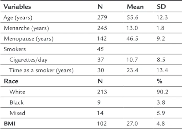
Analyzing the data gathered from the database, the aver-age aver-age of the women included in the study was 55.6 years (SD±12.3), being the youngest patient 20 years old and the oldest, 91 years old. The mean age at menarche and menopause were respectively 13 years (SD±1.8) and 46.5 years (SD±9.2) (Table 1).
TABLE 1 Description of mean values and SD for clinical profile and gynecological history of 279 patients with breast cancer who underwent surgery in a period of six years.
Variables N Mean SD
Age (years) 279 55.6 12.3
Menarche (years) 245 13.0 1.8
Menopause (years) 142 46.5 9.2
Smokers 45
Cigarettes/day 37 10.7 8.5
Time as a smoker (years) 30 23.4 13.4
Race N %
White 213 90.2
Black 9 3.8
Mixed 14 5.9
BMI 102 27.0 4.8
N: number; SD: standard deviation; BMI: body mass index.
medical records, the patients reported to the doctor that they became pregnant with their irst child on average at the age of 23 years (SD±5.6), and the duration of breastfeeding in the sum of pregnancies was, on average, 20 months (SD±12). Twenty ive patients reported us-ing contraception at the irst gynecological consultation described in their records, and only four patients used hormone therapy, representing respectively 8.9% and 1.4% (Table 1).
Also as part of the sociodemographic review, with re-spect to skin color, 90.2% (n=213) of patients said they were white, 6% (n=14) were mixed race, and 4% (n=9) Af-rican descendants. The smokers, 24% (n=67), smoked for an average of 23 years, at least 10 cigarettes/day. At diag-nosis and treatment of breast cancer, women weighed an average of 68 kilos, with a body mass index (BMI) of 27kg/ m² (Table 1).
It is known that family history of breast cancer is an important risk factor for cancer. Thus, in this study, the authors found that 37% (n=103) of patients had a irst-degree relative with breast cancer, and 9.8% (n=27) report-ed having a irst degree relative with cancer in other re-productive organ (vulvar, vaginal, cervical, endometrial or ovarian cancer).
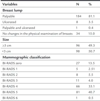
In the irst consultation, as described in the outpatient medical record, 81.1% (n=184) of patients had a palpable mass on breast examination; 3.5% (n=8) had an ulcerated lesion; and 15% (n=34) showed no abnormality on physi-cal examination. The association of nodular and ulcerat-ed lesion was present in one patient only (0.4%). The au-thors found that 41.7% (n=81) of the lumps were located in the upper outer quadrant. Of these lumps, 49.5% (n=96) were in the left breast, 47.8% (n=93) in the right breast, and only 2.7% (n=5) of the women had bilateral deformation when inspected. 50.7% (n=98) of the palpable lumps mea-sured more than three centimeters, as shown in the de-scription of the physical examinations (Table 2).
In the review of the 279 charts, more mammogram results were found, which are shown in Table 2. Of 199 mammograms described in the records, classiications according to the Breast Imaging Reporting and Data Sys-tem (BI-RADS®) and mentioned in written were: 40%
BI-RADS 5; 33% BI-BI-RADS 4; and 13% BI-BI-RADS 0 (zero). Only one patient had a BI-RADS category 6 on mammogra-phy. Out of the mammograms described as BI-RADS 4 and 5, the image of a lump was present in 79.7% (n=158), while microcalciications were reported in 18.2% (n=36) of the expert opinions, and the combination of lump and microcalciications was seen in 2.1% (n=5) of the re-sults.
TABLE 2 Numerical and percentage description of the clinical and mammographic characteristics of patients with breast cancer treated at the university hospital in six years.
Variables N %
Breast lump
Palpable 184 81.1
Ulcerated 8 3.5
Palpable and ulcerated 1 0.4
No changes in the physical examination of breasts 34 15.0
Size
≥3 cm 96 49.3
<3 cm 98 50.7
Mammographic classification
BI-RADS zero 27 13.5
BI-RADS 1 5 2.51
BI-RADS 2 8 5.5
BI-RADS 3 11 4.0
BI-RADS 4 66 33.1
BI-RADS 5 81 40.7
BI-RADS 6 1 0.5
N: number; %: percentage; cm: centimeter; BI-RADS: Breast Imaging Reporting and Data System.
In medical appointments after diagnosis of breast cancer, 97.6% (n=272) of the 279 patients underwent three addi-tional tests (bone scintigraphy, total abdominal ultra-sound and chest X-ray), totaling 652 tests for breast can-cer staging in the period assessed. Only 5.7% (n=16) of patients underwent CT scan after the three additional tests, for more information. Through these staging tests, doctors were able to diagnose probable metastases in 30.1% (n=84) of patients. Of these, 45.2% (n=38) were bone metastases, 31% (n=26), liver, 15.5% (n=13) lung, and only 8.3% (n=7) were brain metastases (Table 3).
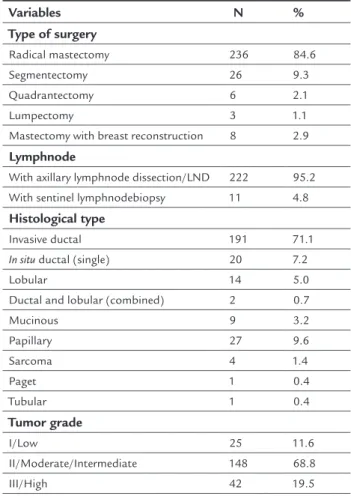
As seen in Table 3 regarding type of surgical proce-dure performed at HUSM, radical mastectomy represent-ed 84.7% (n=236) of the cases. Segmentectomy, quadran-tectomy and lumpectomy were performed in 9.3% (n=24), 2.1% (n=5) and 1.1% (n=3) of the patients, respectively. Mastectomy associated with breast reconstruction in a single surgical event was performed in 2.9% of patients (n=7). The authors also noted that 92.5% (n=222) of the procedures were axillary dissections, and only 4.8% (n=11) were sentinel lymph node dissections.
The postoperative pathological indings in the med-ical records showed that 71.1% (n=191) of tumors were invasive ductal carcinomas; 7.2% (n=20) were single duc-tal carcinomas in situ; 5% (n=14) lobular carcinomas; and
two last histological types. Other histological types, cit-ed in Table 3, were found in the pathological examina-tion of the surgical specimens, namely: mucinous in 3.2% (n=9); papillary in 9.6% (n=27); 1.4% (n=4) of sarcoma; 0.4% (n=1) cases of tubular carcinoma; and also 0.4% (n=1) cases of Paget’s disease.
TABLE 3 Numerical and percentage description of the surgical treatment of 279 patients with breast cancer, from the central region of the state of Rio Grande do Sul, Brazil.
Variables N %
Type of surgery
Radical mastectomy 236 84.6
Segmentectomy 26 9.3
Quadrantectomy 6 2.1
Lumpectomy 3 1.1
Mastectomy with breast reconstruction 8 2.9
Lymphnode
With axillary lymphnode dissection/LND 222 95.2
With sentinel lymphnodebiopsy 11 4.8
Histological type
Invasive ductal 191 71.1
In situ ductal (single) 20 7.2
Lobular 14 5.0
Ductal and lobular (combined) 2 0.7
Mucinous 9 3.2
Papillary 27 9.6
Sarcoma 4 1.4
Paget 1 0.4
Tubular 1 0.4
Tumor grade
I/Low 25 11.6
II/Moderate/Intermediate 148 68.8
III/High 42 19.5
N: number; %: percentage; LND: axillary lymph node dissection.
With respect to immunohistochemical (IHC) studies, the authors noted that 77.8% (n=182) were positive for estrogen receptors and 22.2% (n=52) were negative. IHC for proges-terone receptors revealed 61.5% (n=157) of positive results and 38.5% (n=98), negative. Markers for c-erbB-2/Her2, were negative in 63% (n=145), weak/moderately positive in 19% (n=44), and strongly positive in 17.8% (n=41).
DISCUSSION
The therapeutic approach of breast cancer involves sur-gery, chemotherapy, radiotherapy, hormone therapy, and professional and psychosocial support for the women
af-fected and their families. Usually, treatments combine two or more approaches, since individual characteristics, clinical, psychological or social in nature, are also consid-ered. The goal is to achieve better quality of life for wom-en during treatmwom-ent and survival.6,9
While analyzing some of the demographic, gyneco-logical and obstetric information of the women in this study, the authors found that the mean age (55.6 years) at the time of the irst visit to the mastology clinic – with diagnosis suggestive of breast cancer – was similar to those of other Brazilian references.1,4,11
Relatively rare before 35 years, age is an important risk factor, especially around 50 years.1 Incidence
increas-es rapidly until the age of 50, and after that, the increase is slower. This behavioral change in relation to age is known in the literature as “Clemmesens hook”, and has been attributed to menopause.12
The age of menarche was generally 13 years and meno-pause, 46. These are important aspects that, at irst, would be regarded as risk factors for breast cancer. However, in contrast, the literature indicates that the frequency is higher in women with early menarche and late meno-pause.1,3,13 The average age of menarche in girls in
south-ern Brazil is close to 12 years,14 while menopause
usual-ly occurs at 48.15
Regarding obstetric history, the authors found that most had three pregnancies and breastfed for a total pe-riod of twenty months, including all pregnancies. Preg-nancy and breastfeeding are considered protective factors against breast cancer.9 In this study, however, the authors
noted that, although no test in this respect has been con-ducted, these elements did not seem to play a protective role against the disease. It is possible that, in this study, the genetic factor had a greater effect as 37% (n=103) of the medical records showed that the patients mentioned a irst-degree relative with breast cancer. A positive fam-ily history of breast cancer is one of the main elements related to the development of the disease.7,16
These facts support the need for strong, consistent and permanent education programs for women, especially those with risk factors for the disease. Debunking myths, work-ing with the truth, reinforcwork-ing and insistwork-ing on health ed-ucation can represent a signiicant trigger to modify be-haviors leading to personal physical integrity and self-care, and to achieve goals of early diagnosis, which unfortunate-ly seem not to have occurred in the study population.
malig-nant breast tumors are related, according to the literature, with the urbanization process, showing an increased risk of disease among women with higher socioeconomic status, overweight or obese, and physically inactive.16,17 Thus, the
protective factors are opposed to these lifestyle habits and women should be encouraged and educated to promote health towards the development of better eating habits and physical exercise to maintain appropriate body weight.16,17
In the clinical evaluation, it was clear that most pa-tients already on their irst visit to the mastology outpa-tient clinic had tumors of signiicant sizes, that is, three centimeters on average. This leads to a delayed diagnos-tic reasoning considering tumor size, and impoverishes the prognosis and quality of milder treatments. The re-sults found were similar to those in the national litera-ture, indicating that the reality of our setting is not far from others, at least in Brazil.18-20
Although being considered a cancer with relatively good prognosis if diagnosed and treated in time, breast cancer mortality rates remain high in Brazil, probably be-cause the disease is still diagnosed in advanced stages.1
Worldwide, the average survival rate after ive years is 61%, while in developed countries the number reaches 73% and in developing countries, 57%.1,3,4
Clinical breast examination remains an important aid in the active detection of breast cancer.16 This examination
must be performed insistently among women of popula-tion groups considered at high risk for breast cancer (fam-ily history of breast cancer in irst-degree relatives, and oth-ers), at least annually for women aged 40 to 49 years.4,7,21
Nevertheless, a piece of information found in the research draws attention: 15% (n=34) of all women studied – with histopathological diagnosis of breast cancer –, did not have changes in the physical examination of their breasts, ac-cording to the medical records.
In Santa Maria, 39% (n=109) of patients in irst con-sultation at the mastology outpatient clinic bring with them the results of a previously performed mammogram. During the review of the medical records, 199 more of these tests were found, which was key because the authors could see that 18.2% (n=36) of mammograms classiied as BIRADS 4 or 5 showed only microcalciications and the patients had no palpable nodules. This highlights the importance of mammography as a screening radiograph-ic examination of non palpable breast tumors.
The Brazilian National Cancer Institute, INCA, esti-mated that mammography coverage in 15 cities of Bra-zil and the Federal District ranged between 37 and 76% in the year 2002-2003.9,22 As seen in this publication, the
fact that mammograms are not widely performed can
ex-plain the late diagnosis and high mortality rates still ob-served in the country.3,9 Considering that 62% of women
in Southeastern Brazil older than 60 years have never un-dergone a mammogram, it is clear that we need to pay at-tention to these women, irst through screening.23,24 As a
matter of fact, screening should also be prioritized in the region of Santa Maria.
The method necessary to continue the treatment of patients, in addition to the mandatory staging tests, is called histopathology. As expected, the histological type of breast tumor most prevalent in this study was invasive ductal 71.1% (n=191). These numbers are in agreement with the literature.17,18 After the diagnosis, surgical
tech-niques for the treatment of the disease include both con-servative and radical, with axillary lymphadenectomy.25
The surgical technique of choice is determined according to histological type and tumor size, breast size, experi-ence and preferexperi-ence of the surgeon, age and choice of the patient and also the standardized treatment protocols of the service.25
It has been mentioned that most of the patients at HUSM had a palpable mass, larger than three centime-ters and located in the left breast. Thus, most of them un-derwent radical mastectomy. Conservative surgery was predominantly used in cases with initial staging at diag-nosis, and accounted for only 12.5% (n=35) of patients. These aspects will be discussed below.
Modiied radical mastectomy is the removal of the mammary gland and axillary lymph node dissection, pre-serving both pectoral muscles.26 This procedure is
indi-cated in the following preferred conditions: presence of 3 cm or larger tumors, not attached to the muscle; in tients with recurrence after conservative treatment; pa-tients with any conditions that may render them ineligi-ble for conservative treatment or those who do not agree to preserve the breast.26 In this study, the authors found
that 84.7% (n=236) of the women who had their treat-ment in HUSM underwent this type of surgery. This sug-gests the presence of more advanced stages of the disease, and thus worse prognosis. These data are corroborated by the literature, since the percentages found are similar to those seen in Santa Maria.1,3,4
As for conservative surgery in the treatment of breast cancer, we emphasize that it can be performed in several ways. Quadrantectomy and lumpectomy are the most common techniques. They differ depending on the amount of glandular tissue removed and the safety margin asso-ciated to the tumor.27 During the study period, only 2.1%
Conservative surgery is well accepted due to the min-imization of damage caused by surgery. However, it de-pends on early diagnosis, which is linked to educational and socio-economic factors among women, and eficien-cy of health services as a whole. Effort will be needed for us to have patients in the near future with less advanced tumors treated at the HUSM mastology service.
A therapeutic measure still very much discussed is that of axillary dissection, a surgical procedure consid-ered physically limiting, especially for women who use their arms as a working tool.28 The results of the study
showed that axillary dissection was the technique used in 92.5% (n=222) of the surgical procedures, which points out that the Santa Maria service treated patients in more advanced stages of the disease.
Since it was founded, in 1970, the HUSM is one of the few hospitals located in central Rio Grande do Sul that treats exclusively patients from the Brazilian Uniied Health System, SUS. It is, therefore, a reference for clini-cal, surgical and oncological treatment of both inpatient and outpatient admissions, caring for women with breast cancer in the central part of southern Brazil.29
CONCLUSION
The negative indings of the study concerned the authors of this research. They did not expect the results to be so alarming. We know that measures have been taken to im-prove care for women with breast cancer at HUSM and the mastology service. The institution has sought, in ad-dition to providing a more ethical and effective care to patients, to comply with Law No 12.732/2012, in force
since May 2013, which establishes that patients with ma-lignant cancer are entitled to undergo treatment at SUS within up to 60 days from the date of diagnosis conir-mation through pathology report or a lesser period, de-pending on the therapeutic necessity as seen in the med-ical records.
Study limitations include the fact that any research that is developed based on data gathered from medical records – often poorly written – may fail to collect impor-tant information missing in the documents.
ACKNOWLEDGMENTS
We thank the Scientiic Initiation Scholarship and Re-search Support Program at PROIC-HUSM.
RESUMO
Peril clínico e mamográico das pacientes com câncer de mama, tratadas cirurgicamente.
Objetivo: analisar o peril epidemiológico, clínico e
ma-mográico de mulheres com câncer de mama atendidas no ambulatório de mastologia do Hospital Universitário de Santa Maria (UFSM) e submetidas à cirurgia de mama no período de janeiro de 2007 a dezembro de 2012.
Método: estudo de prevalência realizado de modo
trans-versal, após ter sido aprovado pelo Comitê de Ética em Pesquisa. Foram realizadas revisões dos prontuários das pacientes. As informações obtidas foram transpassadas para um programa de análise estatístico, o Minitab 14.1.
Resultados: o peril das pacientes, encontrado no estudo, mostrou que elas eram, na maioria, naturais ou proceden-tes de Santa Maria (respectivamente 11,1%, n=16, e 26,3%, n=68). Elas tinham 55,6 anos (DP±12,3), eram brancas (90,2%, n=213), gestaram, amamentaram, não eram taba-gistas, mas estavam com sobrepeso (IMC médio de 27 kg/ m²). No exame físico do primeiro atendimento, essas pa-cientes, conforme descrito no prontuário, tinham nódu-lo palpável (81,1%, n=184) com mais de 3 cm na mama es-querda, precisamente no quadrante lateral superior (41,4%, n=81). Na mamograia (39%, n=109), esse nódulo foi clas-siicado como BI-RADS® 5 (40%, n=81). No exame
histo-patológico, o nódulo foi diagnosticado como câncer do tipo ductal invasor (71,1%, n=191). A cirurgia foi, em ge-ral, uma mastectomia radical (84,7%, n=236) com esvazia-mento axilar (92,5%, n=222).
Conclusão: concluiu-se que algumas das características
epidemiológicas, clínicas e mamográicas citadas acima assemelharam-se com a literatura revisada. No entanto, essas pacientes apresentavam câncer de mama em estádio avançado e foram submetidas a uma técnica cirúrgica não conservadora.
Palavras-chave: saúde da mulher, neoplasias da mama,
peril de saúde, epidemiologia descritiva, mastectomia.
REFERENCES
1. INCA. Estimativa 2010: incidência de câncer no Brasil. Instituto Nacional de Câncer. - Rio de Janeiro: INCA; 2009.
2. Ferlay J, Shin HR, Bray F, Forman D, Mathers C, Parkin DM, et al. Estimates of worldwide burden of cancer in 2008: Globocan 2008. Int J Cancer. 2010;127(12):2893-917.
3. INCA. Estimativas 2008: incidência de câncer no Brasil. Brasil. Ministério da Saúde. Secretaria de Atenção à Saúde. Instituto Nacional de Câncer. Coordenação de Prevenção e Vigilância de Câncer. Rio de Janeiro: INCA; 2007. 4. INCA. Controle do câncer de mama: documento de consenso. Instituto Nacional de Câncer (INCA). Rio de Janeiro: INCA; Ministério da Saúde; 2004
5. Souza A, Andrade AN. “Corpos marcados e fé na vida...” Mastectomia e Políticas Públicas de Saúde da Mulher. Rev Psicol Política. 2008;8(5):157-78. 6. INCA. Instituto Nacional de Câncer José Alencar Gomes da Silva.
7. Silva PA da, Riul SS. Câncer de mama: fatores de risco e detecção precoce. Rev Bras Enferm (Brasília). 2011;64(6):1016-21.
8. Maluf MFM, Mori JL, Barros ACDS. Planejamento familiar em mulheres de alto risco de câncer de mama. Rev Bras Cancerol. 2008;54(4):359-65. 9. Thuler LC. Considerações sobre a prevenção do câncer de mama feminino.
Rev Bras Cancerol. 2003;49(4):227-38.
10. Bim CR, Pelloso SM, Carvalho MDB, Previdelli ITS. Diagnóstico precoce do câncer de mama e colo uterino em mulheres do município de Guarapuava, PR, Brasil. Rev Esc Enferm USP. 2010;44(4):940-6.
11. Agência Nacional de Saúde Suplementar (Brasil). Manual técnico de promoção da saúde e prevenção de riscos e doenças na saúde suplementar Agência Nacional de Saúde Suplementar. Rio de Janeiro: ANS; 2006.. 12. Hakama M. The peculiar age speciic incidence curve for cancer of the breast:
Clemmesens hook. Acta Pathol Microbiol Scand. 1969;75(3):370-4. 13. Smeltzer SC, Bare BG. Brunner & Suddarth: tratado de enfermagem
médicocirúrgica. 9ª ed. Rio de Janeiro: Guanabara Koogan; 2005. 14. Horta RL, Santos I. Idade da menarca em Pelotas: estudo piloto. Rev AMRIGS.
1991;35(2):83-7.
15. Pedro AO, Pinto Neto AM, Paiva LHSC, Osis MJ, Hard E, Idade de ocorrência da menopausa natural em mulheres brasileiras: resultados de um inquérito populacional domiciliar. Cad Saúde Pública. 2003;19(1):7-25.
16. INCA/MS, 2002. Prevenção e Controle de Câncer. Rev Bras Cancerol. 2002;48(3):317-32.
17. NCA/MS – Instituto Nacional do Câncer/Ministério da Saúde. Estimativa 2012: incidência de câncer no Brasil. Rio de Janeiro: INCA; 2012. 18. Moraes AB, Zanini RR, Turchiello MS, Riboldi J, Medeiros LR. Estudo da
sobrevida de pacientes com câncer de mama atendidas no hospital da Universidade Federal de Santa Maria, Rio Grande do Sul, Brasil. Cad Saúde Pública. 2006;22(10):2219-28.
19. Pinheiro AB, Lauter DS, Medeiros GC, Cardozo IR, Menezes LM, Souza RMB, et al. Câncer de mama em mulheres jovens: análise de 12.689 Casos. Rev Bras Cancerol. 2013;59(3):351-9.
20. Borges JBR, Soriano PM, Barros N, Souza AZ, Barros ACD, Cerri GG, et al. Avaliação por Doppler colorido do carcinoma da mama: correlação com dados clínicos e histopatológicos. Radiol Bras. 2004;37(5):323-8. 21. Brasil. Ministério da Saúde. Plano de ação para o controle dos cânceres do
colo do útero e da mama 2005-2007. Diretrizes Estratégicas Ministério Da Saúde. Brasília (DF): Ministério da Saúde; 2005.
22. Brasil. Ministério da Saúde. Secretaria de Vigilância em Saúde. Secretaria de Atenção à Saúde. Instituto Nacional de Câncer. Coordenação de Prevenção e Vigilância. Inquérito domiciliar sobre comportamentos de risco e morbidade referida de doenças e agravos não transmissíveis: Brasil, 15 capitais e Distrito Federal, 2002-2003. Rio de Janeiro: INCA; 2004.
23. Instituto Brasileiro de Geograia e Estatística (IBGE). Diretoria e Pesquisa. Departamento de População e Indicadores Sociais. População residente - Censo 2000: Brasil, Unidades da Federação e Municípios, 2000. Available at: http://www.ibge.gov.br/.
24. Departamento de Informática do SUS. Informações de saúde. Estatísticas vitais-mortalidade e nascidos vivos. Available at: http://www.datasus.gov.br. 25. Alves PC, Silva APS, Santos MCL, Fernandes AFC. Conhecimento e expectativas
de mulheres no pré-operatório da mastectomia. Rev Esc Enferm USP. 2010;44(4):989-95.
26. Franco J, Santos R, Castro K, Malfacini S, Santoro C. Tratamento cirúrgico do câncer de mama. In: Franco J. Mastologia-formação do especialista. Rio de Janeiro: Atheneu; 1997.
27. Souza GA. Cirurgia no câncer de mama: tratamento radical x tratamento conservador. Femina. 1999;27(7):587-9.
28. Gonçalves SRDOS, Arrais FMA, Fernandes AFC. As implicações da mastectomia no cotidiano de um grupo de mulheres. Rev Rene. 2007;8(2):9-17. 29. Censo Populacional 2011. Instituto Brasileiro de Geograia e Estatística