Objective: To evaluate bone mass by quantitative ultrasound of the phalanges in young karate practitioners compared to a control group.
Methods: Sample composed of 162 karate practitioners (52 females) and 326 healthy controls (110 females) aged 6 to 16 years old, in Western Paraná (Southern Brazil). Weight, height, BMI, amplitude‑dependent speed of sound (AD‑SoS) and bone transmission time (BTT) were evaluated. BMI, AD‑SoS and BTT values were converted to Z scores. Mann‑Whitney, chi‑square or Fisher Exact tests and multiple linear regression were applied, with signiicance level set at p≤0.05.
Results: Both genders showed higher values of BTT as Z scores when compared to control group. Females from the control group had higher AD‑SoS values (m/s and Z score) compared to female karate practitioners. When relative and absolute frequencies were assessed according to BTT Z score in both groups, male karate practitioners’ bone mass was shown to be adequate more frequently. In female practitioners, age and weight were independent predictors of AD‑SoS (R2=0.42) and BTT (R2=0.45), respectively. Among male karate practitioners, age was related to 26% of AD‑SoS variances and height was responsible for 36% of BTT variances.
Conclusions: Children and adolescents who practice karate were shown to have more bone mass in comparison to the control group, regardless of gender. BTT was more sensitive for this evaluation.
Keywords: Children; Adolescents; BMI; Finger phalanges; Ultrasonography; Bone density.
Objetivo: Avaliar a massa óssea pela ultrassonograia quantitativa de falanges em jovens praticantes de karatê em relação a um grupo controle.
Métodos: Amostra constituída por 162 praticantes de karatê (52 meninas) e 326 controles escolares (110 meninas) saudáveis, de 6 a 16 anos de idade, do oeste do Paraná. Foram avaliados peso, estatura, índice de massa corporal (IMC), Amplitude Dependent Speed of Sound (AD‑SoS) e Bone Time Transmission (BTT), e os valores de IMC, AD‑SoS e BTT transformados em escore Z. Aplicaram‑se testes de Mann‑Whitney, qui‑quadrado ou Exato de Fisher e regressão linear múltipla, sendo signiicante p≤0,05.
Resultados: Para ambos os sexos, os praticantes de karatê apresentaram valores superiores do escore Z do BTT comparados aos controles. Quanto à AD‑SoS, as meninas do grupo de controle apresentaram valor absoluto e de escore Z superiores aos apresentados pelas praticantes de karatê do mesmo sexo. Ao avaliar a frequência relativa e absoluta de acordo com o escore Z do BTT em ambos os grupos, os meninos praticantes de karatê apresentaram maior frequência de massa óssea adequada. Nas meninas praticantes de karatê, a idade apresentou poder de explicação de 42% na variação da AD‑SoS e o peso de 45% na variação do BTT. Nos meninos praticantes de karatê, a idade apresentou poder de explicação de 26% na variação da AD‑SoS e a estatura 36% na variação do BTT. Conclusões: Nesse grupo de crianças e adolescentes, independen‑ temente do sexo, os praticantes de karatê apresentaram maior massa óssea em relação ao grupo controle, sendo o BTT mais sen‑ sível para essa avaliação.
Palavras‑chave: Crianças; Adolescentes; IMC; Falanges dos dedos da mão; Ultrassonograia; Densidade óssea.
ABSTRACT
RESUMO
*Corresponding author. E‑mail: [email protected] (C.J.O. Barbeta).
aUniversidade Estadual de Campinas (UNICAMP), Campinas, SP, Brazil. bCenter for Investigation in Pediatrics (CIPED), Campinas, SP, Brazil. cUniversidade Federal de Santa Catarina (UFSC), Florianópolis, SC, Brazil.
BONE MASS BY QUANTITATIVE ULTRASOUND
OF FINGER PHALANGES IN YOUNG KARATE
PRACTITIONERS
Massa óssea por ultrassonografia quantitativa de falanges em
jovens praticantes de karatê
Camila Justino de Oliveira Barbeta
a,*, Ezequiel Moreira Gonçalves
b, Keila Donassolo
INTRODUCTION
he maximum bone mass reached by the young adult (peak bone mass) is strongly inluenced by the process of sexual matura-tion, as normal growth and interaction between endogenous (hereditary and endocrine features) and exogenous (nutri-tion and physical activity) factors take place.1,2 In childhood
and adolescence, bone mass increases gradually, reaching 90% of its peak, with predominance of bone formation over bone absorption,3 so it is a turning point for bone response
to physical exercise.4
Studies have shown that athletes have more bone mass compared to non-athletes, especially those who practice high-impact sports, as the occurrence of microfractures in bone tissue stimulate osteogenesis.5,6 Bone mass gain seems to
depend on the sport one plays.6-11 However, speciic physical
exercises (type, intensity, frequency and duration) required to improve it in childhood and adolescence have not been outlined yet.10
Karate6, a high-impact sport, is the most popular martial
art in the world, practiced by children, adolescents, adults, and the elderly.12 It involves basic techniques such as kicks,
punches, and blocks (ofensive and defensive) divided into two styles: Kata (imaginary ight) and Kumite (combat).13
The modality engages several muscle groups with com-plex movements and fast accelerations and decelerations.14
he short-duration attack and defense techniques are char-acterized by execution in maximum intensity with short intervals, which makes it comparable to an intermittent and intense exercise.14
Studies that have evaluated bone mass in karate practi-tioners—either by quantitative ultrasound (QUS) of phalan-ges15 or dual energy X-ray absorptiometry (DXA)—point out
the benefits of this modality for bone health.16,17 However,
they are scarce. Only one of them is known to have included males within a very large age range (from 7 to 61 years) and used QUS of phalanges by the Amplitude-Dependent Speed of Sound (AD-SoS in m/s) parameter.14 Two studies
assessed bone mass in children and adolescents who would practice martial arts (not exclusively karate) using DXA.15,16
Thus, the purpose of this study was to evaluate bone mass of children and adolescents practicing karate by QUS of phalanges based on AD-SoS and Bone Transmission Time (BTT, in μs).
METHOD
his is a cross-sectional case-control study with children and adolescents aging 6 to 16 years of both genders. he case group (karate practitioners) was composed of students enrolled in
Karate in 2014 at selected gym centers of all seven cities of western Paraná (Cascavel, Capanema, Matelândia, Medianeira, São Miguel do Iguaçu, Palotina, and Toledo), totaling 258 (98 females and 160 males), of which 162 (63%) (52 females and 110 males) participated in the study. he control group was assembled from a database of our laboratory with healthy stu-dents enrolled in municipal schools of Cascavel (Paraná), all being evaluated by the same method17 and paired in the
pro-portion of two controls for each case, according to gender, age, weight, height, and body mass index (BMI), with total of 326 participants (110 females and 216 males). Inclusion cri-teria for selection of karate practitioners was: being properly enrolled in the karate program at gym centers of the western region of Paraná, aging between 6 and 16 years, being healthy, not using continuous medication, and having the informed consent form signed by their parents or guardians (absence of signature was the reason 37% of karate practitioners did not participate in the study). Control group selection included the following criteria: attending school in the city of Cascavel (western Paraná), aging between 6 and 16 years, presenting complete data in the research group database, being healthy, not using continuous medication, and having the informed consent form signed by their parents or guardians.
he informed consent was granted by the gym center man-agement, parents and/or caregivers of karate practitioners, school principals, parents and/or caregivers of subjects in con-trol groups. his research was approved by the Research Ethics Committees of Universidade Federal de Santa Catarina (con-trol group: 131/2006) and Faculdade Assis Gurgacz (karate practitioners: 191/2013, control group: 220/2008). Data were collected at the gym center or at the schools.
he weight (kg) was evaluated with a digital scale of the brand Tanita® graduated with 100 g; height (cm), was mea-sured with a Seca® wall-mounted stadiometer graduated with 1 mm. From these data, BMI (kg/m2) was calculated. he BMI
values were transformed in Z score and sorted according to the International Obesity Task Force (IOTF):18
normal+over-weight (female <2.19; male <2.29) and obese (female ≥2.19; male ≥2.29). his BMI category grouping (normal+overweight group and obese group) was based on the physiological point of view that overweight does not exert positive inluence on one’s bone mass, approaching normal threshold, but obesity does impact bone mass both positively (weight) and negatively (inlammatory processes).19
inger. Parameters evaluated were AD-SoS (m/s) and BTT (μs). AD-SoS is the measure of range between the irst signal transmit-ted and the last one received, inluencing soft tissue; BTT relects bone properties regardless of efects on soft tissue, thus being considered more accurate for this evaluation. he absolute val-ues of AD-SoS and BTT measures were converted to Z scores, as in the reference study by Barkmann et al.20 Based on these
results, all subjects were sorted into two groups: below expecta-tion (Z scores ≤-2.0) or adequate (Z scores >-1.99). Upon QUS evaluation, intra- and inter-observer variation coeicients in our research group were 0.6 and 1.5%, respectively.17
he Shapiro-Wilk test was used to verify data normality. As they did not present a normal distribution, the variables were shown as median, minimum and maximum values. Mann-Whitney test was used to compare groups according to gender, while the chi-square or Fischer’s exact tests were used to compare the frequencies of individuals with adequate bone mass and BMI. he Spearman’s test was applied to assess correlation between bone parameters, age, and anthropomet-ric measures (weight, height, BMI, BMI Z-score). After data transformation, stepwise multiple linear regression was applied to evaluate the efects of each independent variable on bone parameters. P-value ≤0.05 was adopted as signiicance level. Afterwards, sample efect was studies based on the power of each test and using the Standard Power Calculation method and the R test, with level of signiicance set at α = 0.05 for all tests. SPSS program version 20.0 was used to process statistics.
RESULTS
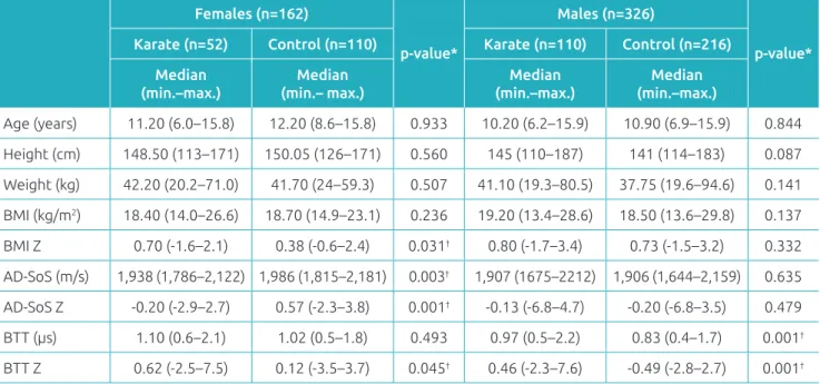
Table 1 shows overall characteristics of karate practitioners and control groups according to gender. Female karate prac-titioners had higher BMI and BTT Z scores when compared to controls; Control group, on its turn, had superior AD-SoS and AD-SoS Z scores. Male karate practitioners presented BTT and BTT Z score higher than the values presented by the con-trol group. All other variables were similar in both groups in relation to gender.
Table 2 shows the frequency of young karate practitioners and control subjects sorted by gender and according to their bone mass and BMI classiications. Only male karate sub-jects had normal bone mass more commonly, as shown by BTT Z score when compared to same-gender subjects in con-trol group (Table 2).
When it comes to correlations between bone and anthro-pometric parameters, none was found between BMI and AD-SoS of males practicing karate. All other variables had moderate to high/signiicant correlations, usually above 0.40. Exceptions were BMI and AD-SoS in females practicing karate, weight and AD-SoS in males practicing karate; BMI and BTT in males practicing karate, BMI and AD-SoS and MTB in male subjects in control group; although positive, such correlations were low (Table 3).
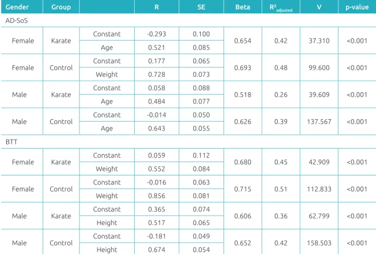
Table 4 shows results of the multiple linear regression model for AD-SoS and BTT variables of both genders. With the excep-tion of females who practiced karate, age was the variable that best
Females (n=162)
p‑value*
Males (n=326)
p‑value* Karate (n=52) Control (n=110) Karate (n=110) Control (n=216)
Median (min.–max.)
Median (min.– max.)
Median (min.–max.)
Median (min.–max.)
Age (years) 11.20 (6.0–15.8) 12.20 (8.6–15.8) 0.933 10.20 (6.2–15.9) 10.90 (6.9–15.9) 0.844
Height (cm) 148.50 (113–171) 150.05 (126–171) 0.560 145 (110–187) 141 (114–183) 0.087
Weight (kg) 42.20 (20.2–71.0) 41.70 (24–59.3) 0.507 41.10 (19.3–80.5) 37.75 (19.6–94.6) 0.141
BMI (kg/m2) 18.40 (14.0–26.6) 18.70 (14.9–23.1) 0.236 19.20 (13.4–28.6) 18.50 (13.6–29.8) 0.137
BMI Z 0.70 (‑1.6–2.1) 0.38 (‑0.6–2.4) 0.031† 0.80 (‑1.7–3.4) 0.73 (‑1.5–3.2) 0.332
AD‑SoS (m/s) 1,938 (1,786–2,122) 1,986 (1,815–2,181) 0.003† 1,907 (1675–2212) 1,906 (1,644–2,159) 0.635
AD‑SoS Z ‑0.20 (‑2.9–2.7) 0.57 (‑2.3–3.8) 0.001† ‑0.13 (‑6.8–4.7) ‑0.20 (‑6.8–3.5) 0.479
BTT (µs) 1.10 (0.6–2.1) 1.02 (0.5–1.8) 0.493 0.97 (0.5–2.2) 0.83 (0.4–1.7) 0.001†
BTT Z 0.62 (‑2.5–7.5) 0.12 (‑3.5–3.7) 0.045† 0.46 (‑2.3–7.6) ‑0.49 (‑2.8–2.7) 0.001†
Table 1 Overall features of the sample comprising karate practitioners and control subjects according to gender.
explained AD-SoS variation, with explanatory power between 26 and 42%. Similar results were obtained in BTT for both groups, and best-prediction variables were weight for females (r2 = 0.45
for karate group, r2 = 0.51 for control groups) and height for
males (r2 = 0.36 for karate group, r2 = 0.42 for control group).
DISCUSSION
In our study, young karate practitioners of both genders had more bone mass, adjusted for age and gender (BTT Z-score) and compared to the control group. Females’ weight and males’ height were the best prediction anthropometric data.
In the literature, only a study by Drozdzowska et al.14
evaluated bone mass using QUS in karate practitioners. However, it only included males aged 7 to 61 years, and the bone mass was evaluated by AD-SoS only. Results showed that the duration, the frequency, and period of beginning of activity were determining for bone mass gain, and that is why adults were shown to beneit more from karate practice. In our study, there was also an increase in bone mass with daily practice of exercises and techniques related to karate among the elderly, but such beneit was also seen in children and adolescents of both genders.
Although the results of our study indicate higher AD-SoS among karate practitioners as opposed to control group, one must note that BTT is more sensitive for bone mass evaluation when assessed by QUS in relation to AD-SoS and, for this reason, this parameter was included in the study. Such vari-ables show the ultrasound velocity in the bone, but AD-SoS is the interval measured between the irst signal transmitted and the last one received with inluence on soft tissues. BTT, on its turn, shows bone properties regardless of the confound-ing efect of soft tissue.20 he diference between AD-SoS and
BTT may explain the higher values of ADS-SoS (absolute value and Z-score) found in females of the control group as opposed to karate practitioners, once subjects in karate group presented higher BMI scores—which indicates the inluence of soft tissue on the evaluation. However, the BTT Z-score was higher in the karate group compared to control group for both genders.
Karate engages several muscle groups with complex move-ments and rapid acceleration and deceleration movemove-ments by
Gender Z‑Score Karate n (%)
Control
n (%) p‑value
AD‑SoS
F <‑2.00 6 (11.5) 5 (4.5) 0.099*†
>‑1.99 46 (88.5) 105 (99.5)
M <‑2.00 19 (17.3) 27 (12.5) 0.242*‡
>‑1.99 91 (82.7) 189 (87.5)
BTT
F <‑2.00 1 (1.9) 4 (3.6) 1.000**†
>‑1.99 51 (98.1) 106 (96.4)
M <‑2.00 1 (0.9) 18 (8.3) 0.050**‡
>‑1.99 109 (99.1) 198 (91.7)
BMI
F <2.19 52 (100) 109 (99.1) 1.000**†
≥2.19 0 (0.0) 1 (0.9)
M <2.29 100 (90.9) 206 (95.4) 0.112*‡
≥2.29 10 (9.1) 10 (4.6)
Table 2 Relative and absolute frequencies with regard
to Z‑score, Amplitude‑Dependent Speed of Sound, Bone Time Transmission and body mass index values according to gender, karate practice, and control group.
AD‑SoS: Amplitude‑Dependent Speed of Sound; BTT: Bone Transmission Time; BMI: body mass index; *chi‑square; **Fischer’s test; †Sample power at 26%; ‡Sample power at 46%.
Females Males
Karate Control Karate Control
AD‑SoS (m/s) BTT (µs) AD‑SoS (m/s) BTT (µs) AD‑SoS (m/s) BTT (µs) AD‑SoS (m/s) BTT (µs)
Age 0.68** 0.69** 0.69** 0.71** 0.49** 0.60** 0.62** 0.62**
Height (cm) 0.59** 0.66** 0.72** 0.74** 0.48** 0.64** 0.61** 0.65**
Weight (kg) 0.48** 0.64** 0.71** 0.76** 0.39** 0.56** 0.56** 0.58**
BMI (kg/m2) 0.27** 0.50** 0.42** 0.51** 0.07 0.20* 0.32** 0.33**
Spermann’s test: *p<0.05; **p<0.01; BTT: Bone Transmission Time; AD‑SoS: Amplitude‑Dependent Speed of Sound; BMI: body mass index.
Table 3 Correlations of Amplitude‑Dependent Speed of Sound and Bone Transmission Time with age, height,
displacement techniques and diferent postures.14 he
short-du-ration attack and defense techniques are characterized by execu-tion in maximum intensity with short intervals, which makes the modality comparable to an intermittent and intense exer-cise.14 Allied to maximum intensity, these techniques reinforce
the indings of the present study, according to which bone mass gain and stress take place through constant executions. In 2008, Koropanovski et al. established that upper limb
tech-niques are often predominant (89.1%) compared to lower limb techniques (8.4%).21 Punching techniques are more eicient,
with greater chance of reaching the target, compared kicking techniques. his may explain the wider use of the upper limbs during karate.22 hese indings are similar to ours: the
predom-inant use of upper limbs in karate and techniques engaging them lead to a higher bone mass index.
Recently, Nasri et al.16 assessed the efects of combat sports
(judo, karate, kyokushinkai karate, boxing, and kung fu) on bone mineral density as measured by DXA in adolescents16,17
and reported greater bone mass in athletes compared to group control. In 2001, Andreolli et al.23 evaluated bone mineral
density in male young adults (judo, karate, water polo prac-titioners and non-pracprac-titioners) and described greater bone mineral density by DXA in karate and judo practitioners com-pared to practitioners of aquatic polo and non-practitioners of such sports, drawing attention to the diference in bone health pointed out regardless of evaluation method and age group. QUS rather than DXA technique was used in our study, and we also stated bone mass gain in karate practitioners compared to control group, which means that the method is suitable for bone tissue assessment with the beneit of easy applicability, handling and portability.
In 2011, Tenforde and Fredericson6 made a review of articles
on bone mineral density assessed by DXA in athletes aged 10 to 30 years and reported a higher density related to the prac-tice of high-impact sports (gymnastics, hurdles, judo, karate, volleyball and other involving jumping), as well as sports with
Gender Group R SE Beta R²adjusted V p‑value
AD‑SoS
Female Karate
Constant ‑0.293 0.100
0.654 0.42 37.310 <0.001
Age 0.521 0.085
Female Control Constant 0.177 0.065 0.693 0.48 99.600 <0.001
Weight 0.728 0.073
Male Karate Constant 0.058 0.088 0.518 0.26 39.609 <0.001
Age 0.484 0.077
Male Control Constant ‑0.014 0.050 0.626 0.39 137.567 <0.001
Age 0.643 0.055
BTT
Female Karate Constant 0.059 0.112 0.680 0.45 42.909 <0.001
Weight 0.552 0.084
Female Control
Constant ‑0.016 0.063
0.715 0.51 112.833 <0.001
Weight 0.856 0.081
Male Karate Constant 0.365 0.074 0.606 0.36 62.799 <0.001 Height 0.517 0.065
Male Control Constant ‑0.181 0.049 0.652 0.42 158.503 <0.001
Height 0.674 0.054
Table 4 Results of the multiple linear regression model for Amplitude‑Dependent Speed of Sound and Bone
Transmission Time variables among females (F) and males (M) by group, according to anthropometric variables.
frequent but not constant impact (soccer, basketball, racquet sports, aerobics, and speed skating) compared to non-impact sports (swimming, water polo, and cycling). Although QUS and DXA techniques that evaluate diferent bone mass data, there is a certain correlation between their results that has already been demonstrated in diferent studies and conirmed by Baroncelli
et al.20 herefore, the indings of the present study, in which
QUS of phalanges was used, more speciically to evaluate BTT, may be comparable to the abovementioned studies on bone mineral density and DXA in karate practitioners.6,16,17,24
Löfgren et al.4 conducted an intervention study with children
of both genders aging 7 to 9 years. hey reported that children submitted to the intervention program presented higher bone mass index upon DXA, without risk of fractures, compared to the control group, conirming that doing exercises, such as karate or other type of physical activity of impact, is funda-mental for the health of children and adolescents.
In Brazil, studies using QUS of phalange in healthy chil-dren and adolescents of both genders showed the inluence of age, weight and height on the increase of bone mass.1,25-27
Such investigations showed that increase in bone mass was inluenced by age, puberty, and height1, and that AD-SoS and
Ultrasound Bone Proile Index (UBPI) were higher according to age and puberty.25 Increase in AD-SoS was dependent on
lean and fat masses26 and the AD-SoS and UBPI were shown
to be higher according to age, puberty and height27. In 2014,
Krahenbühl et al.28 made a systematic review of articles
relat-ing to bone mass assessed by QUS in children and adolescents and found that both AD-SOS and BTT increased with age, just like anthropometric measures of weight and height; these indings are similar to those reported in studies that have been conducted with diferent ethnicities. In this sense, the relation of variables with bone parameters is explained by normal growth for pediatric age, and they can vary from person to person. As noted in our study, age and height have inluence on this.
Consequently, increase in bone mass in children and adoles-cents related to age and puberty is expected and has been well documented by Lappe et al.2 he authors evaluated bone mineral
density of 1,743 children and adolescents of both genders aged between 6 and 16 years and described the efects of weight-lift-ing exercises on bone mass durweight-lift-ing puberty. Also important to note is that puberty is comprised of physical changes, such as weight gain and increase in height, inluenced by the hormonal stimuli of each gender, predominating androgenic stimulation in males, with gain of lean mass and height, and estrogenic in females, with gain of weight and fat mass.28 his explains the
indings of or study, that is, the link between bone mass and anthropometric features, especially the BTT Z-score related to weight in girls and height in boys.
However, some of the limitations of our study are worth mentioning, non-evaluation of puberty stages and of duration and intensity of karate practice included. It is, though, a pio-neering study on bone mass assessed by QUS, with a signii-cant number of children and adolescents practicing karate in relation to a control group.
Bone mass evaluation—in karate practitioners initially when performing basic techniques and, later on, during classes and progress of bands—can help maintain the health of the practitioner and, therefore, to improve their quality of life, conirming the importance and beneits of combat sports in pediatric age. In this study, BTT was shown to be the most adequate and useful parameter to conduct evaluations with karate practitioners due to its greater sensitivity for bone tissue assessment. Considering the lack of evidence on this matter in the scientiic literature, this study may be used as example for further research on combat sports (high impact), to elucidate their beneits for bone health. Conclusion is that karate prac-titioners in this group of children and adolescents, regardless of gender, were shown to present higher bone mass index com-pared to the control group.
Funding
his study did not receive funding.
Conflict of interests
he authors declare no conlict of interests.
REFERENCES
1. Santos KD, Petroski EL, Ribeiro RR, Guerra Junior G. Bone quantity and quality in Brazilian female schoolchildren and adolescents. J Bone Miner Metab. 2009;27:507‑12. 2. Lappe JM, Watson P, Gilsanz V, Hangartner T, Kalkwarf HJ,
Oberield S, et al. The longitudinal efects of physical activity and dietary calcium on bone mass accrual across stages of pubertal development. J Bone Miner Res. 2015;30:156‑64. 3. Mora S, Gilsanz V. Establishment of peak bone mass.
Endocrinol Metab Clin North Am. 2003;32:39‑63.
4. Löfgren B, Dencker M, Nilsson JÅ, Karlsson MK. A 4 year exercise program in children increases bone mass without increasing fracture risk. Pediatrics. 2012;129:e1468‑76. 5. Greene DA, Naughton GA. Adaptive skeletal responses
to mechanical loading during adolescence. Sports Med. 2006;36:723‑32.
7. Gruodytė R, Jürimäe J, Cicchella A, Stefanelli C, Passariello C, Jürimäe T. Adipocytokines and bone mineral density in adolescent female athletes. Acta Paediatr. 2010;99:1879‑84. 8. Ito IH, Mantovani AM, Agostinete RR, Costa Junior P, Zanuto
EF, Christofaro DG, et al. Practice of martial arts and bone mineral density in adolescents of both sexes. Rev Paul Pediatr. 2016;34:210‑5.
9. Kohrt WM, Bloomield SA, Little KD, Nelson ME, Yingling VR. American College of Sports Medicine Position Stand: physical activity and bone health. Med Sci Sports Exerc. 2004;36:1985‑96.
10. Gracia Marco L, Moreno LA, Ortega FB, León F, Sioen I, Kafatos A, et al. Levels of physical activity that predict optimal bone mass in adolescents: the HELENA study. Am J Prev Med. 2011;40:599‑607.
11. Koropanovski N, Berjan B, Bozic PR, Pazin N, Sanader A, Jovanovic S, et al. Anthropometric and physical performance proiles of elite karate kumite and kata competitors. J Hum Kinet. 2011;30:107‑14.
12. Imamura H, Yoshimura Y, Uchida K, Nishimura S, Nakazawa AT. Maximal oxygen uptake, body composition and strength of highly competitive and novice karate practitioners. Appl Human Sci. 1998;17:215‑8.
13. Milanez VF, Dantas JL, Christofaro DG, Fernandes RA. [Heart rate response during a karate training session]. Rev Bras Med Esporte. 2012;18:42‑5.
14. Drozdzowska B, Münzer U, Adamczyk P, Pluskiewicz W. Skeletal status assessed by quantitative ultrasound at the hand phalanges in karate training males. Ultrasound Med Biol. 2011;37:214‑9.
15. Nasri R, Hassen Zrour S, Rebai H, Najjar MF, Nefeti F, Bergaoui N, et al. Grip strength is a predictor of bone mineral density among adolescent combat sport athletes. J Clin Densitom. 2013;16:92‑7.
16. Nasri R, Zrour SH, Rebai H, Nefeti F, Najjar MF, Bergaoui N, et al. Combat sports practice favors bone mineral density among adolescent male athletes. J Clin Densitom. 2015;18:54‑9.
17. Gonçalves EM, Ribeiro RR, Carvalho WR, Moraes AM, Roman EP, Santos KD, et al. Brazilian pediatric reference data for quantitative ultrasound of phalanges according to gender, age, height and weight. PLoS One. 2015;10:e0127294.
18. Cole TJ, Lobstein T. Extended international (IOTF) body mass index cut ofs for thinness, overweight and obesity. Pediatr Obes. 2012;7:284‑94.
19. Barkmann R, Rohrschneider W, Vierling M, Tröger J, de TF, Cadossi R, et al. German pediatric reference data for quantitative transverse transmission ultrasound of inger phalanges. Osteoporos Int. 2002;13:55‑61.
20. Baroncelli GI. Quantitative ultrasound methods to assess bone mineral status in children: technical characteristics, performance, and clinical application. Pediatr Res. 2008;63:220‑8.
21. Koropanovski N, Dopsaj M, Jovanovic S. Characteristics of pointing actions of top male competitors in karate at world and European level. Braz J Biomotricity. 2008;2:241‑51. 22. Chaabène H, Franchini E, Miarka B, Selmi MA, Mkaouer B,
Chamari K. Time motion analysis and physiological responses to karate oicial combat sessions: is there a diference between winners and defeated karatekas? Int J Sports Physiol Perform. 2014;9:302‑8.
23. Andreoli A, Monteleone M, Van Loan M, Promenzio L, Tarantino U, Lorenzo A. Efects of diferent sports on bone density and muscle mass in highly trained athletes. Med Sci Sports Exerc. 2001;33:507‑11.
24. Ribeiro RR, Guerra Junior G, Azevedo Barros Filho A. Bone mass in schoolchildren in Brazil: the efect of racial miscegenation, pubertal stage, and socioeconomic diferences. J Bone Miner Metab. 2009;27:494‑501. 25. Carvalho WR, Gonçalves EM, Ríbeiro RR, Farias ES, Carvalho
SS, Guerra Júnior G. Inluence of body composition on bone mass in children and adolescents. Rev Assoc Med Bras. 2011;57:662‑7.
26. Moraes AM, Gonçalves EM, Barbeta VJ, Guerra Júnior G. Cross sectional study of the association of body composition and physical itness with bone status in children and adolescents from 11 to 16 years old. BMC Pediatr. 2013;13:117. 27. Krahenbühl T, Gonçalves EM, Costa ET, Barros Filho A. Factors
that inluence bone mass of healthy children and adolescents measured by quantitative ultrasound at the hand phalanges: a systematic review. Rev Paul Pediatr. 2014;32:266‑72. 28. Loomba Albrecht LA, Styne DM. Effect of puberty on
body composition. Curr Opin Endocrinol Diabetes Obes. 2009;16:10‑5.