Copyright © 2015 Revista Latino-Americana de Enfermagem This is an Open Access article distributed under the terms of the Creative Commons Attribution Non-Commercial License (CC BY-NC).
This license lets others distribute, remix, tweak, and build upon your work non-commercially, and although their new works must also acknowledge you and be non-commercial, they don’t have to license their derivative works on the same terms.
Corresponding Author: Carla Renata Silva Andrechuk
Universidade Estadual de Campinas. Faculdade de Enfermagem Rua Tessália Vieira de Camargo, 126
Cidade Universitária “Zeferino Vaz” CEP: 13083-887, Campinas, SP, Brasil E-mail: [email protected]
1 Paper extracted from master’s thesis “Sleep, daytime sleepiness and risk for obstructive sleep apnea in patients with acute myocardial
infarction”, presented to Faculdade de Enfermagem, Universidade Estadual de Campinas, Campinas, SP, Brazil.
2 Doctoral student, Faculdade de Enfermagem, Universidade Estadual de Campinas, Campinas, SP, Brazil. Scholarship holder from Coordenação
de Aperfeiçoamento de Pessoal de Nível Superior (CAPES), Brazil.
3 PhD, Associate Professor, Faculdade de Enfermagem, Universidade Estadual de Campinas, Campinas, SP, Brazil.
High risk for obstructive sleep apnea in patients with acute myocardial
infarction
1Carla Renata Silva Andrechuk
2Maria Filomena Ceolim
3Objectives: to stratify the risk for obstructive sleep apnea in patients with acute myocardial infarction, treated at a public, tertiary, teaching hospital of the state of São Paulo, Brazil, and to identify related sociodemographic and clinical factors. Method: cross-sectional analytical study with 113 patients (mean age 59.57 years, 70.8% male). A specific questionnaire was used for the sociodemographic and clinical characterization and the Berlin Questionnaire for the stratification of the risk of obstructive sleep apnea syndrome. Results: the prevalence of high risk was 60.2% and the outcome of clinical worsening during hospitalization was more frequent among these patients. The factors related to high risk were body mass index over 30 kg/m2, arterial hypertension and waist circumference indicative of cardiovascular risk, while older age (60 years and over) constituted a protective factor. Conclusion: considering the high prevalence of obstructive sleep apnea and its relation to clinical worsening, it is suggested that nurses should monitor, in their clinical practice, people at high risk for this syndrome, guiding control measures of modifiable factors and aiming to prevent the associated complications, including worsening of cardiovascular diseases.
Descriptors: Sleep; Myocardial Infarction; Sleep Apnea Syndromes; Nursing.
Rev. Latino-Am. Enfermagem
2015 Sept.-Oct.;23(5):797-805 DOI: 10.1590/0104-1169.0511.2617
www.eerp.usp.br/rlae
Rev. Latino-Am. Enfermagem 2015 Sept.-Oct.;23(5):797-805.
Introduction
Sleep associated disorders, such as the Obstructive Sleep Apnea Syndrome (OSAS), have been recognized as cardiovascular risk factors(1-4). Obstructive Sleep Apnea Syndrome consists of multiple episodes of upper airway obstruction that occur throughout a night’s sleep. These episodes are followed by reduction in oxygen saturation and multiple awakenings at night, resulting in the chronic impairment of sleep(5). The prevalence of OSAS recently found in residents of São Paulo was 32.8%, with predicting events being the age between 60 and 80 years, obesity and male(6). International studies have shown that the prevalence of OSAS ranged from 34% to 79% in patients hospitalized with Acute Myocardial Infarction (AMI), assessed by standardized instruments and polysomnography (PSG)(7-9). Although these patients met criteria consistent with clinical suspicion of OSAS, this had not been diagnosed prior to the hospitalization(8,10). In another study, this disorder constituted an aggravating factor during the hospitalization(11).
The diagnosis of suspected OSAS is performed through the anamnesis and clinical examination, and
conirmed by PSG, which is still dificult to access with
restricted indication in Brazil, due to the complexity of its performance and its high cost(12). The clinical evaluation may therefore prove to be a useful screening tool for people at high risk of OSAS. The clinical evaluation to identify subjects at risk for OSAS should include the Body Mass Index (BMI), as obesity can aggravate this disorder(2,13), as well as the measurement of the neck circumference, which also relates to the severity of OSAS(14) and the increase of which has been associated with cardiovascular risk(15). The presence of Excessive Daytime Sleepiness (EDS)(8) should also be evaluated, as this constitutes one of the more frequent symptoms
of OSAS. There are also speciic instruments for the
screening of OSAS which identify risk factors for this disorder. These constitute useful tools that can be added to the routine health evaluation.
This study was developed considering that the sum of risk factors greatly increases the cardiovascular risk and that it is for nurses, in their work routine, to identify as many of these factors as possible to intervene early and prevent diseases. The aim of this study was to stratify the risk for OSAS in patients with AMI, attended
in a public tertiary teaching hospital of the state of São Paulo, Brazil, and to identify the sociodemographic and clinical factors related to high risk for this disorder.
Method
This was an observational, analytical and cross-sectional study, conducted from October 2013 to March 2014. The study was approved by the Research Ethics Committee of the Faculty of Medical Sciences of the State University of Campinas - UNICAMP (CAAE: 09731112.4.0000.5404). The subjects that met the selection criteria signed an Informed Consent Form (ICF), prepared in accordance with the requirements of Resolution 466/2012, of the National Health Council.
The estimated sample size was 121 patients, among whom 15% were expected to present worsening of the clinical progression (n=18). This calculation was performed for a broader study entitled “Sleep, daytime sleepiness and risk of obstructive sleep apnea in patients with acute myocardial infarction”, from which this work was taken. The methodology proposed for the sample size calculation for an unpaired t-test was used, with reference to a similar study, which compared the scores obtained from the sleep quality evaluation instrument for two subject groups, divided according to their clinical evolution (improvement or worsening)(16); in the sample calculation a signiicance level of 5% and power of 80% were also considered.
The inal study sample (n=113) corresponds to 93.4%
of the total calculated. The study included 113 patients hospitalized with a clinical diagnosis of ST segment elevation myocardial infarction (STEMI) or non-ST segment elevation myocardial infarction (NSTEMI), aged 18 years or over and hospitalization in the study locations (coronary care unit or cardiology ward of a university hospital in the state of São Paulo) within 72 hours of admission to the hospital. The exclusion criteria adopted were: previous hospitalization with discharge date less than 30 days previously and poor
clinical progression within the irst 72 hours, which
could impair the performance of the interview and the responses to the instruments.
Andrechuk CRS, Ceolim MF.
were: gender (male and female), age (up to 60 years, 60 years or over), marital status (married or stable union, single, widowed, separated), schooling (in complete years), family income (in minimum wages), current employment status (active or inactive). The clinical variables of interest were BMI calculated from reported weight and height (greater than 30 kg/m2, equal to or less than 30 kg/m2), smoking habit (non smoker, smoker or ex-smoker), alcohol consumption (cardiovascular risk present, when the amount of ingested ethanol exceeded 30 mg/dl daily for men and 15mg/dL for women, or not)(17), practice of physical activity (yes or no), waist circumference (cardiovascular risk present when the value waist circumference was greater than 94cm for men and 80cm for women)(18), neck circumference (NC) (cardiovascular risk present when the NC was greater than 43 cm for men and 38 cm for women)(19), hypertension (yes or no), hypercholesterolemia (yes or no), diabetes mellitus (yes or no), type (STEMI or NSTEMI) and previous AMI (yes or no).
Patients were monitored until the end of the hospitalization regarding their clinical progression, which was categorized as improved or worsened. Events considered as clinical worsening were: reinfarction,
angina, stroke or cardiovascular death. The stratiication
of the risk of OSAS was evaluated using the Berlin Questionnaire (BQ) which includes 10 items, divided into three categories: presence and frequency of snoring behavior, daytime sleepiness or fatigue, history of hypertension and/or obesity. Categories 1 and 2 are considered positive when the score is equal to or greater than 2 points. Category 3 is positive if the response to item 10 (presence of hypertension) is yes or the BMI is
greater than 30 kg/m². The result are stratiied as high
risk for OSAS when two or more categories are positive and low-risk when one or no category is positive(20).
Data were entered into an electronic spreadsheet (Excel®), transferred and analyzed using the Statistical Analysis System (SAS), version 9.2, software, with the assistance of a statistician of the institution. Descriptive and inferential statistics were used. The association between the risk categories for OSAS and the clinical progression was evaluated using Fisher’s exact test. Univariate and multivariate logistic regression analysis,
with the stratiication of risk for OSAS being the dependent
variable (low or high), was employed to identify the
sociodemographic and clinical factors related to the risk of OSAS. The stepwise variable selection method was
used and the signiicance level was 5% (p≤.05).
Results
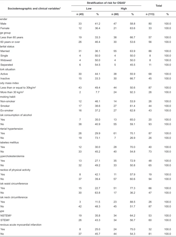
The study included 113 patients, of whom 68
(60.2%) were classiied at high risk for OSAS. Categories
1 (presence of snoring and apneas) and 3 (hypertension and/or obesity) showed higher proportions of positive responses (64.6% and 80.5%, respectively) than category 2 (daytime sleepiness, 23.9%). The patients presented a mean age of 59.7 (standard deviation 12.3) years, a mean of 5.7 (standard deviation 4.4) years of formal study and a mean family income of 3.7 (standard deviation 3.2) times the minimum wage. The mean BMI was 26.9 (standard deviation 4.6) kg/ m2, the mean waist circumference was 97.5 (standard deviation 11.4) cm for the men and 94.0 (standard deviation 12.7) cm for the women, and the mean neck circumference was 40.4 (standard deviation 3.3) cm for the men and 35.9 (standard deviation 3.5) cm for the women. Table 1 presents the sociodemographic and clinical variables distributed according to the
stratiication of risk for OSAS.
The risk for OSAS was signiicantly associated with
the worsening of the clinical progression. It was found that among the 14 patients with worsened progression,
85.7% were classiied as high risk for OSAS, while
among the 99 patients with clinical improvement a high risk for OSAS was present in 56.6% (p=.043 by Fisher’s exact test).
The factors related to high risk for OSAS, identiied
Rev. Latino-Am. Enfermagem 2015 Sept.-Oct.;23(5):797-805.
Table 1 - Distribution of sociodemographic and clinical variables according to stratiication of risk for obstructive sleep
apnea syndrome, as measured by the Berlin Questionnaire. Campinas, SP, Brazil, 2013-2014
Sociodemographic and clinical variables*
Stratification of risk for OSAS†
Total
Low High
n (45) % n (68) % n (113) %
Gender
Male 33 41.2 47 58.8 80 100.0
Female 12 36.4 21 63.6 33 100.0
Age group
Less than 60 years 19 33.3 38 66.7 57 100.0
60 years or over 26 46.4 30 53.6 56 100.0
Marital status
Married 31 36.1 55 63.9 86 100.0
Single 4 50.0 4 50.0 8 100.0
Widowed 4 50.0 4 50.0 8 100.0
Separated 6 54.5 5 45.5 11 100.0
Work situation
Active 30 44.1 38 55.9 68 100.0
Inactive 15 33.3 30 66.7 45 100.0
Body mass index
Less than or equal to 30kg/m2 43 49.4 44 50.6 87 100.0
More than 30 kg/m2 2 7.7 24 92.3 26 100.0
Smoking habit
Non-smoker 12 46.1 14 53.9 26 100.0
Smoker 17 38.6 27 61.4 44 100.0
Ex-smoker 16 37.2 27 62.8 43 100.0
Risk consumption of alcohol
Yes 7 35.0 13 65.0 20 100.0
No 38 40.9 55 59.1 93 100.0
Arterial hypertension
Yes 26 29.9 61 70.1 87 100.0
No 19 73.1 7 26.9 26 100.0
Diabetes mellitus
Yes 12 30.0 28 70.0 40 100.0
No 33 45.2 40 54.8 73 100.0
Hypercholesterolemia
Yes 13 27.1 35 72.9 48 100.0
No 32 49.2 33 50.8 65 100.0
Practice of physical activity
Yes 8 42.1 11 57.9 19 100.0
No 37 39.4 57 60.6 94 100.0
Risk waist circumference
Yes 15 22.7 51 77.3 66 100.0
No 30 63.8 17 36.2 47 100.0
Risk neck circumference
Yes 3 11.5 23 88.5 26 100.0
No 42 48.3 45 51.7 87 100.0
Type
NSTEMI‡ 19 35.8 34 64.2 53 100.0
STEMI§ 26 43.3 34 56.7 60 100.0
Previous acute myocardial infarction
Yes 8 25.0 24 75.0 32 100.0
No 37 45.7 44 54.3 81 100.0
*n=113
†Obstructive sleep apnea syndrome
Andrechuk CRS, Ceolim MF.
Table 2 - Factors related to high risk for obstructive sleep apnea syndrome in patients with acute myocardial infarction Campinas, SP, Brazil, 2013-2014
Sociodemographic and clinical variables Crude OR* IC 95%† p Adjusted OR‡ CI 95%† p
Gender
Male 1.00 (ref)
Female 1.23 0.53;2.84 .629
Age group
Less than 60 years 1.00 (ref) 1.00 (ref)
More than or equal to 60 years 0.58 0.27;1.24 .156 0.27 0.09; 0.81 .019
Marital status
Married 1.00 (ref)
Single 0.56 0.13; 2.41 .540
Widower 0.56 0.13; 2.41
Separated 0.47 0.13; 1.67
Work situation
Active - -
-Inactive - -
-Body mass index
Less than or equal to 30kg/m2 1.00 (ref) 1.00 (ref)
More than 30 kg/m2 11.73 2.61; 52.68 .001 6.92 1.19; 40.11 .031
Smoking habit
Non-smoker 1.00 (ref)
Smoker 1.36 0.51; 3.63 .748
Ex-smoker 1.45 0.54; 3.89
Risk consumption of alcohol
Yes 1.28 0.47; 3.51 .627
No 1.00 (ref)
Arterial hypertension
Yes 6.37 2.39; 16.97 .0001 16.62 4.04; 68.37 .0001
No 1.00 (ref) 1.00 (ref)
Diabetes mellitus
Yes 1.93 0.85; 4.36 .116
No 1.00 (ref)
Hypercholesterolemia
Yes 2.61 1.17; 5.82 .018
No 1.00 (ref)
Practice of physical activity
Yes 0.89 0.33; 2.43 .823
No 1.00 (ref)
Risk waist circumference
Yes 6.00 2.62; 13.73 .0001 6.00 2.08; 17.32 .001
No 1.00 (ref) 1.00 (ref)
Risk neck circumference
Yes 7.16 2.00; 25.60 .002
No 1.00 (ref)
Type
NSTEMI§ 1.37 0.64; 2.92 .417
STEMI|| 1.00 (ref)
Previous AMI¶
Yes 2.52 1.01; 6.28 .046
No 1.00 (ref)
*Crude odds ratio (odds ratio, according to the univariate logistic regression analysis) †Conidence interval
‡Adjusted odds ratio (odds ratio, according to the univariate logistic regression analysis) §Non-ST segment elevation myocardial infarction
Rev. Latino-Am. Enfermagem 2015 Sept.-Oct.;23(5):797-805.
Discussion
In this study, 60.2% of the patients presented a high risk for OSAS according to the BQ. These results highlight the importance of nurses identifying this risk in their clinical practice, mainly considering the association with worse clinical progression of the AMI,
as veriied in this study. The need is emphasized for
training of nurses for the recognition of the factors associated with the risk for OSAS, as well as for the management of screening instruments for people at high risk, their interpretation and the ways to refer cases of high suspicion for more conclusive tests. A study that aimed to determine the knowledge of nurses, to identify and assess the risk of adult OSAS, showed that after an educational program, these professionals were better prepared for this purpose(21).
Some authors have shown that approximately 35% of CVD patients have OSAS(9) and among AMI patients the prevalence is higher, 34.2% to 69%(4,7-8), data similar to those found in this study. Other authors studying the adult population, reported 3% to 7% prevalence of OSAS, with some factors such as age, male gender, obesity, family history, menopause, craniofacial abnormalities, smoking and high alcohol consumption implying increased risk(22). It is understood that the large difference could be explained due to the present study investigating a clinical population, in which all patients presented CVD, which culminated in an AMI.
It should be noted that, in the majority of studies, the occurrence of OSAS is evaluated objectively using PSG, although, there are results that highlight the reliability of the subjective measurement obtained through the BQ. In one study carried out with 158
patients hospitalized due to their irst episode of AMI,
the authors showed 54 (equivalent to 34.2%) subjects with high suspicion of OSAS, using the BQ. Of these, 53 patients were referred to PSG and the diagnosis was positive in 48, or 90.6% of those suspected(8). The association with clinical worsening observed in this study corroborates data from two other national studies, in which patients hospitalized with acute coronary syndrome and high risk for OSAS presented a worse clinical progression throughout the hospitalization compared to patients with low risk(11,23). The high risk for OSAS therefore appears to be a factor for worsening of the condition of patients hospitalized with AMI.
Four of the 15 variables examined in this study were independently associated with a high risk for OSAS: hypertension, BMI greater than 30 kg/m2, risk waist circumference, and aged 60 years or more, with the latter, unlike the others, shown as a protective factor.
The association with the irst two was expected, since the
presence of hypertension and BMI greater than 30 kg/ m2 are positive criteria in one of the three BQ categories. It should be noted that some of the variables identiied
have been highlighted in the literature as causes and others as consequences of the presence of OSAS. There are study results that contribute to support the assertion that OSAS is implicated in the occurrence of CVD, and that various risk factors are shared between the two conditions, such as older age and obesity, especially abdominal obesity(7,22,24). Conversely, hypertension has been consistently highlighted as a consequence of OSAS, and is considered one of the most important risk factors for CVD(1,22).
The subjects aged 60 years or over presented less chance of high risk for OSAS compared to those under the age of 60, which contradicts the results found in the literature(6,22,25). It can be proposed that the patients aged 60 and over were not able to accurately estimate the data relating to their own snoring and daytime sleepiness, and that their responses underestimated this information. On the other hand, it was observed that the patients of the higher age group presented BMIs
signiicantly lower in relation to the younger age group,
which may have contributed for these results.
In this study the patients with hypertension presented almost 17 times greater chance of high risk for OSAS, when compared to the patients without hypertension. Although it is not possible to identify causal relationships in a cross-sectional study, it is worth considering that the hypertension in these subjects was a consequence of the OSAS. In another study, conducted with patients affected by AMI, hypertension was found
in 92.6% of the subjects identiied at high risk for OSAS
through the BQ(8).
As observed in this study, other recent studies
conirm the relationship between anthropometric
measurements of waist circumference, neck circumference and BMI, and the high risk for OSAS or even
Andrechuk CRS, Ceolim MF.
OSAS than those who presented the measure below this value. Another study, involving 120 patients with AMI, showed a 5.7 times greater chances of presenting OSAS among the individuals with this risk factor(7). Increased waist circumference is indicative of abdominal obesity, which is also a risk factor for CVD.
This study found a signiicant relationship between
BMI greater than 30kg/m2, which indicates obesity, and high risk for OSAS. Other authors also reported the same relationship in patients with AMI, in agreement with the data presented here(4,7-8). In addition to the higher prevalence of OSAS among obese people, its severity is greater in these subjects, as demonstrated in a study, with 112 patients, which compared the characteristics of OSAS in obese and non-obese subjects. The authors found that obese patients presented a mean of 28.42 episodes per hour of sleep disruption and a mean O2 saturation of 89.68% in one night evaluated through PSG, while the non-obese subjects showed 17.84 interruptions per hour of sleep and a mean O2 saturation of 94.59%(13). The episodes of hypoxemia and sleep fragmentation that characterize OSAS, are proposed as possible mechanisms underlying the complex association between the syndrome and CVD. Such events lead to endothelial dysfunction, oxidative stress, systemic
inlammation, coagulation-ibrinolysis imbalance
and increased sympathetic activity, among other pathophysiological events, predisposing to CVD(3). These results suggest that obese people should be monitored and evaluated more frequently for the risk of OSAS.
Other factors were signiicantly related to OSAS
in the univariate analysis, however, did not remain in
the inal model: hypercholesterolemia, increased neck
circumference and previous AMI. Hypercholesterolemia was associated, in the univariate analysis, to an almost three times higher chance of risk for OSAS in the subjects studied. As well as hypertension, hypercholesterolemia is a known risk factor for CVD and may be considered a consequence of OSAS, by contributing to trigger the previously mentioned pathophysiological events
responsible for inlammation and endothelial
dysfunction(1). Authors, however, suggest the need to better clarify the interaction between dyslipidemia and OSAS(1). A seven times greater chance of high risk for OSA among individuals whose neck circumference indicated cardiovascular risk, compared to those in which this measure was less than the risk index was found in the univariate analysis. The presence of a strong
correlation between waist circumference and neck circumference, in this study, may have contributed to
the maintenance of only one of the factors in the inal
model. In another study with AMI patients there was no
statistical signiicance between risk neck circumference
and the presence of OSAS(7). Other authors, however, found an association between OSAS severity and neck circumference, regardless of visceral fat(14), and a study, with 4,201 participants aged between 20 and 85 years, showed that neck circumference was independently associated with cardiometabolic risk factors(15). These data suggest the importance of obtaining this measurement when evaluating the risk for OSAS and the risk for CVD.
The main limitations of this study should be noted. The cross-sectional design prevented the evaluation
of the inluence of the variables over time; the risk for
OSAS was assessed through self-reports, and there was no validation by objective measures, such as PSG; and the sample size was slightly lower than estimated in the sample calculation, which may have compromised the
statistical signiicance of the indings.
Conclusion
The evaluation of 113 patients during hospitalization due to AMI, allowed the conclusion that there was an increased prevalence of high risk for OSA (60.2%) in these people. The factors related to high risk for OSAS were BMI greater than 30kg/m2, arterial hypertension and waist circumference indicative of cardiovascular risk. Being aged 60 years or over proved to be a protective factor. The evolution to clinical worsening was more frequent in patients with high risk for OSAS. Various aspects contribute to alert health professionals, especially nurses, to the importance of identifying in their clinical practice patients at risk for OSAS. The prevention of future injuries can be obtained through multidisciplinary team strategies that aim to control the syndrome or its deleterious effects. It is the responsibility of the nurse to track the subjects at high risk, adopt control measures of the related factors, and monitor the results of the instituted therapeutics.
References
Rev. Latino-Am. Enfermagem 2015 Sept.-Oct.;23(5):797-805.
2. Fava C, Montagnana M, Favaloro EJ, Guidi GC, Lippi G. Obstructive sleep apnea syndrome and cardiovascular diseases. Semin Thromb Hemost. 2011;37(3):280-97. 3. Zamarrón C, Valdés CL, Alvarez-Sala R. Pathophysiologic mechanisms of cardiovascular disease in obstructive sleep apnea syndrome. Pulm Med. 2013;2013:521087. doi:10.1155/2013/521087
4. Sert Kuniyoshi FH, Singh P, Gami AS, Garcia-Touchard A, van der Walt C, Pusalavidyasagar S, et al. Patients with obstructive sleep apnea exhibit impaired endothelial function after myocardial infarction. Chest. 2011;140(1):62-7.
5. Torres-Alba F, Gemma D, Armada-Romero E, Rey-Blas JR, López-de-Sá E, López-Sendon JL. Obstructive sleep apnea and coronary artery disease: from pathophysiology to clinical implications. Pulm Med. 2013;2013:768064. doi:10.1155/2013/768064
6. Tufik S, Santos-Silva R, Taddei JA, Bittencourt LR. Obstructive sleep apnea syndrome in the Sao Paulo Epidemiologic Sleep Study. Sleep Med. 2010;11(5):441-6.
7. Ben Ahmed H, Boussaid H, Hamdi I, Boujnah MR. Prévalence et facteurs prédictifs du syndrome d’apnée obstructive du sommeil au décours de l’infarctus du myocarde. Ann Cardiol Angeiol. 2014;63(2):65-70. 8. Szymanski FM, Filipiak KJ, Hrynkiewicz-Szymanska A, Karpinski G, Opolski G. Clinical Characteristics of Patients with Acute Coronary Syndrome at High Clinical Suspicion for Obstructive Sleep Apnea Syndrome. Hellenic J Cardiol. 2013;54(5):348-54.
9. Shah N, Redline S, Yaggi HK, Wu R, Zhao CG, Ostfeld R et al. Obstructive sleep apnea and acute myocardial infarction severity: ischemic preconditioning? Sleep Breath. 2013;17(2):819-26.
10. Lee CH, Khoo SM, Tai BC, Chong EY, Lau C, Than Y, et al. Obstructive sleep apnea in patients admitted for acute myocardial infarction. Prevalence, predictors, and effect on microvascular perfusion. Chest. 2009;135(6):1488-95.
11. Correia LC, Souza AC, Garcia G, Sabino M, Brito M, Maraux M, et al. Obstructive sleep apnea affects hospital outcomes of patients with non-ST-elevation acute coronary syndromes. Sleep. 2012;35(9):1241-5.
12. Kushida CA, Littner MR, Morgenthaler TI, Alessi CA, Bailey D, Coleman J et al. Practice parameters for the indications for polysomnography and related procedures: an update for 2005. Sleep. 2005;28(4):499-521.
13. Garg R, Singh A, Prasad R, Saheer S, Jabeed P, Verma R. A comparative study on the clinical and polysomnographic pattern of obstructive sleep apnea among obese and non-obese subjects. Ann Thorac Med. 2012;7(1):26-30.
14. Kawaguchi Y, Fukumoto S, Inaba M, Koyama H, Shoji T, Shoji S, et al. Different impacts of neck circumference and visceral obesity on the severity of obstructive sleep apnea syndrome. Obesity. (Silver Spring). 2011;19(2):276-82.
15. Zhou JY, Ge H, Zhu MF, Wang LJ, Chen L, Tan YZ, et al. Neck circumference as an independent predictive contributor to cardio-metabolic syndrome. Cardiovasc Diabetol. 2013;12(1):76-82.
16. Furlani R, Ceolim MF. Sleep quality of women with gynecological and breast cancer. Rev. Latino-Am. Enfermagem. 2006;14(6):872-9.
17. Sociedade Brasileira de Cardiologia/Sociedade Brasileira de Hipertensão / Sociedade Brasileira de Nefrologia. VI Diretrizes Brasileiras de Hipertensão. Arq Bras Cardiol. 2010; 95(1 supl 1):1-51.
18. Associação Brasileira para o Estudo da Obesidade e da Síndrome Metabólica. Diretrizes brasileiras de obesidade 2009/2010 / ABESO - Associação Brasileira para o Estudo da Obesidade e da Síndrome Metabólica. - 3.ed. - Itapevi, SP : AC Farmacêutica, 2009.
19. Davidson TM, Patel MR. Waist circumference and sleep disordered breathing. Laryngoscope. 2008;118(2):339-47. 20. Vaz AP, Drummond MP, Mota C, Severo M, Almeida J, Winck JC. Tradução do Questionário de Berlim para língua Portuguesa e sua aplicação na identificação da SAOS numa consulta de patologia respiratória do sono. Rev Port Pneumol. 2011;17(2):59-65.
21. Valerio TD, Heaton K. The effects of an online educational program on nurse practitioners’ Knowledge of obstructive sleep apnea in adults. J Am Assoc Nurse Pract. 2014;26(11):603-11.
22. Punjabi NM. The epidemiology of adult obstructive sleep apnea. Proc Am Thorac Soc. 2008;5(2):136-43. 23. Jesus EV, Dias-Filho EB, Mota BM, Souza L, Marques-Santos C, Rocha JB, et al. Suspicion of obstructive sleep apnea by Berlin Questionnaire predicts events in patients with acute coronary syndrome. Arq Bras Cardiol. 2010;95(3):313-20.
Andrechuk CRS, Ceolim MF.
25. Young T, Finn L, Peppard PE, Szklo-Coxe M, Austin D, Nieto FJ, et al. Sleep disordered breathing and mortality: eighteen-year follow-up of the Wisconsin sleep cohort. Sleep. 2008;31(8):1071-8.
Received: Oct 22nd 2014