Intraoperative lactate measurements are not
predictive of death in high risk surgical patients
A medida do lactato arterial intraoperatório não é determinante
de mortalidade em pacientes cirúrgicos de alto risco
INTRODUCTION
About 234 million surgeries are performed yearly.(1) Major surgeries
postoperative complications cause morbidity and mortality.(2,3) The
mor-tality in high risk surgical patients ranges between 9.7% in the USA and 35.9% in the United Kingdom.(4)
Data confirm that unfavorable postoperative outcomes in high risk surgery are a global concern.(5-7) Even in patients who survive beyond
hospitalization, complications remain as key determinants for short sur-vival.(7) Therefore, the search for tools able to improve these patients
outcome is very relevant.
Several reports indicate that postoperative major and high risk surgery complications are strongly correlated with oxygen supply disorders, which in
João Manoel Silva Junior1, Amanda
Maria Ribas Rosa Oliveira2, Bruno
Ricciardi Silveira3, Ulisses Pinto
Ferreira3, Rodrigo Natal Albretht3,
Tiago Bertacini Gonzaga3, Ederlon
Rezende2
1. Physician of the Anesthesiology and Intensive Care Service of the Hospital do Servidor Público Estadual “Francisco Morato de Oliveira” HSPE-FMO – São Paulo (SP), Brazil.
2. Physician of the Intensive Care Service of the Hospital do Servidor Público Estadual “Francisco Morato de Oliveira” HSPE-FMO - São Paulo (SP), Brazil.
3. Resident Physicians of the Hospital do Servidor Público Estadual “Francisco Morato de Oliveira” HSPE-FMO – São Paulo (SP), Brazil.
ABSTRACT
Objectives: An increased lactate level is classically considered a marker for poorer prognosis, however little information is available on intraopera-tive lactate’s kinetics and its connection with prognosis. his study aimed to evaluate the time when perioperative lactate is most relevant for prognosis.
Methods: This was an observa-tional prospective study conducted in a tertiary hospital. Patients with requested intensive care unit postop-erative stay, aged ≥ 18 years, under-going major surgery were included. Palliative surgery patients and those with heart and/or severe liver failure were excluded. Arterial lactate levels were measured immediately before the surgery start (T0), after anesthe-sia induction (T1), 3 hours after the surgery start (T2), intensive care unit admission (T3) and 6 hours after the intensive care unit admission (T4).
Results: Sixty seven patients were included. The mean lactate values for the patients’ T0, T1, T2 and T4 were 1.5 ± 0.8 mmol/L, 1.5 ± 0.7 mmol/L, 1.8 ± 1.2 mmol/L, 2.7 ± 1.7 mmol/L and 3.1 ± 2.0 mmol/L, re-spectively. The hospital mortality rate was 25.8%, and surviving and non-surviving patients lactate values in the intensive care unit were 2.5 ± 1. and 4.8 ± 2.8 mmol/L (P < 0.0001), re-spectively. The other times measure-ments showed no statistically signifi-cant differences between the groups.
Conclusions: In surgical patients, intraoperative arterial lactate levels failed to show a predictive value; how-ever during the postoperative period, this assessment was shown to be use-ful for hospital mortality prediction.
Keywords: Intraoperative period; Intraoperative care; Perfusion; Lactates/ administration & dosage; Prognosis; Time factors
Received from the Intensive Care Service of the Hospital do Servidor Público Estadual “Francisco Morato de Oliveira” HSPE-FMO – São Paulo (SP), Brazil.
Submitted on July 5, 2010 Accepted on August 31, 2010
Author for correspondence:
João Manoel Silva Jr.
Rua Pedro de Toledo, 1800 - 6º andar – Vila Clementino
Zip Code: 04039-901 - São Paulo (SP), Brasil.
turn are related to microvascular low impairment.(8-10)
When cells oxygen availability is limited, anaerobic metabolism takes place, causing metabolic acidosis. This metabolic acidosis may be quantified by means of arterial gas contents assessment, analysis of the base difference and serum lactate concentration.(11)
Therefore, lactate is a key hypoperfusion indicator, and has been studied for several decades,(12) showing
increased serum levels to be strongly correlated with poorer patients’ prognosis.(11)
In some instances, as during sepsis, trauma, or shock, the use of serum lactate as surrogate of tissue hypoxia is established.(13-15) However, little
periopera-tive arterial lactate kinetics in high risk surgical pa-tients information is available.(16)
Therefore this study was aimed to evaluate the perioperative lactate measurement and to assess its best discriminative time for surviving versus non-sur-viving patients.
METHODS
After approved by the institution’s Ethics Commit-tee, this study was conducted in a tertiary hospital surgical center and intensive care unit (ICU).
This was a prospective study. The inclusion criteria were: ≥18 years-old patients who signed the informed consent form before the surgery, undergoing surgery with requested ICU postoperative stay and at least two of the following:
- severe cardiorespiratory comorbidities (coronary insufficiency, chronic obstructive pulmonary disease, previous stroke),
- surgery scheduled for neoplasm resection (esoph-agectomy, total gastrectomy) longer than 8 hours,
- above 70 years-old, with evidence of physiological reserve impairment involving at least one vital organ,
- acute renal failure (blood urea nitrogen > 100 mg/dL or creatinine > 3 mg/dL),
- advanced vascular disease, or aortal involvement, - massive acute intraoperative blood loss predict-ed and
- severe nutritional disorders patients.
Palliative surgery, low life expectancy, liver failure (Child B or C) and functional class IV or heart failure with less than 30% echocardiogram ejection fraction, were excluded.
The study primary endpoint was to evaluate the in-traoperative lactate levels and hospital mortality, while the secondary endpoint was to evaluate postoperative
complications such as organ dysfunction, i.e., circula-tory shock (vasoactive drugs required for more than 1 hours although volume resuscitation), worsened pul-monary oxygen exchange [oxygen partial pressure/in-spired oxygen fraction (PaO2/FiO2 < 200) ratio], renal failure (50% creatinine increase or urinary output less than 400 mL in 24 hours), mental confusion (behav-ioral change, memory loss or psychomotor agitation) and platelet dysfunction (30% platelets count drop from baseline) up to 24 hours postoperatively. Addi-tionally, infection during the ICU stay, hospital length of stay and intraoperative hyperlactatemia risk factors, such as blood transfusions and vasoactive drugs.
Later, the patients were divided into two groups, survivors and non-survivors. In addition, the compli-cations were assessed and correlated with the arterial lactate collection times.
The intraoperative therapy was conducted as deter-mined by the surgical team, while in the postoperative period, by the intensivist. All patients were followed until their discharge from the hospital.
By the inclusion were evaluated the scores Multiple-Organ Dysfunction Syndrome (MODS),(17) Acute
Physi-ology And Chronic Health Evaluation (APACHE II),(18)
and Physiologic and Operative Severity Score for the enUmeration of Mortality and Morbidity (POSSUM score),(19) using the variables’ worst ratings scores.
Addi-tionally, before surgery (T0), after anesthesia induction (T1), 3 hours from the surgery start (T2), ICU admis-sion (T4), and 6 hours post- ICU admisadmis-sion (T5) were collected arterial gasometry and lactate samples. It is im-portant to remark that in this institution the patients are transferred to the ICU immediately after the surgery, as both surgical center and ICU are located in the same area. It is thus expected that the ICU admission lactate is similar to the end-of-surgery lactate values.
Statistical analysis
he included patients’ demographic, clinical and physiological characteristics were described. For the categorical variables description, the frequencies and percentages were calculated. he quantitative variables were described by central trend and dispersion measures (mean and standard deviations, median and percentiles).
the lactate samples drawing times.
The predictive ability to discriminate survivors from non-survivors was assessed using Receiver Oper-ating Characteristic (ROC) curves.
RESULTS
Sixty seven patients were included during a 6 months period, being 37 male and 30 female patients, mean age 65.6 ± 12.2 years. Most underwent elective surgeries (Table 1).
Hospital mortality was 25.8% (17 patients). Survi-vor and non-surviSurvi-vor patients’ lactate dosages 6 hours after the ICU admission were 2.5 ± 1.3 and 4.8 ± 2.8 mmol/L, respectively (P < 0.0001). The other times measurements failed to show statistically significant inter-groups differences (baseline, P = 0.46; surgery start, P = 0.57; 3 hours of surgery, P = 0.51, ICU ad-mission, P = 0.23) (Figure 1).
ICU – intensive care unit.
Figure 1 - Perioperative lactate values comparison for survi-vor and non-survisurvi-vor patients.
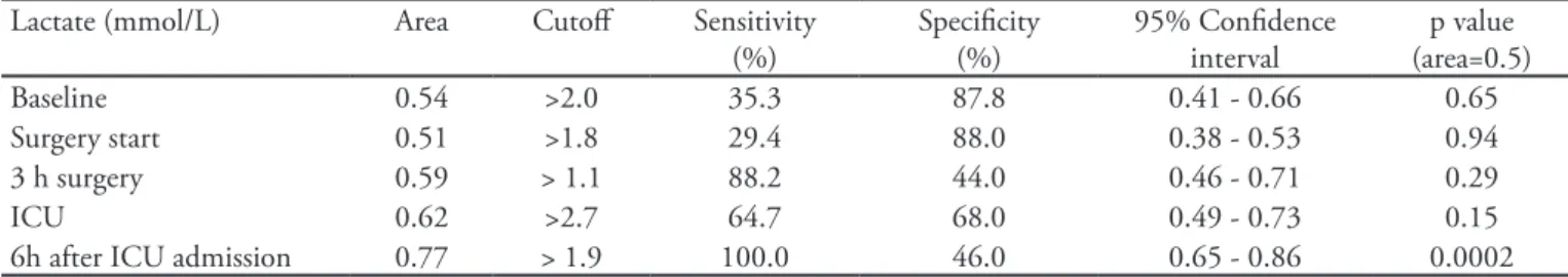
Also in the ROC curve sensitivity and specificity analysis, the intraoperative lactate values had hospital mortality discriminative power. Yet, the ICU admis-sion and 6 hours after the ICU admisadmis-sion values dis-criminative power, was better (Table 2).
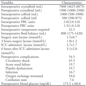
During the surgery, 50% of the patients had blood transfusion, and 32.5% were given vasoactive drugs; 54.5% of the complications were in the postoperative period, with circulatory shock predominance. The lactate values were comparatively higher in the post-operative period (Tables 3 and 4).
Table 1- Patients’ baseline characteristics
Variables Characteristics
Age (years) 65.6 ± 12.2
Male gender 56.1
APACHE II 16.9 ± 5.6
MODS 3.0 (1.0-4.0)
POSSUM 37.4 ± 7.6
Elective surgery 95.5
Surgical specialty
Gastrointestinal 78.8
Vascular 7.6
Chest 4.5
Orthopedic 3.0
Neurological 3.0 Gynecological 1.5
Urological 1.5
ASA
I 6.6
II 68.9
III 24.6
Surgery time (hours) 6.9 ± 2.4 Preoperative lactate (mmol/L) 1.5 ± 0.8 Preoperative base diference (mmol/L) -0.7 (-3.0- 0.6) Preoperative blood glucose (mg/dl) 120.0 ± 54.2 ICU length of stay (days) 3.0 (2.0 - 4.0) Hospital length of stay (days) 20.0 (12.5 – 27.5)
APACHE II - Acute Physiology And Chronic Health Evaluation II; MODS - Multiple-Organ Dysfunction Syndrome; POSSUM - Phy-siologic and Operative Severity Score for the enUmeration of Mortality and Morbidity; ASA - American Society of Anesthesiologists; ICU – intensive care unit. Results expressed as percents, mean ± standard deviation or median (25-75% percentiles).
Table 2 - ROC for arterial lactate drawing times versus hospital mortality
Lactate (mmol/L) Area Cutof Sensitivity (%)
Speciicity (%)
95% Conidence interval
p value (area=0.5)
Baseline 0.54 >2.0 35.3 87.8 0.41 - 0.66 0.65
Surgery start 0.51 >1.8 29.4 88.0 0.38 - 0.53 0.94
3 h surgery 0.59 > 1.1 88.2 44.0 0.46 - 0.71 0.29
ICU 0.62 >2.7 64.7 68.0 0.49 - 0.73 0.15
6h after ICU admission 0.77 > 1.9 100.0 46.0 0.65 - 0.86 0.0002
DISCUSSION
One of the major challenges for physicians respon-sible for severely ill patients is the tissue perfusion control. Its changes lead to several organs dysfunction, and increased mortality. Early tissue hypoxia identifi-cation, for a prompt therapy start, is fundamental to improve these patients prognosis.(20)
Currently, few parameters are available in clinical practice for tissue hypoxia assessment, such as urinary output, base difference and blood lactate, however these measures only indicate that hypoperfusion is al-ready ongoing, and may be late tools to guide hemo-dynamic resuscitation start.(21)
This study has shown that the up to 3 hours intra-operative lactate was not able to discriminate progno-sis, while the ICU admission and 6 hours from admis-sion measurements may predict unfavorable outcome.
The serum lactate level has been classically ac-cepted as anaerobic metabolism and tissue hypoxia indicator.(22,23) The blood lactate kinetics-associated
pathways are complex, but this does not void this in-dicator as an excellent surgical patients prognosis
pre-Table 4- Lactate trend versus intraoperative hyperlactatemia and outcomes risk factors
Variables Baseline Surgery start 3 hours surgery ICU 6 hours after ICU admission Intraoperative transfusion (Yes) 1.6±0.9 1.5±0.8 2.0±1.4 3.1±1.9 3.6±2.4
Intraoperative transfusion (No) 1.4±0.5 1.5±0.5 1.6±0.9 2.3±1.2 2.6±1.3
p value 0.3 0.89 0.15 0.04 0.02
Intraoperative vasopressors (Yes) 1.5±0.8 1.5±0.7 2.0±1.5 3.3±2.1 4.4±2.9 Intraoperative vasopressors (No) 1.4±0.6 1.4±0.5 1.6±1.1 2.2±1.2 2.4±1.1
p value 0.73 0.65 0.29 0.02 0.00
Postoperative complications (Yes) 1.4±0.8 1.4±0.6 1.8±1.3 3.0±1.9 3.7±2.4 Postoperative complications (No) 1.6±0.8 1.6±0.7 1.8±1.1 2.3±1.2 2.4±1.0
p value 0.25 0.33 0.87 0.09 0.01
Shock (Yes) 1.3±0.7 1.4±0.7 1.9±1.3 3.1±1.8 4.0±2.5
Shock (No) 1.7±0.8 1.5±0.7 1.8±1.1 2.4±1.5 2.4±1.1
p value 0.11 0.38 0.72 0.10 0.00
Renal failure (Yes) 1.4±0.7 1.3±0.5 1.5±0.4 3.4±2.0 4.4±3.0 Renal failure (No) 1.5±0.8 1.529±0.7 1.9±1.3 2.6±1.5 2.8±1.6
p value 0.61 0.25 0.30 0.12 0.01
Platelet dysfunction (Yes) 1.4±0.6 1.2±0.4 1.6±0.8 3.4±2.2 4.4±3.4 Platelet dysfunction (No) 1.5±0.8 1.5±0.7 1.8±1.3 2.6±1.5 2.8±1.4
p value 0.53 0.15 0.56 0.11 0.01
Infection (Yes) 1.6±1.0 1.4±0.6 1.7±0.5 3.8±2.3 5.3±3.3
Infection (No) 1.5±0.7 1.5±0.7 1.8±1.3 2.5±1.4 2.7±1.3
p value 0.74 0.81 0.81 0.02 0.00
Oxygen exchange worsened (Yes) 1.7±0.9 1.7±0.8 2.2±1.4 3.1±1.9 3.0±2.4 Oxygen exchange worsened (No) 1.5±0.5 1.5±0.5 1.8±0.9 2.7±1.2 3.1±1.3
p value 0.53 0.45 0.40 0.51 0.94
Confusion state (Yes) 1.2±0.5 1.3±0.2 1.4±0.3 3.2±1.5 3.4±1.9 Confusion state (No) 1.5±0.8 1.5±0.7 1.8±1.2 2.7±1.7 3.1±2.0
p value 0.37 0.50 0.45 0.48 0.71
he values represent mean ± standard deviation; ICU – intensive care unit.
Table 3- Patients’ characteristics during surgery and posto-peratively
Variables Characteristics
Intraoperative crystalloid (mL) 7000 (4625-8875) Postoperative crystalloid (mL) 1900 (1000-2500) Intraoperative colloid (mL) 1000 (500-1000) Postoperative colloid (ml) 500 (500-875) Intraoperative PBC units 2.0(2.0-3.0) Postoperative PBC units 1.5(1.0-2.0) Intraoperative vasopressors required 32.5 Intraoperative luid balance (mL) 800 (175-1420) Surgery start lactate (mmol/L) 1.5±0.7 3 hours surgery lactate (mmol/L) 1.8±1.2 ICU admission lactate (mmol/L) 2.7±1.7 6 hours after ICU admission lactate
(mmol/L)
3.1±2.0
Postoperative complications 54.5 Circulatory shock 45.5 Acute renal failure 19.7 Platelet dysfunction 19.7
Infection 16.7
Oxygen exchange worsened 10.6
Confusion state 7.6
Postoperative blood glucose (mg/dL) 175.5 ± 60.0
by the other hand the clearence determined the prog-nosis. In heart surgery, a postoperative 3 mmol/L value determined increased morbidity and mortality risks.(34) Almeida et al.(29) only found ICU admission
values above 3.2 mmol/L to discriminate mortality, and did not evaluate the marker trends.
Attempting to improve the method accuracy, we identified studies evaluating lactate levels develop-ment as a prognostic factor, and in one of then this was named “lactime”(35), i.e. the time when lactate
was high, and could demonstrate that lactic metabol-ic acidosis is the best multi-organ dysfunction and death predictor, and that the initial lactate measure-ment, and in another study, the early lactate clear-ance,(26) were shown to be correlated with improved
prognosis, with worsened survival in patients with delayed lactate normalization following an interven-tion.(36) A more recent study, conducted in 21
Brazil-ian ICUs, evidenced that ICU admission lactate is an early risk of death for multi-organs and systems dysfunction determinant in a high risk surgical pa-tients population.(37)
These investigations provide strong evidence to support the role of lactate as a guide for hypoperfu-sion and surgical patients’ prognostic indicator only in the postoperative time.
However, some limiting aspects should be consid-ered for this study. First, it was not evaluated if the reanimation measures for hyperlactatemia had any impact on survival; however as this study was aimed to assess the time when lactate has the best surgical patient prognosis value, this discussions on lactate-guided therapy, in addition to be controversial, would exceed the initial aim.(38)
Second, it should be noted that this study only involved a small population, and larger studies are warranted to better evaluate if these results reliabil-ity. Also, additional lactate measurements, in different times of surgery, could bring additional information; however this does not void our findings, as the times selected for lactate samples draws correspond to cru-cial times during surgery, and the intervals between the draws were short, considering a mean surgery time of 6 hours and the measurements made at the begin-ning, third hour and end of the procedure.
Finally, the time from the operation room to the ICU admission could influence the results; however, no patient had intercurrences during this time, and the transportation time was short, as the surgical cen-ter and the ICU are very close.
dictor. Normal serum lactate concentration is below 2 mmol/L at rest, and above 4 mmol/L levels indicate systemic inflammatory response syndrome (SIRS) and increased mortality, even in patients with blood pres-sure considered normal.(24)
Tissue hypoxia increases lactate levels by increased anaerobic glycolysis aimed to keep cell energy produc-tion as close as possible to normal, and this is com-mon in the shock settings.(11)
Additionally, in early shock, the increased blood lactate levels were associated with reduced oxygen of-fer, thus, tissue hypoxia and increased mortality.(13,25)
However, it should be remarked that in normal situ-ations the liver is able to increase the lactate metabo-lism, leading to a delay of some hours before lactate levels increase can be detected in hypoxemic situations and anaerobic metabolism,(26) which may, in part,
jus-tify this study findings.
During circulatory shock, lactate levels may also increase during hypoperfusion. The increased produc-tion may occur, for instance, when the glucose me-tabolism exceeds the mitochondrial oxidative capacity due to catecholamines administration, pyruvate desid-rogenase disorders, and respiratory alkalosis. Yet, in liver dysfunction, its excretion may be reduced.(27)
Therefore, during the surgical procedure, specially in major surgery, more vasopressors are used, and the liver clearance is reduced by lower oxygen consump-tion due to anesthesia.(28,29) Thus, immediate
postop-erative patients have higher serum lactate levels,(29) in
addition to that after anesthesia the restored full cell function could provide a washout phenomenon,(30)
which is reported following circulatory resuscitation. Additionally, some already inflamed organs may increase lactate levels. Studies have shown that the lung and muscle are important lactate producers,(31)
and important lactate levels increase may be seen in pulmonary injury patients. De Backer et al.(32) have
shown increased lactate production in acute pulmo-nary injury patients, with acute inflammatory process. Thus, the postoperative period is the main organs flammation time, and this may be correlate with in-creased lactate during this phase, and its mortality and complications prognostic value, as found in this sample, corroborating its lack of intraoperative prog-nostic value.
Meregalli et al.(33) have shown that a postoperative
CONCLUSION
In surgical patients, intraoperative arterial lactate failed to show prognostic value; however, when evalu-ated in the ICU and 6 hours after ICU admission, arterial lactate was useful to determine hospital mor-bidity and mortality.
RESUMO
Objetivos: Classicamente, lactato elevado é considerado como marcador de pior prognóstico, entretanto poucos dados existem a respeito da cinética do lactato no periodo intraope-ratório e sua associação com o prognóstico. O objetivo deste estudo foi avaliar em qual momento do período perioperatório o valor do lactato apresenta maior importância prognóstica.
Métodos: Estudo prospectivo observacional de um hos-pital terciário. Foram incluídos pacientes com solicitação de pós-operatório em unidade de terapia intensiva com idade ≥18 anos, submetidos a cirurgias de grande porte. Pacientes de cirurgias paliativas, com insuiciência cardíaca e/ou
hepá-tica grave foram excluídos. Valores de lactato arterial foram mensurados imediatamente antes do início da cirurgia (T0), após indução anestésica (T1), após 3hs de cirurgia (T2), na admissão da unidade de terapia intensiva (T3) e após 6 h da admissão na unidade de terapia intensiva (T4).
Resultados: Foram incluídos 67 pacientes. Os valores médios do lactato dos pacientes no T0, T1, T2, T3 e T4 fo-ram respectivamente 1,5 ± 0,8mmol/L, 1,5 ± 0,7mmol/L, 1,8 ± 1,2mmol/L, 2,7 ± 1,7mmol/L e 3,1 ± 2,0mmol/L. A taxa de mortalidade hospitalar foi 25,8% e as dosagens de lactato dos pacientes sobreviventes e dos não sobreviventes 6 h após admissão na unidade de terapia intensiva foram 2,5 ± 1,3 e 4,8 ± 2,8 mmol/L (p<0,0001), respectivamente. As medidas nos demais períodos não demonstraram diferenças estatisticamente signiicativas dentre estes grupos.
Conclusões: Em pacientes cirúrgicos o lactato arterial no período intraoperatório não apresentou valor prognós-tico, entretanto quando avaliado no pós-operatório, ele foi melhor para determinar mortalidade hospitalar.
Descritores: Período intraoperatório; Cuidados intra-operatórios; Perfusão; Lactatos/administração & dosagem; Prognóstico; Fatores de tempo
REFERENCES
1. Weiser TG, Regenbogen SE, hompson KD, Haynes AB, Lipsitz SR, Berry WR, Gawande AA. An estimation of the global volume of surgery: a modelling strategy based on available data. Lancet. 2008;372(9633):139-44.
2. Pearse RM, Harrison DA, James P, Watson D, Hinds C, Rhodes A, et al. Identiication and characterisation of the high-risk surgical population in the United Kingdom. Crit Care. 2006;10(3):R81.
3. Jhanji S, homas B, Ely A, Watson D, Hinds CJ, Pearse RM. Mortality and utilisation of critical care resources amongst high-risk surgical patients in a large NHS trust. Anaesthesia. 2008;63(7):695-700.
4. Bennett-Guerrero E, Hyam JA, Shaei S, Prytherch DR, Sutton GL, Weaver PC, et al. Comparison of P-POSSUM risk-adjusted mortality rates after surgery between patients in the USA and the UK. Br J Surg. 2003;90(12):1593-8. 5. Haynes AB, Weiser TG, Berry WR, Lipsitz SR, Breizat AH,
Dellinger EP, Herbosa T, Joseph S, Kibatala PL, Lapitan MC, Merry AF, Moorthy K, Reznick RK, Taylor B, Gawande AA; Safe Surgery Saves Lives Study Group. A surgical safety checklist to reduce morbidity and mortality in a global population. N Engl J Med. 2009;360(5):491-9.
6. Juul AB, Wetterslev J, Gluud C, Kofoed-Enevoldsen A, Jensen G, Callesen T, Nørgaard P, Fruergaard K, Bestle M,
Vedelsdal R, Miran A, Jacobsen J, Roed J, Mortensen MB, Jørgensen L, Jørgensen J, Rovsing ML, Petersen PL, Pott F, Haas M, Albret R, Nielsen LL, Johansson G, Stjernholm P, Mølgaard Y, Foss NB, Elkjaer J, Dehlie B, Boysen K, Zaric D, Munksgaard A, Madsen JB, Øberg B, Khanykin B, Blemmer T, Yndgaard S, Perko G, Wang LP, Winkel P, Hilden J, Jensen P, Salas N; DIPOM Trial Group. Efect of perioperative beta blockade in patients with diabetes undergoing major non-cardiac surgery: randomised placebo controlled, blinded multicentre trial. BMJ. 2006;332(7556):1482.
7. Khuri SF, Henderson WG, DePalma RG, Mosca C, Healey NA, Kumbhani DJ; Participants in the VA National Surgical Quality Improvement Program. Determinants of long-term survival after major surgery and the adverse efect of postoperative complications. Ann Surg. 2005;242(3):326-41; discussion 341-3.
8. Shoemaker WC, Montgomery ES, Kaplan E, Elwyn DH. Physiologic patterns in surviving and nonsurviving shock patients. Use of sequential cardiorespiratory variables in deining criteria for therapeutic goals and early warning of death. Arch Surg. 1973;106(5):630-6.
9. Jhanji S, Lee C, Watson D, Hinds C, Pearse RM. Microvascular low and tissue oxygenation after major abdominal surgery: association with post-operative complications. Intensive Care Med. 2009;35(4):671-7.
Nácul F, et al. Epidemiologia e desfecho de pacientes cirúrgicos não cardíacos em unidades de terapia intensiva no Brasil. Rev Bras Ter Intensiva. 2008;20(4):376-84.
11. Silva Júnior JM, Rezende E, Campos EV, Sousa JMA, Silva MO, Amendola CP, Almeida SLS. Não é possível predizer o lactato arterial elevado utilizando a mensuração da diferença de base em pacientes com sepse grave na fase precoce de reanimação. Rev Bras Ter Intensiva. 2005;17(3):157-61. 12. Weil MH, Aii AA. Experimental and clinical studies on lactate
and pyruvate as indicators of the severity of acute circulatory failure (shock). Circulation. 1970;41(6):989-1001.
13. Bakker J, Cofernils M, Leon M, Gris P, Vincent JL. Blood lactate levels are superior to oxygen-derived variables in predicting outcome in human septic shock. Chest. 1991;99(4):956-62.
14. Rivers E, Nguyen B, Havstad S, Ressler J, Muzzin A, Knoblich B, Peterson E, Tomlanovich M; Early Goal-Directed herapy Collaborative Group. Early goal-directed therapy in the treatment of severe sepsis and septic shock. N Engl J Med. 2001;345(19):1368-77.
15. Husain FA, Martin MJ, Mullenix PS, Steele SR, Elliott DC. Serum lactate and base deicit as predictors of mortality and morbidity. Am J Surg. 2003;185(5):485-91.
16. Bakker J, de Lima AP. Increased blood lacate levels: an important warning signal in surgical practice. Crit Care. 2004;8(2):96-8.
17. Marshall JC, Cook DJ, Christou NV, Bernard GR, Sprung CL, Sibbald WJ. Multiple organ dysfunction score: a reliable descriptor of a complex clinical outcome. Crit Care Med. 1995;23(10):1638-52.
18. Knaus WA, Draper EA, Wagner DP, Zimmerman JE. APACHE II: a severity of disease classiication system. Crit Care Med. 1985;13(10):818-29.
19. Prytherch DR, Whiteley MS, Higgins B, Weaver PC, Prout WG, Powell SJ. POSSUM and Portsmouth POSSUM for predicting mortality. Physiological and Operative Severity Score for the enUmeration of Mortality and morbidity. Br J Surg. 1998;85(9):1217-20.
20. Lobo SM, Salgado PF, Castillo VG, Borim AA, Polachini CA, Palchetti JC, et al. Efects of maximizing oxygen delivery on morbidity and mortality in high-risk surgical patients. Crit Care Med. 2000;28(10):3396-404.
21. Rezende E, Silva JM Jr, Isola AM, Campos EV, Amendola CP, Almeida SL. Epidemiology of severe sepsis in the emergency department and diiculties in the initial assistance. Clinics (Sao Paulo). 2008;63(4):457-64.
22. Cowan BN, Burns HJ, Boyle P, Ledingham IM. he relative prognostic value of lactate and haemodynamic measurements in early shock. Anaesthesia. 1984;39(8):750-5.
23. Huckabee WE. Abnormal resting blood lactate. I. he signiicance of hyperlactatemia in hospitalized patients. Am J Med. 1961;30:840-8.
24. Aduen J, Bernstein WK, Khastgir T, Miller J, Kerzner R, Bhatiani A, et al. he use and clinical importance of a
substrate-speciic electrode for rapid determination of blood lactate concentrations. JAMA. 1994;272(21):1678-85. 25. Dragosavac D, Dragosavac S, Bilevicius E, Terzi RGG, Araújo
S. Prognostic value of blood lactate and APACHE II in septic patients. Rev Bras Ter Intensiva. 2001;13(2):81-5.
26. Nguyen HB, Rivers EP, Knoblich BP, Jacobsen G, Muzzin A, Ressler JA, Tomlanovich MC. Early lactate clearance is associated with improved outcome in severe sepsis and septic shock. Crit Care Med. 2004;32(8):1637-42.
27. Kirschenbaum LA, Astiz ME, Rackow EC. Interpretation of blood lactate concentrations in patients with sepsis. Lancet. 1998;352(9132):921-2.
28. Rivers EP, Ander DS, Powell D. Central venous oxygen saturation monitoring in the critically ill patient. Curr Opin Crit Care. 2001;7(3):204-11.
29. Almeida SLS, Amendola CP, Horta VM, Sousa E, Gusmão CAB, Silva Júnior JM, Rezende E. Hiperlactatemia à admissão na UTI é um determinante de morbimortalidade em intervenções cirúrgicas não cardíacas de alto risco. Rev Bras Ter Intensiva. 2006;18(4):360-5.
30. Leavy JA, Weil MH, Rackow EC. ‘Lactate washout’ following circulatory arrest. JAMA. 1988;260(5):662-4. 31. Bellomo R, Kellum JA, Pinsky MR. Transvisceral lactate luxes
during early endotoxemia. Chest. 1996;110(1):198-204. 32. De Backer D, Creteur J, Zhang H, Norrenberg M, Vincent
JL. Lactate production by the lungs in acute lung injury. Am J Respir Crit Care Med. 1997;156(4 Pt 1):1099-104. 33. Meregalli A, Oliveira RP, Friedman G. Occult hypoperfusion is
associated with increased mortality in hemo dynamically stable, high-risk, surgical patients. Crit Care. 2004;8(2):R60-5. 34. Maillet JM, Le Besnerais P, Cantoni M, Nataf P, Rufenach
A, Lessana A, Brodaty D. Frequency, risk factors, and outcome of hyperlactatemia after cardiac surgery. Chest. 2003;123(5):1361-6.
35. Bakker J, Gris P, Cofernils M, Kahn RJ, Vincent JL. Serial blood lactate levels can predict the development of multiple organ failure following septic shock. Am J Surg. 1996;171(2):221-6.
36. Arnold RC, Shapiro NI, Jones AE, Schorr C, Pope J, Casner E, Parrillo JE, Dellinger RP, Trzeciak S; Emergency Medicine Shock Research Network (EMShockNet) Investigators. Multicenter study of early lactate clearance as a determinant of survival in patients with presumed sepsis. Shock. 2009;32(1):35-9.
37. Lobo SM, Rezende E, Knibel MF, Silva NB, Páramo JA, Nácul FE, et al. Early Determinants of Death Due to Multiple Organ Failure After Noncardiac Surgery in High-Risk Patients. Anesth Analg. 2010 [Epub ahead of print]. 38. Jansen TJ, van Bommel J, Schoonderbeek J, Sleeswijk Visser