Received on 02/23/2011. Approved on 12/14/2011. The authors declare no confl ict of interest. Faculdade de Odontologia, Universidade de São Paulo – USP.
1. Master’s degree in Oral and Maxillofacial Surgery and Traumatology, Universidade de São Paulo – USP 2. PhD, Professor of the Department of Maxillofacial Surgery, Prosthesis and Traumatology, USP
3. Associate Professor, Department of Maxillofacial Surgery, Prosthesis and Traumatology, USP; Habilitation Thesis in Oral and Maxillofacial Surgery and Traumatology, USP
4. Full Professor, Department of Dentistry, USP; Habilitation Thesis in Oral Pathology, USP
Correspondence to:Profa. Dra. Maria da Graça Naclério-Homem. Av. Professor Lineu Prestes, 2227 – Cidade Universitária. CEP: 05508-000. São Paulo, SP, Brasil. E-mail: mgracanh@usp.br
Bisphosphonate-related osteonecrosis of the jaw
Mariana Aparecida Brozoski1, Andreia Aparecida Traina2, Maria Cristina Zindel Deboni3, Márcia Martins Marques4, Maria da Graça Naclério-Homem3
ABSTRACT
Bisphosphonates (BPs) have been used for the management of bone metabolic diseases. Currently their therapeutic use has increased, as also have their adverse effects, one of the most important being the bisphosphonate-related osteonecrosis of the jaw (BRONJ), a complication of diffi cult treatment and solution. Until now, the physiopathology of BRONJ remains unclear, and its treatment is uncertain. Although the literature provides several treatment options, there is no defi ned protocol. We present a review about BRONJ, focusing on its pathogenesis and its reported forms of treatment.
Keywords: osteonecrosis, alendronate, jaw.
© 2012 Elsevier Editora Ltda. All rights reserved.
INTRODUCTION
Bisphosphonates (BPs) have been used since 1960 for the treatment of conditions, such as bone metastases, lung cancer, multiple myeloma, Paget’s disease, and calcium metabolism disorders.1,2 Their therapeutic use has increased mainly for the treatment and prevention of osteoporosis and osteopenia. It is estimated that, from May 2003 to April 2004, approximately 22 million prescriptions of alendronate were issued in the USA.2 BPs have been considered the most prescribed drug for the treatment of osteoporosis worldwide.2
BPs alter the mechanism of bone resorption and remodel-ing, and, thus, have a therapeutic action on the above-cited conditions.1 With the increased use of BPs and its longer duration, the fi rst reports on complications associated with their use appeared, the most common being myalgia and esophagitis.3,4 In 2003, bisphosphonate-related osteonecrosis of the jaw (BRONJ) was fi rst reported, with the demonstration of 36 bone lesions of the mandible and/or maxilla in patients on pamidronate or zoledronate, the lesions being attributed to a severe unknown side effect.5 Since then, BRONJ has been recognized as an entity with a signifi cant impact on the quality of life of the patients on those drugs.5
The variety of clinical signs and symptoms of BRONJ, its etiology, preventive measures, effects of BPs discontinuation, and indicators of prognosis remain undefi ned. In addition, the effectiveness and effi cacy of the treatment for BRONJ have not been properly characterized.
MECHANISM OF ACTION OF BISPHOSPHONATES
BPs are non-metabolizable analogues of inorganic pyro-phosphates, used as an additive for toothpaste to reduce the formation of dental calculus by inhibiting calcium precipitation.1,6
When used as pharmacological agents, BPs have funda-mental biological effects on the calcium metabolism, inhibiting bone calcifi cation and resorption.1 They have two mechanisms of action related to anti-osteoclastic and anti-angiogenic activi-ties.7 The plasma half-life of BPs is approximately 10 years, and prolonged use can result in a substantial accumulation on the skeleton.8
on the bone. From the molecular viewpoint, BPs are postulated to modulate the function of osteoclasts, reacting with a surface receptor or with an intracellular enzyme.6
Regarding the BPs’ antiresorptive activity, one of the most important factors is the inhibition of osteoclastic activity. This function is related to their therapeutic action in the treatment of osteoporosis and skeletal system cancer.1
The reduction in bone resorption by BPs can be explained by considering that the metabolites of non-nitrogenous com-pounds are toxic to osteoclasts, leading them to death. On the other hand, nitrogenous compounds block the differentiation of osteoclasts and stimulate osteoblasts to produce inhibit-ing factors of osteoclasts. This results in a reduction in bone resorption. As bone metabolism is based on resorption and deposition, bone remodeling is jeopardized. However, the bone tissue continues to mineralize, and becomes fragile, brittle, and less elastic.8
CONCEPT
In 2007, the American Association of Oral and Maxillofacial
Surgeons (AAOMS) characterized BRONJ as an area of
exposed bone in the maxillofacial region that does not repair in eight weeks and affects patients who are receiving or have received BPs systemically and who had no history of radiation therapy to the maxillomandibular complex.9
ETIOPATHOGENESIS
So far, the etiopathogenesis of BRONJ remains uncertain. It is worth noting that BPs act at the following levels: physical-chemical, tissue, cellular, and molecular.1,6,7 Studies6,8 have reported that BRONJ is secondary to the mechanisms of ac-tion of BPs involving anti-osteoclastic and anti-angiogenic activities, which alter bone metabolism, inhibiting bone resorption and reducing bone turnover. In addition, it is worth noting the anatomical peculiarities of the maxillary and mandibular bones, separated from the oral cavity by a thin mucosa, a barrier that can be easily broken by physi-ological activities, such as mastigation.8 These peculiarities are more marked in the mandible than in the maxilla,8,10–12 which could explain the higher prevalence of BRONJ in the former.
The mouth is colonized by a large number of bacteria, and the maxillary bones are frequently involved in septic processes of periodontal or pulpar origin.9 In the presence of BP accumulation capable of decreasing bone metabolism, tissue repair following an induced or a physiological trauma
does not occur properly, leading to the exposure of a necrotic bone area to the oral environment.8 Thus, the hypothesis that best explains the development of BRONJ would be an alteration in bone turnover associated with the particular characteristics of the maxillary bones, such as their mucosal coating, frequent risk of infection, and constant potential for trauma.8,13 Some authors have discussed the appearance of BRONJ and infection by Actinomyces, and have reported several cases associating bone necrosis and osteomyelitis caused by that microorganism.14
The following predisposing factors for the development of BRONJ have been reported: BP type, BP administration route, BP use duration, concomitant administration of other drugs (mainly corticosteroids, chemotherapeutic drugs, and estrogen),12,15 and invasive dental procedures.16–18
Anti-angiogenic and chemotherapeutic drugs, such as thalidomide or bevacizumab, have been suggested as factors that can predispose to BRONJ or increase the risk of develop-ing BRONJ.19
Some studies have reported that, when using zoledronic acid for controlling bone metastases, approximately six doses of intravenous BP per month are associated with the risk of developing BRONJ. For BPs orally administered, such as alendronate, three years or 156 week doses would be required for the development of BRONJ. According to the authors, such difference is due to the low lipid solubility of BPs orally administered, which results in an intestinal absorption of only 0.63% of the drug. Orally administered BPs are accumulated slowly in the bones, and the clinical exposure of the necrotic bone does not occur before three years of BP administration, its incidence and severity increasing with each additional year of BP use.8,12,20
The BP administration route can be associated with the oc-currence of BRONJ. In patients using the intravenous route, the prevalence is of 1%–10%, while in those using the oral route the prevalence is of 0.00007%–0.04%.10 There is no doubt that the risk of BP users developing BRONJ is greater when the drug is intravenously administered as compared with the oral route.10 Both the American Dental Association (ADA)2 and AAOMS21 have confi rmed that such risk is dose/time-dependent. This fact, however, is based only on the clinical observations of the authors.
BPs for oncological treatment for over a year, and had received previous treatment with chemotherapy or steroids.
Some theories try to explain that the lack of epithelial repair of intraoral exposed bone secondary to the use of BPs can be attributed to the toxicity of BPs on the epithelial tis-sue caused by the high concentrations of those drugs in the bone tissue.23
DIAGNOSIS AND CLINICAL CHARACTERISTICS
The diagnosis of BRONJ is primarily based on the pa-tient’s history and clinical examination. Most of the time, patients have necrotic bone exposure ranging from a few millimeters to larger areas, which can be asymptomatic for weeks, months, or years. Usually, the lesion becomes symptomatic when infl ammation or infection of adjacent tissues occurs, and in 60% of the cases pain in the exposed bone is reported.9,24
The fi rst signs and symptoms reported are deep pain in the bone and dental mobility with no relation to periodontal diseases, dental traumas, or other lesions, such as increased volume, erythema, ulceration, and sinus fi stula.12
The incidence of BRONJ is higher in the mandible than in the maxilla (2:1, respectively), in areas of thin mucosa over-lying bony prominences, such as tori and mylohyoid ridge. The amount of exposed bone varies. Initially it is a single-point that can remain or progress to a larger exposure.8,10–12 Radiographically, thickening of lamina dura and periodontal ligament in the alveolar bone can be observed at the starting point of BRONJ12 (Figure 1).
Patients receiving the drug orally require a longer pe-riod of drug use to develop exposed bone, which is usually smaller than that of patients receiving the drug systemically. The symptoms are less intense, and might improve with BP discontinuation.17
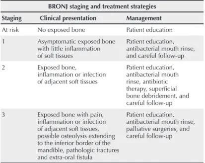
Table 1 shows the clinical staging of BRONJ and the treat-ment proposed by the AAOMS.21
The different stages of BRONJ can be observed in Figures 2, 3, and 4.
Figure 1
Computed tomography of the mandible – axial view showing bone sequestration.
Table 1
Clinical staging of BRONJ and the treatment proposed by the AAOMS
BRONJ staging and treatment strategies
Staging Clinical presentation Management
At risk No exposed bone Patient education
1 Asymptomatic exposed bone with little infl ammation of soft tissues
Patient education, antibacterial mouth rinse, and careful follow-up
2 Exposed bone,
infl ammation or infection of adjacent soft tissues
Patient education, antibacterial mouth rinse, antibiotic therapy, superfi cial bone debridement, and careful follow-up
3 Exposed bone with pain, infl ammation or infection of adjacent soft tissues, possible osteolysis extending to the inferior border of the mandible, pathologic fractures and extra-oral fi stula
Patient education, antibacterial mouth rinse, palliative surgeries, and careful follow-up
Adapted from Ruggiero SL et al.21
Figure 2
Figure 3
Symptomatic exposed bone in the mandible.
Figure 4
Exposed bone in the mandible extending to its inferior border, in the lingual region.
TREATMENT AND PREVENTION
The microorganisms most frequently found in exposed bone are: Actinomyces, Veillonella, Eikenella, Moraxella,
Fusobacterium, Bacillus, Staphylococcus, Streptococcus,and
Selenomonas. All of them are sensitive to penicillin, which is, thus, the drug of choice for the non-surgical treatment of BRONJ.7,12,24
The major goal of prevention for patients at risk for BRONJ or of treatment for those who have BRONJ is to preserve their quality of life, controlling pain and infections, and avoiding the development of new necrotic areas.8
The risk is associated with the accumulation of drug doses during years of treatment. Patients should undergo
careful dental evaluation, including radiographic exams, and be instructed about the possibility of developing BRONJ. Whenever any surgical procedure is required, some authors2,8 have suggested that the patients should sign a written in-formed consent.
The treatment of patients receiving intravenous BPs should be focused on reducing the risk for BRONJ, mini-mizing the need for surgical procedures. In such cases, they should be carefully instructed about oral hygiene practices. Preferentially, prior to beginning BPs therapy, patients should be assessed clinically and radiographically. Dental treatment including dental restorations, endodontic treatment and surgical procedures should be performed prior to initiating BPs therapy.2,8
BRONJ's treatment comprises the following: pain control, antibiotic therapy, mouth rinse, BP discontinuation, hyper-baric chamber therapy, lasertherapy,25 and surgical debride-ment.11,26,27 Such measures, however, do not always achieve the resolution of the clinical fi ndings – prevention is always the best option.2,8
The serum CTx test (C-terminal telopeptide of type I col-lagen, or ITCP), a marker of bone resorption that assesses the elimination of specifi c fragments produced by type I collagen hydrolysis, can be used as a parameter to assess the risk of developing BRONJ.17
There is a direct exponential relation between the duration of BPs use and the exposed bone size. Patients with CTx levels lower than 150 pg/mL should contact the attending physician and consider discontinuation of the BP (drug holiday) for a period of 4–6 months. After that period, the test should be repeated, and, if the CTx level is still lower than 150 pg/mL, the literature17 recommends extending the “drug holiday” for a period of 6–9 months. When CTx levels are not greater than 150 pg/mL and a “drug holiday” is not possible, the instruc-tions to patients about the risk of developing BRONJ should be emphasized. The search for a non-invasive form of treatment should always be recommended.17,24
It is important to distinguish and emphasize that BRONJ due to orally administered BPs seems to be less frequent, less severe, and responds better to a “drug holiday” and surgical debridement.8,15 Patients receiving oral BPs seem to have a better chance of improvement with a “drug holiday”.15
The statement that the discontinuation of BP for three
months prior to surgery, as recommended by the AAOMS21
their substantial accumulation in the skeleton.8 Thus, a long “drug holiday” would be required to eliminate the drug from the body. This “drug holiday” is not always possible, because of the benefi ts BPs provide for the prevention of osteoporosis and treatment of bone metastases.
FINAL CONSIDERATIONS
The communication of the BP prescribing physician with the patient’s dentist is essential to try to establish a preventive treatment for BRONJ prior to starting therapy with BPs.
REFERENCES REFERÊNCIAS
1. Fleisch H. Bisphosphonates: mechanisms of action. Endocr Rev 1998; 19(1):80–100.
2. ADA. Dental management of patients receiving oral bisphosphonate
therapy: expert panel recommendations. J Am Dent Assoc 2006; 137(8):1144–50.
3. Hillner BE, Ingle JN, Berenson JR, Janjan NA, Albain KS, Lipton A
et al. American Society of Clinical Oncology guideline on the role of bisphosphonates in breast cancer. American Society of Clinical Oncology Bisphosphonates Expert Panel. J Clin Oncol 2000; 18(6):1378–91.
4. de Groen PC, Lubbe DF, Hirsch LJ, Daifotis A, Stephenson W,
Freedholm D et al. Esophagitis associated with the use of
alendronate. N Engl J Med 1996; 335(14):1016–21.
5. Marx RE. Pamidronate (Aredia) and zoledronate (Zometa) induced
avascular necrosis of the jaws: a growing epidemic. J Oral Maxillofac Surg 2003; 61(9):1115–7.
6. Ruggiero SL, Mehrotra B, Rosenberg TJ, Engroff SL. Osteonecrosis
of the jaws associated with the use of bisphosphonates: a review of 63 cases. J Oral Maxillofac Surg 2004; 62(5):527–34.
7. Sedghizadeh PP, Kumar SK, Gorur A, Schaudinn C, Shuler CF, Costerton JW. Identifi cation of microbial biofi lms in osteonecrosis of the jaws secondary to bisphosphonate therapy. J Oral Maxillofac Surg 2008; 66(4):767–75.
8. Ruggiero SL, Woo SB. Biophosponate-related osteonecrosis of the
jaws. Dent Clin North Am 2008; 52(1):111–28.
9. AAOMS. American Association of Oral and Maxillofacial Surgeons
position paper on bisphosphonate-related osteonecrosis of the jaws. J Oral Maxillofac Surg 2007; 65(3):369–76.
10. Mariotti A. Bisphosphonates and osteonecrosis of the jaws. J Dent Educ 2008; 72(8):919–29.
11. Marx RE, Sawatari Y, Fortin M, Broumand V. Bisphosphonate-related exposed bone (osteonecrosis/osteopetrosis) of the jaws: risk factors, recognition, prevention, and treatment. J Oral Maxillofac Surg 2005; 63(11):1567–75.
12. Sawatari Y, Marx RE. Bisphosphonates and bisphosphonate induced osteonecrosis. Oral Maxillofac Surg Clin North Am 2007; 19(4):487–98.
13. Migliorati CA, Schubert MM, Peterson DE, Seneda LM. Bisphosphonate-associated osteonecrosis of mandibular and maxillary bone: an emerging oral complication of supportive cancer therapy. Cancer 2005; 104(1):83–93.
14. Naik NH, Russo TA. Bisphosphonate-related osteonecrosis of the jaw: the role of actinomyces. Clin Infect Dis 2009; 49(11):1729–32. 15. Grant BT, Amenedo C, Freeman K, Kraut RA. Outcomes of placing dental implants in patients taking oral bisphosphonates: a review of 115 cases. J Oral Maxillofac Surg 2008; 66(2):223–30.
16. King AE, Umland EM. Osteonecrosis of the jaw in patients receiving
intravenous or oral bisphosphonates. Pharmacotherapy 2008; 28(5):667–77.
17. Marx RE, Cillo JE Jr., Ulloa JJ. Oral bisphosphonate-induced osteonecrosis: risk factors, prediction of risk using serum CTx testing, prevention, and treatment. J Oral Maxillofac Surg 2007; 65(12):2397–410.
18. Pazianas M, Miller P, Blumentals WA, Bernal M, Kothawala P. A review of the literature on osteonecrosis of the jaw in patients with osteoporosis treated with oral bisphosphonates: prevalence, risk factors, and clinical characteristics. Clin Ther 2007; 29(8):1548–58. 19. Aragon-Ching JB, Ning YM, Chen CC, Latham L, Guadagnini JP,
Gulley JL et al. Higher incidence of Osteonecrosis of the Jaw (ONJ)
in patients with metastatic castration resistant prostate cancer treated with anti-angiogenic agents. Cancer Invest 2009; 27(2):221–6. 20. Ruggiero SL, Fantasia J, Carlson E. Bisphosphonate-related
osteonecrosis of the jaw: background and guidelines for diagnosis, staging and management. Oral Surg Oral Med Oral Pathol Oral Radiol Endod 2006; 102(4):433–41.
jaws-22. Abu-Id MH, Warnke PH, Gottschalk J, Springer I, Wiltfang J,
Acil Y et al. “Bis-phossy jaws” – high and low risk factors for
bisphosphonate-induced osteonecrosis of the jaw. J Craniomaxillofac Surg 2008; 36(2):95–103.
23. Reid IR, Bolland MJ, Grey AB. Is bisphosphonate-associated osteonecrosis of the jaw caused by soft tissue toxicity? Bone 2007; 41(3):318–20.
24. Woo SB, Hande K, Richardson PG. Osteonecrosis of the jaw and bisphosphonates. N Engl J Med 2005; 353(1):99–102.
25. Vescovi P, Merigo E, Manfredi M, Meleti M, Fornaini C,
Bonanini M et al. Nd:YAG laser biostimulation in the treatment
of bisphosphonate-associated osteonecrosis of the jaw: clinical experience in 28 cases. Photomed Laser Surg 2008; 26(1):37–46. 26. Lee CY, David T, Nishime M. Use of platelet-rich plasma in the
management of oral bisphosphonate-associated osteonecrosis of the jaw: a report of 2 cases. J Oral Implantol 2007; 33(6):371–82. 27. Magopoulos C, Karakinaris G, Telioudis Z, Vahtsevanos K,
Dimitrakopoulos I, Antoniadis K et al. Osteonecrosis of the jaws
