Received on 05/30/2011. Approved on 12/14/2011. The authors declare no confl ict of interest. Financial Support: CAPES, CNPQ and FINEP. Ethics Committee: 2007740.
Exercise Research Laboratory, Physical Education School, Universidade Federal do Rio Grande do Sul – UFRGS.
1. Specialist in Kinesiology, UFRGS; Master degree candidate in Geriatrics, Pontifícia Universidade Católica do Rio Grande do Sul – PUC-RS 2. Master degree in Human Motion Sciences, Universidade Federal do Rio Grande do Sul – UFRGS; Researcher and PhD candidate, UFRGS 3. Master degree in Human Motion Sciences, UFRGS
4. PhD in Kinesiology, University of Calgary; Adjunct Professor, UFRGS
Correspondence to:Cintia Helena Ritzel. Departamento de Cirurgia Ortopédica e Traumatológica, Escola de Medicina – UFRGS. Rua Ramiro Barcellos, 2400 - Santana. CEP: 90035-003. Porto Alegre, RS, Brasil. E-mail: [email protected]
Work and power of the knee fl exor and
extensor muscles in patients with osteoarthritis
and after total knee arthroplasty
Denise Bastiani1, Cintia Helena Ritzel2, Silvia Manfrin Bortoluzzi3, Marco Aurelio Vaz4
ABSTRACT
Introduction: The infl ammatory manifestations of knee osteoarthritis (OA) lead to muscle inhibition and hypotrophy, resulting in a reduction in total muscle work and muscle power. Total knee arthroplasty (TKA) is the most adequate sur-gery for the treatment of advanced OA. However, its effects on muscle functional behavior have not been well understood.
Objective: To compare the total work and power of the knee fl exor and extensor muscles in patients with OA (20) and in patients post-TKA (12) at two angular velocities (60°/sec and 240°/sec). Methods: An isokinetic Biodex dynamometer was used to assess muscle power and total work during isokinetic contractions. Two-way ANOVA for repeated measures was used to compare total muscle work and muscle power between the groups (SPSS software, version 13.0; signifi cance level, P < 0.05). Results: There was no difference between the OA and TKA groups for the total work of both knee exten-sors and fl exors at the two angular velocities (P ≥ 0.05). In addition, no difference was observed in the muscle power of the knee extensors and fl exors (P ≥ 0.05). Conclusion: Total work and power were similar in the OA and TKA groups, suggesting that TKA did not improve functional capacity, which was similar in both groups.
Keywords: knee osteoarthritis, total knee arthroplasty, dynamometer.
© 2012 Elsevier Editora Ltda. All rights reserved.
INTRODUCTION
Population ageing is a reality in Brazil, as is worldwide. Consequently, an increase in diseases associated with advanced age, especially the chronic degenerative ones, has been ob-served.1 As age advances, there is a reduction in skeletal muscle
mass (sarcopenia), and, thus, muscle weakness and increased joint overload occur.2,3
Due to changes in muscle mechanics and joint overload, adaptations occur, leading to joint instability, strength change, and muscle inhibition, and, consequently, to increased vulner-ability to muscle lesions and fatigue.3,4 This can result in
over-load change and joint degeneration due to the reduced ability of the muscle system to absorb repeated impacts, which has
been suggested to be a risk factor for the development and/or worsening of osteoarthritis (OA).4–6
OA is the most prevalent joint disease and also the major cause of pain and physical disability in the elderly popula-tion.5–7 It is a degenerative disease that leads to joint cartilage
loss, marginal osteophyte formation, and changes in the liga-ments, synovial membranes, meniscus, and subchondral bone.8
Although the causes of OA are not yet well understood, items such as biomechanical stresses, biochemical changes in carti-lage and synovial membrane, and genetic factors are important in its pathogenesis.9,10
domestic activities.11 In OA of the knee, edema and synovial
thickening lead to a refl ex inhibition of the quadriceps muscle, causing subsequent muscle hypotrophy. Pain, hypotrophy, and failure in voluntary quadriceps muscle activation have been suggested as causes of strength reduction in both individuals with knee OA and the elderly, generating important functional impact.4,9,12–14
Surgery is indicated for the more advanced stages of disease, in which greater cartilage and bone degenerations are evident, and where there is already impairment of the three knee joint surfaces, joint function alterations and pain.15
Patients with grade II and III OA and progressive impairment of autonomy in daily life activities, in addition to failure in conservative treatment, should be referred to the orthopedist, who will indicate surgical treatment.16 The most used
proce-dure in such cases is total knee arthroplasty (TKA),17 in which
the joint is totally replaced by a total prosthesis. It is mainly aimed at pain reduction, functional rehabilitation, and quality of life improvement.18–22 Most patients undergoing TKA have
excellent clinical results; however, some dysfunctions related to functional problems, not always clinically or radiographi-cally evident, might persist.17,18,22,23
Quadriceps muscle weakness after TKA has been reported by several authors as normal.19,23,24 Many studies have reported
a reduction in the quadriceps muscle strength, in the volun-tary muscle activation, and in the cross-sectional area (CSA) after TKA as compared with pre-operative values.22,23,25 Even
authors who have reported an improvement in quadriceps muscle strength in individuals after TKA have not observed total recovery as compared with healthy individuals in the same age group.18,26,27
The infl ammatory manifestations, defi cit in voluntary muscle activation, and hypotrophy of patients with knee OA and those undergoing TKA are believed to lower total muscle work,28,29 leading to a reduction in torque, muscle power30 and
resistance. This generates important functional capacity loss, predisposing such individuals to fatigue.12
The reduction in total muscle work and the consequent muscle power loss due to osteoarticular diseases usually inter-feres with the individual’s autonomy and quality of life.2,29–31
Despite the impact on functional results, quadriceps muscle work and power have not been typically assessed in post-oper-ative studies of TKA. Thus, those functional variables should be investigated in studies of populations with knee OA and TKA. In addition, the adaptive processes of musculoskeletal tissues to joint degeneration and replacement of the damaged joint by a prosthesis need to be better established. Any effort to clarify the mechanisms related to that disease aimed at reducing
or delaying its effects is highly important. Thus, the present study aimed at comparing muscle performance regarding total work and power of the knee fl exor and extensor muscles in individuals with knee OA and after TKA.
MATERIAL AND METHODS
Sample
This study sample comprised 32 individuals of both genders, over the age of 50 years, divided into the following two groups: the OA group, comprised by 20 individuals with grade II, III, and IV OA of the knee (Dejour classifi cation, 1991);15 and the
TKA group, comprised by 12 individuals in the post-operative period of TKA (range of one to three years).
The individuals were selected intentionally based on the clinical assessment by a physician specialized in Orthopedics and Traumatology (Service of Orthopedics and Traumatology of the Hospital São Lucas, PUC-RS). For the individuals with OA, X-rays (in the anteroposterior and lateral views, monopo-dal stance) were requested to confi rm the OA diagnosis and grading. The TKA group comprised only individuals who had undergone surgery one to three years before, based on a previous history of primary knee OA. In addition, they should all have undergone the same surgical technique, received the same prosthesis model (MB-VI, Metabio), and had the same post-operative period.
The exclusion criteria were as follows: previous history of hip joint surgery and/or revision knee surgery; severe pain assessed by use of the visual analogue scale of pain (VASP); limitation of range of motion of the knee; and cardiac, neuro-logical, musculoskeletal, and metabolic changes that hindered the execution of maximal voluntary contraction tests.32 The
Humac® recommendations were also respected.33
Participants were assessed regarding their functionality de-gree (Western Ontario and McMaster Universities Osteoarthritis Index - WOMAC Index)34,35 and pain intensity (VASP).36
This study was approved by the Ethics Committee in Research of the Universidade Federal do Rio Grande do Sul (protocol #2007740). After being instructed about the pro-cedures that would be performed, the participants provided written informed consent, assuring their rights according to resolution #196/96 of the Brazilian Health Board.
Instrumentation
To assess total work and muscle power, the isokinetic
dyna-mometer Biodex System 3 Pro(Biodex Medical Systems,
Procedures
The individuals were positioned in the dynamometer ac-cording to the manufacturer’s instructions. Then, they underwent a session of warm-up and familiarization, with one set of ten submaximal repetitions of knee flexion and extension at the velocity of 120º/sec. After a two-minute rest, total muscle work and muscle power were assessed with a set of five repetitions at the velocity of 60º/sec, and a set of 20 repetitions at the velocity of 240°/sec.37,38
Between the sets, a two-minute interval was observed to minimize the effects of fatigue. During the isokinetic tests, the same verbal stimulus for maximal effort was provided to all participants.39
At the end of the assessment protocol, stretching was per-formed and ice was applied to the knee joint for 20 minutes, aiming at preventing possible muscle discomfort due to the unusual maximal effort. All individuals received a program of home exercises for muscle reinforcement and stretching to aid with their functional recovery.
Statistical analysis
Quantitative variables were described as mean, standard deviation, median, skewness, and kurtosis. To assess data normality and homogeneity, the Shapiro Wilk and Levene tests were performed, respectively. Based on this analysis, the data were considered normal, and parametric statistical analysis was adopted.
Two-way analysis of variance (two-way ANOVA) for repeated measures was used for comparing the differences between the groups regarding the variables total muscle work and muscle power. The post-hoc Bonferoni test was used to identify the differences between the groups. One-way ANOVA with post-hoc Bonferoni test were used to compare the sample characterization variables between the groups.
The SPSS (Statistical Package for the Social Sciences) software, version 13.0, was used for statistical analysis, and the signifi cance level adopted for all analyses was P < 0.05.
RESULTS
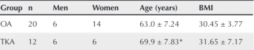
The sample comprised 32 individuals divided into two groups. The OA group consisted of 20 individuals (14 women and six men) with ages ranging from 52 to 76 years (63.0 ± 7.24). The TKA group consisted of 12 individuals (six women and six men) with ages ranging from 56 to 81 years (69.9 ± 7.83). Table 1 shows the characteristics of the sample concerning age and body mass index (BMI). The groups differed regarding
age (P = 0.028), which was higher in the TKA group. No difference in BMI was observed between the groups (P = 0.493).
Figures 1 and 2 show the results of total work of the knee extensor and fl exor muscles obtained for the two groups at the two angular velocities assessed. Comparing the groups, differences were observed between the total work performed by neither the extensor muscles at the velocities of 60°/sec (P = 0.198) and 240°/sec (P = 0.125), nor the fl exor muscles at the velocities of 60°/sec (P = 0.180) and 240°/sec (P = 0.081). Both groups had similar behaviors regarding the total work performed by the knee muscles at the angular velocities of 60°/sec and 240°/sec.
Similarly to total muscle work, OA and TKA groups also showed similar behaviors regarding muscle power at both an-gular velocities, and differences were observed for neither the knee extensor muscles at the velocities of 60°/sec (P = 0.297) and 240°/sec (P = 0.163), nor the knee fl exor muscles at the velocities of 60°/sec (P = 0.300) and 240°/sec (P = 0.121), as shown in Figures 3 and 4.
Figure 1
Total work of the knee extensor muscles of the OA and TKA groups at the angular velocities of 60°/sec (P = 0.198) and 240°/sec (P = 0.125).
To
tal w
o
rk (J)
900
600
300
0
Total work of the knee extensors
OA TKA
Velocity (°/sec)
60 240
Table 1
Characterization of the sample regarding age and body mass index (mean ± SD)
Group n Men Women Age (years) BMI
OA 20 6 14 63.0 ± 7.24 30.45 ± 3.77
TKA 12 6 6 69.9 ± 7.83* 31.65 ± 7.17
Signifi cant difference (P = 0.022) was found in the total WOMAC index between the OA and TKA groups, with greater values for the OA group. In general, the pain values were close to zero, mainly at the end of the evaluations. When comparing pain between the groups, a signifi cant difference in the mean pain value was observed, with lower values in the TKA group (P < 0.05), as shown in Table 2. However, difference regarding pain was observed neither between the pre-test and post-test period, nor between groups.
Figure 2
Total work of the knee fl exor muscles of the OA and TKA groups at the angular velocities of 60°/sec (P = 0.180) and 240°/sec (P = 0.081).
To
tal w
o
rk (J)
900
600
300
0
OA TKA
Velocity (°/sec)
60 240 Total work of the knee fl exors
Figure 3
Power of the knee extensor muscles of the OA and TKA groups at the angular velocities of 60°/sec (P = 0.297) and 240°/sec (P = 0.163).
P
o
w
er (W
a
s)
100
50
0
OA TKA
Velocity (°/sec)
60 240 Power of the knee extensors
Figure 4
Power of the knee fl exor muscles of the OA and TKA groups at the angular velocities of 60°/sec (P = 0.300) and 240°/s (P = 0.121).
P
o
w
er (W
a
s)
100
50
0
OA TKA
Velocity (°/sec)
60 240 Power of the knee fl exors
Table 2
Visual analogue scale of pain at three segments: pain, pre-assessment pain, and post-pre-assessment pain (mean)
OA group TKA group P
Pain 0.45 0.04 0.014*
Pre-assessment pain 0.37 0.03 0.207
Post-assessment pain 0.17 0.00 0.258
P = (pre- x post-) 0.352 0.337
-* Signifi cant difference.
DISCUSSION
Several researchers have investigated the alterations in the muscle strength of individuals with OA and TKA by assessing torque.3,13,15,18,26,27 Because peak torque represents performance
at a single point of the range of motion, it might not be a good indicator of total functional capacity. Thus, muscle strength over the entire range of motion (work) and strength per unit of time (power) would be more clinically relevant. However, such variables are not regularly assessed in studies of populations with knee OA and of post-operative results of TKA.31 Thus,
Total muscle work is the action of strength over a specifi c distance, that is, the action of torque during the range of motion. That variable is calculated by use of the area under the curve for torque, which physically is the energy developed by the muscle. A low value can indicate that the muscle function is clinically altered and that the energy expenditure during a range of motion is not adequate or has muscle defi cit.12,32 On the other hand, muscle
power expressed in Watts (W) represents total work divided by contraction time, that is, energy expenditure during a contraction over a certain time. It clinically represents the amount of energy that the muscle can waste over a shorter time to generate torque, and can be altered due to some muscle functional defi cit.12,32
Because knee OA causes a reduction in muscle mass due to refl ex muscle inhibition resulting from pain,10,14 and TKA is
aimed at reverting the effects of pain by removing the tissues degenerated by OA,20,27 the TKA group was expected to show
higher muscle work and power as compared with those of the OA group. However, the results showed no difference between the groups at the angular velocities assessed for both the knee extensor and fl exor muscles.
Byrne et al.22 have investigated the functional alterations in
the lower limb joints resulting from TKA, by comparing muscle work and power between healthy individuals and individuals after undergoing TKA. The work for the knee muscles was lower in the latter group than in the control group, and such defi cit was compensated by the increase in the work performed by the hip extensor muscles.
A study by Walsh et al.25 has assessed the walking velocity,
ability to climb stairs, knee torque, and total work in 29 women one year after TKA and in 40 healthy women. The individuals undergoing TKA showed lower values for all those variables as compared with the control group. Those authors have reported that the strength defi cit in the non-operated upon knee remains up to one year after surgery, which explains the decrease in the work of the extensor muscles. On the other hand, Aquino and Leme40 have found no difference regarding the total work of
the fl exor and extensor muscles between patients undergoing TKA and healthy individuals.
Work and power of the knee muscles have also been as-sessed in studies on other knee musculoskeletal disorders. Meireles et al.12 have assessed knee peak torque, total work,
and power of 50 patients with rheumatoid arthritis (RA) and 50 healthy individuals, comparing them at angular velocities of 60°/sec, 80°/sec, and 300°/sec. All variables of patients with RA were lower than those of controls.
Lennox et al.41 have assessed 16 patients undergoing
uni-lateral patellectomy, and their mean quadriceps muscle power was 60% of that of the contralateral healthy knee. The maximal
torque and mean work produced during eccentric and concen-tric contractions were one-third lower in the operated – side as compared with those of the contralateral muscle. Those authors have reported that muscle strength is not recovered immedi-ately after patellectomy, but with a long-term rehabilitation program it can be restored.
Neeter et al.42 have assessed the muscle power of knee
extensors and fl exors in 23 patients with anterior cruciate ligament (ACL) injury and 44 patients after reconstruction of that same ligament, six months after injury and surgery, respectively. Those authors have compared the values obtained with those of the non-affected sides of those patients and with those of other 13 healthy individuals. In 90% of the patients with ACL injury and in 60% of those whose ACL had been reconstructed, a defi cit in muscle power was observed.
Although the literature is not clear about whether that which is valid for peak torque is valid for work production and mean power,28–30 the data of studies assessing muscle strength only
by use of torque should be analyzed along with the results of the present study.
Some authors, comparing voluntary muscle strength and activation in patients with primary knee OA before and after TKA, have reported a reduction in the quadriceps muscle strength and activation after surgery. They have reported that, despite the reduction in pain and improvement in the range of motion in the knee, quadriceps muscle weakness and reduc-tion in funcreduc-tional capacity were typically present, even one year after surgery.13,21 A reduction in the quadriceps muscle
strength is observed in long-term post-operative assessments, and correlates with functional limitations present prior to the surgical procedure of individuals with knee OA.3 The occurrence
of a muscle strength defi cit after TKA has been reported in some studies as normal.12,15 On the other hand, Berth et al.,18 in a study
with patients assessed three years after TKA, have demonstrated that voluntary muscle activation improved over time, but did not reach the levels of healthy individuals. Anchuela et al.26 and
Lorentzen et al.27 have also reported similar results, with greater
capacity of strength production after surgery.
to the permanence of the functional defi cit. Lennox et al.41
have suggested that if the patients assessed in their study underwent rehabilitation, the success rate of surgery might have been higher.
Total knee arthroplasty is the last resource for individuals with degenerative OA, to eliminate pain and improve function. There are several ways to assess the evolution of individuals undergoing TKA, in addition to assessing total muscle work and muscle power, the individual’s functionality and quality of life can also be assessed by use of questionnaires, such as the WOMAC index.
The scores of the WOMAC index questionnaire differed between the OA and TKA groups. The TKA group scored lower than the OA group for the total WOMAC, pain, stiffness, and functionality. Although the groups assessed did not differ regarding muscle work and muscle power, they did differ in the WOMAC. Thus, placing a total knee prosthesis without subsequent physical therapy does not improve the mechanical properties assessed in this study, but improves the individual’s quality of life and pain.
All individuals were asked about the pain in their injured knee, since the initial interview on the assessment day until the fi nal assessment. A rule representing the VASP in a 0–10 scale was shown to the participants. In general, pain levels were close to zero, mainly by the end of the assessments. Comparing the pain levels between the groups, a signifi cant difference was observed in mean pain, with lower levels for the TKA group. Comparing the pre- and post-test pain levels, a differ-ence was found neither between the groups nor between the assessment times. Several studies have shown a signifi cant improvement in physical and psychological capacities in the post-operative period of TKA as compared with the pre-operative period. In studies showing that improvement, all participants received physical therapeutic care after surgery until total rehabilitation.43,44 Other authors, however, have
found a correlation between the presence of pain, depression, OA, and alteration in functionality.45
Several patients can have problems, such as muscle weak-ness and contracture, reduced range of motion, diffi culty in walking, and limitation of daily life activities, despite replacing the injured joint with a prosthesis. Unfortunately, that reduction in functional capacity has been accepted by several authors as a normal functional defi cit.25,45 Ulrich et al.23 have emphasized
the importance of using some techniques to identify functional problems after TKA, so that the treatment can be specifi cally focused on the impairment, leading to success of the clinical results for that population. The use of more appropriate rehabili-tation programs, with exercises that emphasize strong muscle contractions, and clinical tools that facilitate muscle activation, such as biofeedback and neuromuscular electric stimulation, might be necessary to reverse the failure in muscle activation and weakness in patients with OA and after TKA, although the effects of such therapies have not yet been clarifi ed.14,18,30 Such
programs should benefi t patients with orthopedic problems in the years immediately after surgery, and, even more important, should help them to preserve their functional capacity and main-tain their independence for a long period of time.25,46
The literature about the functional results of the TKA, especially regarding total muscle work and muscle power of the knee, is still inconsistent, indicating the need for further studies with that population. Studies about different types of rehabilitation are also relevant in the search for clarifi cation on the OA-related mechanisms and the factors that can potentiate the benefi ts of TKA.
CONCLUSION
REFERENCES REFERÊNCIAS
1. Maciel ACC, Guerra RO. Fatores associados à alteração da mobilidade em idosos residentes na comunidade. Rev Bras Fisioter 2005; 9(1):17–23.
2. Bijlsma JW, Knahr K. Strategies for the prevention and management of osteoarthritis of the hip and knee. Best Pract Res Clin Rheumatol 2007; 21(1):59–76.
3. Herzog W, Longino D. The role of muscles in joint degeneration and osteoarthritis. J Biomech 2007; 40(Suppl 1):S54–63.
4. Bennell KL, Hunt MA, Wrigley TV, Lim BW, Hinman RS. Role of muscle in the genesis and management of knee osteoarthritis. Rheum Dis Clin North Am 2008; 34(3):731–54.
5. Pereira HLA, Ribeiro SLE, Ciconelli RM. Topical anti-infl ammatory drugs in osteoarthritis of the knee. Rev Bras Reumatol 2006; 46(3):188–93.
6. Vasconcelos KSS, Dias JMD, Dias RC. Relação entre intensidade de dor e capacidade funcional em indivíduos obesos com osteoartrite de joelho. Rev Bras Fisioter 2006; 10(2):213–8.
7. Jamtvedt G, Dahm KT, Holm I, Flottorp S. Measuring physiotherapy performance in patients with osteoarthritis of the knee: a prospective study. BMC Health Serv Res 2008; 8:145.
8. National Collaborating Centre for Chronic Conditions. Osteoarthritis: national clinical guideline for care and management in adults. London: Royal College of Physicians, 2008.
9. Lewek MD, Rudolph KS, Snyder-Mackler L. Quadriceps femoris muscle weakness and activation failure in patients with symptomatic knee osteoarthritis. J Orthop Res 2004; 22(1):110–5.
10. Lange AK, Vanwanseele B, Foroughi N, Baker MK, Shnier R, Smith RM et al. Resistive Exercise for Arthritic Cartilage Health (REACH): a randomized double-blind, sham-exercise controlled trial. BMC Geriatr 2009; 9:1.
11. Marx FC, Oliveira LM, Bellini CG, Ribeiro MCC. Tradução e Validação Cultural do Questionário Algofuncional de Lequesne para Osteoartrite de Joelhos e Quadris para a Língua Portuguesa. Rev Bras Reumatol 2006; 46(4):253–60.
12. Meireles SM, Oliveira LM, Andrade MS, Silva AC, Natour J. Isokinetic evaluation of the knee in patients with rheumatoid arthritis. Joint Bone Spine 2002; 69(6):566–73.
13. Mizner RL, Stevens JE, Snyder-Mackler L. Voluntary activation and decreased force production of the quadriceps femoris muscle after total knee arthroplasty. Phys Ther 2003; 83(4):359–65.
14. Stevens JE, Mizner RL, Snyder-Mackler L. Quadriceps strength and volitional activation before and after total knee arthroplasty for osteoarthritis. J Orthop Res 2003; 21(5):775–9.
15. Dejour H, Carret JP. Les gonarthroses. In: 7ème Journées Lyonnaises de Chirurgie du Genou. Lyon: Simep, 1991, pp.775–9.
16. Coimbra IB, Pastor EH, Greve JMD, Puccinelli MLC, Fuller R, Cavalcanti FS et al. Osteoartrite (artrose): tratamento. Rev Bras Reumatol 2004; 44(6):450 –3.
17. Lamb SE, Toye F, Barker KL. Chronic disease management programme in people with severe knee osteoarthritis: effi cacy and moderators of response. Clin Rehabil 2008; 22(2):169–78. 18. Berth A, Urbach D, Awiszus F. Improvement of voluntary quadriceps
muscle activation after total knee arthroplasty. Arch Phys Med Rehabil 2002; 83(10):1432–6.
19. Berman AT, Bosacco SJ, Israelite C. Evaluation of total knee arthroplasty using isokinetic testing. Clin Orthop Relat Res 1991; 271:106–13.
20. Silva M, Shepherd EF, Jackson WO, Pratt JA, McClung CD, Schmalzried TP. Knee strength after total knee arthroplasty. J Arthroplasty 2003; 18(5):605–11.
21. Stevens JE, Binder-Macleod S, Snyder-Mackler L. Characterization of the human quadriceps muscle in active elders. Arch Phys Med Rehabil 2001; 82(7):973–8.
22. Byrne JM, Gage WH, Prentice SD. Bilateral lower limb strategies used during a step-up task in individuals who have undergone unilateral total knee arthroplasty. Clin Biomech (Bristol, Avon) 2002; 17(8):580–5.
23. Ulrich SD, Bhave A, Marker DR, Seyler TM, Mont MA. Focused rehabilitation treatment of poorly functioning total knee arthroplasties. Clin Orthop Relat Res 2007; 464:138–45.
24. Huang CH, Cheng CK, Lee YT, Lee KS. Muscle strength after successful total knee replacement: a 6- to 13-year of follow-up. Clin Orthop Relat Res 1996; 328:147–54.
25. Walsh M, Woodhouse LJ, Thomas SG, Finch E. Physical impairments and functional limitations: a comparison of individuals 1 year after total knee arthroplasty with control subjects. Phys Ther 1998; 78(3):248–58.
26. Anchuela J, Gomes-Pellico L, Ferrer-Blanco M, Slocker M, Rodriguez R. Muscular function and bone mass after knee arthroplasty. Int Orthop 2001; 25(4):253–6.
27. Lorentzen JS, Petersen MM, Brot C, Madsen OR. Early changes in muscle strength after total knee arthroplasty. A 6-month follow-up of 30 knees. Acta Orthop Scand 1999; 70(2):176–9.
28. McGinnis PM. Biomecânica do esporte e exercício. São Paulo: Artmed, 2002.
29. Simão R, Monteiro W, Araújo CGS. Fidedignidade inter e intradias de um teste de potência muscular. Rev Bras Med Esporte 2001; 7(4):118–24.
30. Ferri A, Scaglioni G, Pousson M, Capodaglio P, Van Hoecke J, Narici MV. Strength and power changes of the human plantar fl exors and knee extensors in response to resistance training in old age. Acta Physiol Scand 2003; 177(1):69–78.
31. Charteris J. Effects of velocity on upper to lower extremity muscular work and power output ratios of intercollegiate athletes. Br J Sports Med 1999; 33(4):250–4.
32. Ribeiro FM, Novaes JS, Lemos A, Simão R. Reprodutibilidade inter e intradias do Power Control em um teste de potência muscular. Rev Bras Med Esporte 2006; 12(5):255–8.
33. Humac®/Norm Testing & Rehabilitation System – User’s Guide –
34. Bellamy N, Buchanan WW, Goldsmith CH, Campbell J, Stitt LW. Validation study of WOMAC: a health status instrument for measuring clinically important patient relevant outcomes to anti-rheumatic drug therapy in patients with osteoarthritis of the hip or knee. J Rheumatol 1988; 15(12):1833 –40.
35. Fernandes MI. Tradução e validação do questionário de qualidade de vida específi co para osteoartrose WOMAC (Western Ontário and McMaster Universities) para a língua portuguesa. São Paulo: Universidade Federal de São Paulo, Escola Paulista de Medicina, 2003, pp.1–100.
36. Franklin P, Li W, Drew J, Ayers D. Pain relief and functional improvement after total knee arthroplasty. The Journal of Arthroplasty 2008; 23(2):320.
37. Dias JMD, Arantes PMM, Alencar MA, Faria JC, Machala CC, Camargos FFO et al. Relação isquiotibiais/quadríceps em mulheres idosas utilizando o dinamômetro isocinético. Rev Bras Fisioter 2004; 8(2):111–5.
38. van Loan MD, Sutherland B, Lowe NM, Turnlund JR, King JC. The effects of zinc depletion on peak force and total work of knee and shoulder extensor and fl exor muscles. Int J Sport Nutr 1999; 9(2):125–35.
39. Perrin DH. Isokinetic exercise and assessment. Champaign, IL, Human Kinetics Publishers, 1993, pp.57–65.
40. de Amorin Aquino M, Leme LE. Isokinetic dynamometry in elderly women undergoing total knee arthroplasty: a comparative study. Clinics (São Paulo) 2006; 61(3):215–22.
41. Lennox IA, Cobb AG, Knowles J, Bentley G. Knee function after patellectomy. A 12- to 48-year follow-up. J Bone Joint Surg Br 1994; 76(3):485–7.
42. Neeter C, Gustavsson A, Thomeé P, Augustsson J, Thomeé R, Karlsson J. Development of a strength test battery for evaluating leg muscle power after anterior cruciate ligament injury and reconstruction. Knee Surg Sports Traumatol Arthrosc 2006; 14(6):571–80.
43. Stubbe JH, Gelsema T, Delnoij DM. The Consumer Quality Index Hip Knee Questionnaire measuring patients’ experiences with quality of care after a total hip or knee arthroplasty. BMC Health Serv Res 2007; 7:60.
44. Leite AA, Costa AJG, Lima BAM, Padilha AVL, Albuquerque EC, Marques CDL. Comorbidades em pacientes com osteoartrite: frequência e impacto na dor e na função física. Rev Bras Reumatol 2011; 51(2):118–23.
45. Rooks DS, Huang J, Bierbaum BE, Bolus SA, Rubano J, Connolly CE et al. Effect of preoperative exercise on measures of functional status in men and women undergoing total hip and knee arthroplasty. Arthritis Rheum 2006; 55(5):700–8.