for Vascular Dysfunction in Idiopathic Recurrent
Spontaneous Miscarriage
Priyanka Banerjee
1☯, Sanghamitra Ghosh
β☯, Mainak Dutta
1, Elavarasan Subramani
1, Jaydeep Khalpada
1,
Sourav RoyChoudhury
1, Baidyanath Chakravarty
β, Koel Chaudhury
1*1 School of Medical Science and Technology, Indian Institute of Technology, Kharagpur, India, β Institute of Reproductive Medicine, Kolkata, India
Abstract
Poor endometrial perfusion during implantation window is reported to be one of the possible causes of idiopathic recurrent spontaneous miscarriage (IRSM). We have tested the hypothesis that certain angiogenic and vasoactive factors are associated with vascular dysfunction during implantation window in IRSM and, therefore, could play a contributory role in making the endometrium unreceptive in these women. This is a prospective case-controlled study carried out on 66 women with IRSM and age and BMI matched 50 fertile women serving as controls. Endometrial expression of pro-inflammatory (IL-1 , TNF-α, IFN- , TGF- 1), anti-inflammatory (IL-4, -10), angiogenesis-associated cytokines (IL-β, -6, -8), angiogenic and vasoactive factors including prostaglandin Eβ (PGEβ), vascular endothelial growth factor (VEGF), endothelial nitric oxide synthase (eNOS), nitric oxide (NO) and adrenomedullin (ADM) were measured during implantation window by ELISA. Subendometrial blood flow (SEBF) was assessed by color Doppler ultrasonography. Multivariate analysis was used to identify the significant factor(s) responsible for vascular dysfunction in IRSM women during window of implantation and further correlated with vascular dysfunction. Endometrial expression of pro-inflammatory cytokines and PGEβ were up-regulated and anti-inflammatory and angiogenesis-associated cytokines down-regulated in IRSM women as compared with controls. Further, the angiogenic and vasoactive factors including VEGF, eNOS, NO and ADM were found to be down-regulated and SEBF grossly affected in these women. Multivariate analysis identified IL-10, followed by VEGF and eNOS as the major factors contributing towards vascular dysfunction in IRSM women. Moreover, these factors strongly correlated with blood flow impairment. This study provides an understanding that IL-10, VEGF and eNOS are the principal key components having a contributory role in endometrial vascular dysfunction in women with IRSM. Down-regulation of these factors is also associated with impaired endometrial perfusion which possibly makes the endometrium unreceptive that may eventually cause early pregnancy loss.
Citation: Banerjee P, Ghosh S, Dutta M, Subramani E, Khalpada J, et al. (β01γ) Identification of Key Contributory Factors Responsible for Vascular Dysfunction in Idiopathic Recurrent Spontaneous Miscarriage. PLoS ONE 8(11): e80940. doi:10.1γ71/journal.pone.0080940
Editor: Kwang-Hyun Baek, CHA University, Republic of Korea
Received July β1, β01γ; Accepted October 8, β01γ; Published November 15, β01γ
Copyright: © β01γ Banerjee et al. This is an open-access article distributed under the terms of the Creative Commons Attribution License, which permits unrestricted use, distribution, and reproduction in any medium, provided the original author and source are credited.
Funding: These authors have no support or funding to report.
Competing interests: The authors have declared that no competing interests exist. * E-mail: [email protected]
☯ These authors contributed equally to this work.
Introduction
Recurrent spontaneous miscarriage (RSM), affecting 1-γ% of fertile couples, is defined as spontaneous abortion of three or more clinically diagnosed pregnancies within less than β4 weeks of gestation [1]. Various causative factors such as genetic, endocrine, anatomic, immunological, infectious, environmental, thrombophilic and metabolic are thought to be responsible for RSM. Regardless of extensive research undertaken in this field, etiology of ~50% of these cases still remains unknown and hence poses to be a clinical challenge [β]. An unreceptive endometrium, leading to abnormal
implantation, is believed to be associated with idiopathic recurrent spontaneous miscarriage (IRSM). However, whether the relationship is a causal or casual one remains to be established [γ].
constitutively on all vascular cells and plays a crucial role in angiogenesis. Reduced expression of PECAM-1 is reported in cytotrophoblasts, syncytiotrophoblasts, and extravillous trophoblasts of the abortion materials in women with spontaneous abortion [7]. The endothelium of uterine blood vessels uses NO, produced in the blood vessel by endothelial nitric oxide synthase (eNOS), to signal the surrounding smooth muscles to relax. This results in vasodilation, thereby making the endometrium receptive [8]. Further, increased expression of endometrial eNOS during mid-secretory phase suggests its role in endometrial receptivity and blastocyst implantation [9]. Adrenomedullin (ADM) is another vasoactive peptide which has drawn considerable attention in recent years in view of its involvement in regulating the expression of pinopodes which facilitate endometrium-blastocyst attachment [10]. The influence of these factors on SEBF during implantation window in women with IRSM is not reported so far.
This study is a continuation of our previous work where an aberrant expression of inflammatory and angiogenic factors concomitant with down regulation of anti-inflammatory and angiogenesis-associated cytokines was observed during implantation window in women with IRSM [11]. The expression of vasodilators including ADM, eNOS and NO during implantation period of women with IRSM is investigated in the present study. Pro-inflammatory, anti-inflammatory and angiogenesis-associated cytokines and angiogenic factors are included and taken together, multivariate data analysis applied to identify the significant factor(s) responsible for vascular dysfunction in IRSM women during window of implantation. Further, SEBF is estimated in these women and correlated with the major factor(s) identified.
Materials and Methods
Patient selection
The present study was conducted at the Institute of Reproductive Medicine, Kolkata India, a tertiary care hospital and School of Medical Science and Technology, Indian Institute of Technology, Kharagpur, India.
100 women (age<γ5 years, BMI≤β8), who have had three or more consecutive miscarriages within the first trimester (upto 1β weeks of gestation) with no apparent cause, reporting at the Institute of Reproductive Medicine, for infertility treatment were initially identified for the study (IRSM group). The women were not associated with any other gynecological disorder, and had not received any kind of medication since the last three months. All women included in the study were south Asian of Indian origin.
The following tests were performed to confirm that there was no apparent cause of recurrent pregnancy loss: TSH and anti-thyroid antibody tests, antiphospholipid antibodies tests (anticardiolipin antibodies and Lupus anticoagulants IgG and IgM), TORCH (Toxoplasmosis, Rubella, Cytomegalovirus and Herpes) tests, paternal and maternal chromosomal analysis, hysterosalpingography and hysteroscopy to rule out uterine defects, abnormal fasting level of homocysteine, PCOS, exclusion of diabetes mellitus, and estimation of mid-luteal serum progesterone to exclude luteal phase defect.
Women suffering from recurrent miscarriage due to any other pre-existing medical conditions like antiphospholipid syndrome, hormonal disorder, chromosomal defects, thrombophilia, hyper-homocystinemia, infectious diseases, PCOS, uterine malformation, environmental causes, gestational hypertension and other known causes were excluded. Further, women with male partners having fragmented sperm DNA, sperm meiotic alternations and poor sperm parameters were also excluded from the study. 100 proven fertile women (age and BMI-matched) undergoing sterilization were identified as controls. Women of these groups had parity between β-5 (β.5±0.1β) and normal regular menstrual cycles. They had no history of failed pregnancies and other significant clinical abnormalities.
Out of the total β00 women, 84 subjects had to be excluded; γ4 from the IRSM group and 50 from controls. These women either did not report for (a) serial folliculometry to confirm ovulation during the specified period or for (b) endometrial biopsy collection during day 18-ββ of their menstrual cycle (implantation window) or the (c) endometrial tissue collected from several women was highly flaky or inadequate, rendering these samples unsuitable for the experimental study or (d) did not express interest in participating in the study. Power calculation was subsequently performed on the reduced sample size of 66 IRSM women and 50 controls cases. A power > 99% for this sample size at a significance level α of 0.05 on comparing both the groups was observed.
Ethics statement
The study was approved by the Institute Ethics Committee, Institute of Reproductive Medicine (IRM/IEC/BNC-IHP-γ5/β6-0β-β010) and written informed consent obtained from all couples.
Sample collection
In vitro measurements
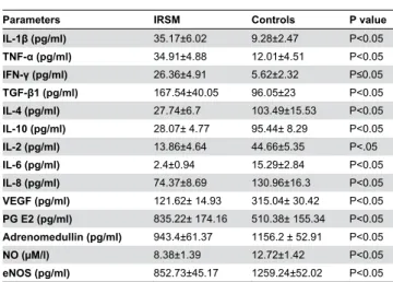
Our earlier report in γ6 IRSM cases and γ0 controls where endometrial expression of various pro-inflammatory, anti-inflammatory, angiogenesis-associated cytokines, and angiogenic factors were compared [11], is extended to a larger sample size in the present study. Thirty additional cases of IRSM and β0 controls are included. In addition to interleukin (IL)-1 , tumor necrosis factor (TNF)-α, interferon (IFN)- , transforming growth factor (TGF)- 1, IL-4, IL-10, IL-β, 6, 8, vascular endothelial growth factor (VEGF), and prostaglandin (PG) Eβ assessment, endothelial nitric oxide synthase (eNOS), nitric oxide (NO), adrenomedullin (ADM) and SEBF are estimated during implantation window in these women. Serum estradiol and progesterone were also measured during the day of tissue collection.
ELISA
Tissue homogenates containing protein concentration of γ0 μg/ml were used for estimating the level of IL-1 , TNF-α, IFN- , TGF- 1, IL-10, -β, -6, -8, VEGF, eNOS and ADM. VEGF and PGEβ were assessed using anti-human rabbit polyclonal antibody to VEGF (ab9570) and PGEβ (ab-βγ18; Abcam, Cambridge, UK), respectively employing quantitative direct enzyme immunoassay technique. IL-1 , TNF-α, IFN- , IL-10, -4, -β, -6, and -8 were measured using human IL-1 (557966), TNF-α (550610), IFN- (55061β), IL-10 (55061γ), IL-4 (550614), IL-β (550611), IL-6 (550799), and IL-8 (550999) ELISA Kit (BD Biosciences, San Jose, CA, USA), respectively. TGF- 1 was analysed using anti-human mouse monoclonal antibody to TGF- 1 (sc-5744γ; Santacruz, Inc. USA). eNOS and ADM were analysed using anti-human rabbit polyclonal antibody to eNOS (sc-8γ11) and ADM (sc-γγ187; Santacruz, Inc. USA), respectively. The principle of the assay is based on the quantitative sandwich enzyme immunoassay technique. The intra- and inter-assay variability was respectively β.5% and γ.11% for IL-1 , β.7% and 5.γ% for TNF-α, γ.β% and 4.γ% for IFN- , <7 and <8% for TGF- 1, γ.7% and β.6% for IL-4, β.1% and 5.8% for IL-10, γ.1% and γ.γ% for IL-β, 4.1% and 7.9% for IL-6, 4% and γ.β% for IL-8, 5.γ% and 7.6% for VEGF, <5% and <14% for ADM, 4.4% and 9.β% for PGEβ, and 8% and 10% for eNOS. Samples with expression level less than the limit of detection were assigned as half of the sensitivity limit.
Nitric oxide (NO) measurement
NO was measured in the tissue as nitrite/nitrate, by the Griess reaction [1β]. 100 µl of tissue homogenate sample was mixed with an equal volume of Griess reagent (0.1% (1-naphthyl) ethylene diamine dihydrochloride, 1% sulfanilamide, and β.5% phosphoric acid) and the resulting absorbance measured at 550 nm in a microplate reader. Background (blank) was determined in each experiment by incubating the reagent without samples. Amount of NO in each sample was determined using a standard curve generated with known concentration of NO and its concentration expressed as µM/l. The inter- and intra-assay viability co-efficient was 4.1% and β.9%, respectively.
SEBF measurement
The endometrial thickness, together with SEBF were evaluated immediately before endometrial curettage during the window of implantation. This was done in both the groups using transvaginal color Doppler and two dimensional Power Doppler using a 7.5 MHz probe (Medison SA 9900, South Korea). The resistance index {RI = (peak systolic velocity – end diastolic velocity)/peak systolic velocity}, pulsatility index {PI = (peak systolic velocity – end diastolic velocity)/mean velocity}, and systolic/diastolic ratio (S/D ratio = systolic velocity / end diastolic velocity) were calculated on three consecutive waveforms. USG was performed in all patients by only one person to avoid inter-observer variation and the sonologist blinded to avoid significant bias. For assessing the reproducibility of Doppler measurements, RI, PI and S/D ratio were measured in 10 patients by the same operator γ times at 10-minute intervals and the coefficient of variation was found to be 5.8%.
Immunohistochemistry
γ-5 µm thick sections obtained from formaldehyde fixed, paraffin-embedded tissue were dehydrated in graded ethanol. After antigen retrieval, slides were incubated for an hour in γ% blocking serum (BSA) in PBS for controlling non-specific binding of primary antibody. The slides were then incubated with goat anti-PECAM 1 (sc-1506; Santacruz biotechnology, INC., Santa Cruz, California, USA). Excess primary antibody was washed with PBS and the sections were again incubated with anti-goat (ab6885) secondary antibody (Abcam, Cambridge, UK) according to the manufacturer’s protocol. Labeled cells were visualized with diaminobenzidine (DAB) and sections counterstained with hematoxylin. Next, the slides were dehydrated using series of alcohol gradient and mounted using distrene, tricresyl phosphate (DPX) and xylene. The slides were then examined under bright field microscope (Carl Zeiss, Jena, Germany). The DAB staining intensities on each slide were individually graded by visual inspection. Semi-quantitative scoring was done independently by two observers to assess the staining intensity which provides a measure of the expression of these molecules. Immunostaining was classified on the basis of intensity of staining ((no staining-0; weak-1 point; moderate-β points; strong-γ points) and percentage/ extent of stained cells (0%-0 point; <10%-1 point; 11%-50%-β points; 51%-80%-γ points; >80%-4 points). A final immunohistochemical score (Score 0-1β) were obtained by multiplying intensity score and extent of stained cells.
Statistical Analysis
performed using SIMCA 1γ.0.β. PCA is an unsupervised multivariate projection method designed to extract and display systematic variations within the data matrix. It is primarily used to detect intrinsic clusters and outlier (observations that are extreme or that do not fit the PCA model) within the data set. Followed by sample clustering using PCA, partial least squares discriminant analysis (PLS-DA) analysis was performed using SIMCA 1γ.0.β. PLS-DA is another method of multivariate statistical analysis which is used for constructing predictive models. Unlike PCA, PLS-DA is a robust form of analysis, directed towards factor space that are associated with high variation in the responses but biased towards directions that are accurately predicted (in this case, sample groups i.e. diseased and controls).
Statistical significance was defined as p≤0.05. Pearson linear correlation was used to identify those angiogenic and vasculogenic factors which correlate highly with the Doppler parameters. Ky Plot version β.0 beta 1γ software was used for this purpose.
Results
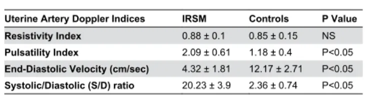
The demographic characteristics are summarized in Table 1. No significant differences were observed in terms of age, BMI, endometrial thickness, serum estrogen and progesterone levels between women with IRSM and controls. USG of the endometrium indicated PI, EDV and S/D ratio to be grossly affected in women with IRSM as compared with controls. RI, however, was found to be comparable between the two groups (Table β; Figure 1).
Levels of the vasoactive factors including eNOS, NO and ADM estimated during implantation window were found to be significantly less in IRSM women as compared with controls. The angiogenic factors, pro-inflammatory, anti-inflammatory and angiogenesis-associated cytokines showed a similar trend as reported in our previous study on a smaller sample size. IL-1 , TNF-α, IFN- , TGF- 1 and PGEβ were found to be up-regulated and IL-4, IL-10, IL-β, 6, 8 and VEGF down-up-regulated in IRSM cases as compared with controls (Table γ). PECAM-1, the marker of angiogenesis and vascular development, exhibited low immunoreactivity levels in women with IRSM in contrast to strong immunoreactivity observed in controls (Table 4; Figure β).
Table 1. Clinical characteristics of women with idiopathic recurrent spontaneous miscarriage (IRSM) and controls during implantation window.
Parameters IRSM Controls P value
Age β8.8±γ.1 β8.56±β.9β NS
BMI β7.8β±1.55 β7.β6±1.9β NS
Endometrial thickness (mm) 8.71±1.55 9.11±1.68 NS
Serum estrogen level (pg/ml) ββ7.0γ±60.4 β05.04±61.8 NS
Serum progesterone level (ng/ml) 18.γ5±γ.58 19.β5±γ.0β NS Unpaired Student's t-test is used to compare the differences between parametric data sets.
doi: 10.1γ71/journal.pone.0080940.t001
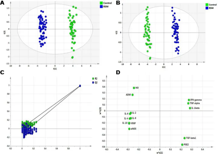
Exploratory PCA was employed to detect intrinsic clustering and possible outliers. Scatter plot of t1 vs tβ indicates unsupervised (unbiased and having no prior knowledge of sample groups) separation trend between IRSM and controls (Figure γA). The group separation was further maximized by PLS-DA (Figure γB). The predictive capability (Qβ) and explained variance (Rβ) were extracted for the PLS-DA model. The model with both Rβ and Qβ well above 0.9 indicated a very good predictive ability. Further, validation of the PLS-DA model was performed using permutation test statistics. This validation is performed to compare the Rβ and Qβ of the original model with the Rβ and Qβ of several models based on data where the order of the class variable is randomly permuted, while the other data matrix is kept intact. Results of permutation test statistics indicated the original model to be far more superior than all the one hundred model generated by random permutation of class variable (Figure γC).
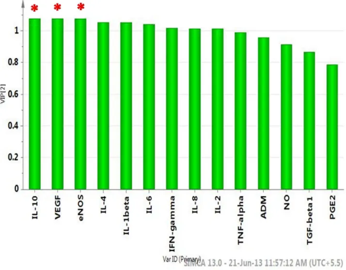
Factors with higher loadings were identified (Figure γD). Further a variable lies away from the plot origin, the stronger impact the variable has on the model. Comparing the loading and scores plot, pro-inflammatory cytokines (IL-1 , TNF-α, IFN-, TGF- 1) and PGEβ showed positive factor 1 loadingIFN-, hence indicating more bias towards IRSM. Similarly, angiogenic and vasoactive factors (VEGF, NO, ADM, eNOS), anti-inflammatory (IL-4 and IL-10) and angiogenesis-associated cytokines (IL-β, 6, 8) showed negative factor 1 loading, indicating bias towards the control group. Factors with high variable importance in projection (VIP) scores are regarded as significant and, therefore, considered for quantitative analysis of variation. IL-10 followed by VEGF and eNOS were identified as the prime factors corresponding to three highest VIP scores (Figure 4). Further, IL-10, VEGF and eNOS, the three major regulatory factors identified, showed significant correlation with the Doppler indices. A significant positive correlation was observed between IL-10 and PI (r = 0.79, P < .001), VEGF with EDV (r = 0.61, P < .001), and eNOS with EDV (r = 0.74, P < .001). The S/D ratio showed a significant negative correlation between VEGF (r = -0.78, P < .001) and eNOS (r = -0.67, P < .001), respectively.
Discussion
A hypothesis that certain angiogenic and vasoactive factors are associated with vascular dysfunction and could play a
Table β. Doppler indices for sub-endometrial blood flow in women with idiopathic recurrent spontaneous miscarriage (IRSM) and controls during implantation window.
Uterine Artery Doppler Indices IRSM Controls P Value Resistivity Index 0.88 ± 0.1 0.85 ± 0.15 NS
Pulsatility Index β.09 ± 0.61 1.18 ± 0.4 P<0.05
End-Diastolic Velocity (cm/sec) 4.γβ ± 1.81 1β.17 ± β.71 P<0.05
Systolic/Diastolic (S/D) ratio β0.βγ ± γ.9 β.γ6 ± 0.74 P<0.05 Unpaired Student's t-test is used to compare the differences between parametric data sets.
contributory role in poor endometrial receptivity in IRSM is proposed. To ascertain this hypothesis, the major factors responsible for angiogenesis and vascularisation and expressed during implantation window in women with IRSM were estimated. Further, multivariate analysis including PCA and PLS-DA was used to determine the key components that should be retained for further analysis.
On plotting t1 × tβ, the plots for IRSM are observed to lie in a principal component plane which is significantly different from the plane where plots for controls are clustered (Figure γA) PLS-DA indicates statistically significant separation between IRSM and control cases (Figure γB). A quantitative measure of the goodness of fit in the PLS-DA model is given by the parameter Rβ, which varies between 0 and 1; with 1 representing a perfect fit and 0, no fit at all. However Rβ is inflationary and approaches unity as model complexity
increases; model complexity is governed by model parameters and number of components. It is, therefore, not sufficient to have a high Rβ value. On the other hand, the goodness of prediction represented by Qβ is less inflationary and does not automatically come close to 1 with increasing model complexity. In short, both Rβ and Qβ are important to assess the validity of the model. Our values corresponding to Rβ near to 0.989 and Qβ close to 0.986 indicate that the model is valid and can predict better than chance. Another validation, showing that the PLS-DA model not only just fits the training set well but also predicts class well for new observations, was performed using permutation test statistics. The permutation test assured the validity of the PLS-DA model with all Rβ and Qβ values calculated from the permuted data lower than the original one in the validation plot. Qβ intercepted the y-axis at −0.1βγ (Figure γC). VIP scores indicate the importance of each Figure 1. Typical βD Doppler ultrasound image of sub-endometrial blood flow measured in (A) controls and (B) IRSM.
doi: 10.1γ71/journal.pone.0080940.g001
Figure β. Immunohistologic images of expression of endometrial PECAM-1/CDγ1 in (A) controls and (B) IRSM.
variable in the projection used in a PLS-DA model and is often used for variable selection. Variables with top three VIP scores, IL-10, VEGF and eNOS were identified as the three prime factors contributing to vascular dysfunction in the IRSM group.
The importance of IL-10, VEGF and eNOS in implantation is well established. IL-10, a potent vascular cytokine, is reported to inhibit inflammation-mediated vascular dysfunction [1γ]. Several studies have associated IL-10 deficiency to recurrent spontaneous abortion [14,15]; however, the mechanisms that may lead to poor IL-10 production at the maternal–fetal interface are not well understood. Moreover, there are no studies reported so far on the endometrial expression of IL-10 during implantation window in women with IRSM. A significant decrease was seen in the level of this cytokine during peri-implantation period in IRSM women as compared to controls. This is supported by the earlier findings of Bates et al. (β00β) where spontaneous abortions have been correlated with decreased production of IL-10 by peripheral blood mononuclear cells as compared with normal pregnancy [16].
The angiogenic factor, VEGF plays an important role during implantation by stimulating endothelial cell proliferation and
Table γ. Expression levels of endometrial pro-inflammatory, anti-inflammatory, angiogenesis associated cytokines, angiogenic and vasoactive factors during the implantation window period in women with idiopathic recurrent spontaneous miscarriage (IRSM) and controls as determined by parametric t-test for unpaired comparison.
Parameters IRSM Controls P value
IL-1 (pg/ml) γ5.17±6.0β 9.β8±β.47 P<0.05
TNF-α (pg/ml) γ4.91±4.88 1β.01±4.51 P<0.05
IFN- (pg/ml) β6.γ6±4.91 5.6β±β.γβ P≤0.05
TGF- 1 (pg/ml) 167.54±40.05 96.05±βγ P<0.05
IL-4 (pg/ml) β7.74±6.7 10γ.49±15.5γ P<0.05
IL-10 (pg/ml) β8.07± 4.77 95.44± 8.β9 P<0.05
IL-β (pg/ml) 1γ.86±4.64 44.66±5.γ5 P<.05
IL-6 (pg/ml) β.4±0.94 15.β9±β.84 P<0.05
IL-8 (pg/ml) 74.γ7±8.69 1γ0.96±16.γ P<0.05
VEGF (pg/ml) 1β1.6β± 14.9γ γ15.04± γ0.4β P<0.05
PG Eβ (pg/ml) 8γ5.ββ± 174.16 510.γ8± 155.γ4 P<0.05
Adrenomedullin (pg/ml) 94γ.4±61.γ7 1156.β ± 5β.91 P<0.05
NO (µM/l) 8.γ8±1.γ9 1β.7β±1.4β P<0.05
eNOS (pg/ml) 85β.7γ±45.17 1β59.β4±5β.0β P<0.05 doi: 10.1γ71/journal.pone.0080940.t00γ
Table 4. Scoring method based on expression patterns of PECAM-1 markers in endometrium during implantation window of women with IRSM and controls by immunohistochemistry.
Marker Scoring Categories IRSM Control p value
PECAM-1 0-β 50 8 P<0.0001
γ-1β 16 4β
doi: 10.1γ71/journal.pone.0080940.t004
increasing vascular permeability [17]. A significant increase in VEGF during implantation period in normal fertile women is documented [18,19]. We observed a significant decrease in the expression of endometrial VEGF in women with IRSM. This finding is attributed to the reduced expression of angiogenesis-associated cytokines in IRSM which interferes with the process of angiogenesis during the preparation of a receptive endometrium [11].
Expression of endometrial eNOS protein and mRNA is known to vary during the menstrual cycle of normal fertile women, with maximal expression during the mid-secretory phase [β0]. It is well established that lack of eNOS-derived nitric oxide is associated with vasospasm and vascular infarction in IRSM [β1,ββ]. In addition, eNOS gene polymorphism has been found in these women and is suggested to be a genetic determinant of the risk of IRSM [ββ,βγ]. This is also reflected in our present findings where down-regulated eNOS emerges to be one of the key factors playing a contributory role in IRSM. A correlation between eNOS and NO is also observed (r = 0.5γ, P < .001). It is hypothesized that reduced eNOS and NO expression observed in IRSM women is possibly due to associated eNOS polymorphism. Contrary to our findings, there is only one report by Najafi et al. (β01β) where a significantly higher endometrial eNOS expression in women with recurrent miscarriage is reported [9]. The group hypothesizes that excessive expression of eNOS causes excess NO generation, which leads to failure of early pregnancy maintenance. Although interesting, these findings need further validation in view of the fact that the sample size is rather small (n=10).
Poor endometrial perfusion is reported to be a possible cause of IRSM [β4]. It is likely that the significant decrease in the expression of IL-10, VEGF and eNOS in IRSM women is associated with vascular dysfunction and poor endometrial receptivity. This possibility of impairment in uterine perfusion motivated us to explore whether a relationship exists between these three major vasoactive factors and SEBF. A high correlation was observed between IL-10 and PI (r = 0.79, P < . 001). VEGF showed significant positive correlation with EDV (r = 0.61, P < .001) and significant negative correlation with S/D ratio (r = -0.78, P < .001). eNOS showed a similar trend, correlating significantly with EDV (r = 0.74, P < .001) and negatively with S/D ratio (r = -0.67, P < .001). Additionally, we observed reduced endometrial expression of PECAM-1 in IRSM which indicates defect in endometrial vascular development and angiogenesis (Figure β). It is evident from these findings that there exists an association between impaired uterine blood flow and angiogenesis and vascularisation-associated factors during peri-implantation period in IRSM women.
Figure γ. Multivariate data analyses of factors responsible for vascular dysfunction. (A) Scores scatter plot t1 vs. tβ resulting after applying PCA to endometrial expression of various angiogenic and vasoactive factors of controls (green) and IRSM (blue), (B) scores scatter plot t1 vs tβ resulting after applying PLS-DA to endometrial expression of various angiogenic and vasoactive factors of controls (green) and IRSM (blue), (C) Results from the permutation test for the IRSM group suggest a valid PLS-DA model. The vertical axis is the Rβ and Qβ values of each model and the horizontal axis shows the correlation between the permuted class vectors and the original class vector. The original class has the correlation 1.0 with itself, defining the high point on the horizontal axis. All Rβ and Qβ values calculated from the permuted data are lower than the original model in the validation plot. Y-axis intercepts: Rβ= (0.0, 0.0189), Qβ= (0.0, −0.1βγ). (D) Loading scatter plot indicates factors IL-1 , TNF-α, IFN- , TGF- 1, PGEβ are upregulated in IRSM and factors IL-β, IL-4, IL-6, IL-8, IL-10, VEGF, ADM, eNOS, NO are upregulated in controls.
Figure 4. Important features identified by PLS-DA and VIP scores. A VIP score is a measure of a variable’s importance in the PLS-DA model. It summarizes the contribution a variable makes to the model. The VIP score of a variable is calculated as a weighted sum of the squared correlations between the PLS-DA components and the original variable. The weights correspond to the percentage variation explained by the PLS-DA component in the model. The number of terms in the sum depends on the number of PLS-DA components found to be significant in distinguishing the classes. The Y axis indicates the VIP scores corresponding to each variable on the X- axis. The red asterisks indicate the factors with the highest VIP scores and thus are the most contributory variables in class discrimination in the PLS-DA model.
Acknowledgements
The authors are grateful to all the volunteers who participated in this study. Dr. Ratna Chattopadhyay from IRM, Kolkata is acknowledged for patient monitoring. The authors also acknowledge Saikat K Jana from IIT Kharagpur for assisting with the experiments.
Author Contributions
Conceived and designed the experiments: PB BNC KC. Performed the experiments: PB SG ES. Analyzed the data: PB MD JK. Contributed reagents/materials/analysis tools: SRC. Wrote the manuscript: PB MD KC.
References
1. Toth B, Jeschke U, Rogenhofer N, Scholz C, Würfel W et al. (β010) Recurrent miscarriage: current concepts in diagnosis and treatment. J Reprod Immunol 85: β5-γβ. doi:10.1016/j.jri.β009.1β.006. PubMed: β0185181.
β. Bates SM (β010) Consultative hematology: The pregnant patient pregnancy loss. Hematology Am Soc Hematol Educ Program β010: 166-17β.
γ. Li TC, Tuckerman EM, Laird SM (β00β) Endometrial factors in recurrent miscarriage. Hum Reprod Update 8: 4γ-5β. doi:10.109γ/humupd/ 8.1.4γ. PubMed: 11866βγ9.
4. Sher G, Fisch JD (β000) Vaginal sildenafil (Viagra): a preliminary report of a novel method to improve uterine artery blood flow and endometrial development in patients undergoing IVF. Hum Reprod 15: 806-809. doi: 10.109γ/humrep/15.4.806. PubMed: 107γ98β4.
5. Ng EH, Chan CC, Tang OS, Yeung WS, Ho PC (β004) Comparison of endometrial and subendometrial blood flow measured by three-dimensional power Doppler ultrasound between stimulated and natural cycles in the same patients. Hum Reprod 19: βγ85-βγ90. doi:10.109γ/ humrep/dehγ84. PubMed: 15γ19γ89.
6. Wang L, Qiao J, Li R, Zhen X, Liu Z (β010) Role of endometrial blood flow assessment with color Doppler energy in predicting pregnancy outcome of IVF-ET cycles. Reprod Biol Endocrinol 18: 1ββ. PubMed: β095559γ.
7. Yurdakan G, Ekem TE, Bahadir B, Gun BD, Kuzey GM et al. (β008) Expression of adhesion molecules in first trimester spontaneous abortions and their role in abortion pathogenesis. Acta Obstet Gynecol Scand 87: 775-78β. doi:10.1080/00016γ4080β17741β. PubMed: 18607815.
8. Chwalisz K, Garfield RE (β000) Role of nitric oxide in implantation and menstruation. Hum Reprod γ: 96-111. PubMed: 11041ββ6.
9. Najafi T, Novin MG, Ghazi R, Khorram O (β01β) Altered endometrial expression of endothelial nitric oxide synthase in women with unexplained recurrent miscarriage and infertility. Reprod Biomed Online β5: 408-414. doi:10.1016/j.rbmo.β01β.07.004. PubMed: ββ8779γ9.
10. Li M, Wu Y, Caron KM (β008) Haploinsufficiency for adrenomedullin reduces pinopodes and diminishes uterine receptivity in mice. Biol Reprod 79(6): 1169–1175. doi:10.1095/biolreprod.108.069γγ6. PubMed: 18716β89.
11. Banerjee P, Jana SK, Pasricha P, Ghosh S, Chakravarty B et al. (β01γ) Proinflammatory cytokines induced altered expression of cyclooxygenase-β gene results in unreceptive endometrium in women with idiopathic recurrent spontaneous miscarriage. Fertil Steril 99: 179-187. doi:10.1016/j.fertnstert.β01β.08.0γ4. PubMed: ββ999790. 1β. Green LC, Wagner DA, Glogowski J, Skipper PL, Wishnok JS et al.
(198β) Analysis of nitrate nitrite and [15N] nitrate in biological fluids. Anal Biochem 1β6: 1γ1–1γ6. doi:10.1016/000γ-β697(8β)90118-X. PubMed: 7181105.
1γ. Didion SP, Kinzenbaw DA, Schrader LI, Chu Y, Faraci FM (β009) Endogenous interleukin-10 inhibits Angiotensin II-induced vascular
dysfunction. Hypertension 54: 619–6β4. doi:10.1161/ HYPERTENSIONAHA.109.1γ7158. PubMed: 196β0507.
14. Raghupathy R, Makhseed M, Azizieh F, Hassan N, Al-Azemi M et al. (1999) Maternal Th1- and Thβ-type reactivity to placental antigens in normal human pregnancy and unexplained recurrent spontaneous abortions. Cell Immunol 196(β): 1ββ-1γ0. doi:10.1006/cimm.1999.15γβ. PubMed: 105β7564.
15. Plevyak M, Hanna N, Mayer S, Murphy S, Pinar H et al. (β00β) Deficiency of decidual IL-10 in first trimester missed abortion: a lack of correlation with the decidual immune cell profile. Am J Reprod Immunol 47: β4β-β50. doi:10.10γ4/j.1600-0897.β00β.01060.x. PubMed: 1β069γ91.
16. Bates MD, Quenby S, Takakuwa K, Johnson PM, Vince GS (β00β) Aberrant cytokine production by peripheral blood mononuclear cells in recurrent pregnancy loss? Hum Reprod 17: β4γ9-β444. doi:10.109γ/ humrep/17.9.β4γ9. PubMed: 1ββ0β4γ8.
17. Hoeben A, Landuyt B, Highley MS, Wildiers H, Van Oosterom AT et al. (β004) Vascular endothelial growth factor and angiogenesis. Pharmacol Rev 56: 549-580. doi:10.11β4/pr.56.4.γ. PubMed: 1560β010. 18. Licht P, Russu V, Lehmeyer S, Wissentheit T, Siebzehnrübl E et al.
(β00γ) Cycle dependency of intrauterine vascular endothelial growth factor levels is correlated with decidualization and corpus luteum function. Fertil Steril 80: 1ββ8-1βγγ. doi:10.1016/ S0015-0β8β(0γ)0β165-4. PubMed: 14607580.
19. Malamitsi-Puchner A, Sarandakou A, Tziotis J, Stavreus-Evers A, Tzonou A et al. (β004) Circulating angiogenic factors during periovulation and the luteal phase of normal menstrual cycles. Fertil Steril 81: 1γββ-1γβ7. doi:10.1016/j.fertnstert.β00γ.10.0β5. PubMed: 151γ6097.
β0. Khorram O (β00β) The role of nitric oxide in implantation. Rev Endcor Metab Disors γ: 145-149. doi:10.10βγ/A:10154590β9γ97.
β1. Su MT, Lin SH, Chen YC (β011) Genetic association studies of angiogenesis- and vasoconstriction-related genes in women with recurrent pregnancy loss: a systematic review and meta-analysis. Hum Reprod Update 17: 80γ-81β. doi:10.109γ/humupd/dmr0β7. PubMed: β164ββ94.
ββ. Tempfer C, Unfried G, Zeillinger R, Hefler L, Nagele F et al. (β001) Endothelial nitric oxide synthase gene polymorphism in women with idiopathic recurrent miscarriage. Hum Reprod 16: 1644-1647. doi: 10.109γ/humrep/16.8.1644. PubMed: 1147γ956.
βγ. Suryanarayana V, Rao L, Kanakavalli M, Padmalatha V, Deenadayal M et al. (β006) Recurrent early pregnancy loss and endothelial nitric oxide synthase gene polymorphisms. Arch Gynecol Obstet β74: 119-1β4. doi: 10.1007/s00404-005-0107-x. PubMed: 16γ6βγ1β.