Inluence of peripheral muscle strength on the
decannulation success rate
Inluência da força da musculatura periférica no sucesso da
decanulação
IntroductIon
Tracheostomy is probably the most frequent surgical procedure performed in critically ill patients. Approximately 10% of critically ill patients requiring mechanical ventilation (MV) support undergo tracheostomy, the main advantage of which is to allow better patient
mobility and easy prolonged MV weaning.(1-3)
Tracheostomy has several advantages versus orotracheal intubation, including shorter MV weaning time, lower airflow resistance, less dead
Cibelle Andrade Lima1, Tiago
Branco Siqueira1, Érica da Fonseca
Travassos1, CatarineMaria Gomes
Macedo1, Andrezza Lemos
Bezerra2, Marçal Durval Siqueira
Paiva Júnior3, Flávio Maciel Dias
Andrade4, Eduardo Eriko Tenório
França5
1. General Intensive Care Unit of Hospital Agamenon Magalhães – HAM – Recife (PE), Brazil.
2. General Intensive Care Unit of Hospital Agamenon Magalhães; Physiotherapy Course at Faculdade Pernambucana de Saúde - Recife (PE), Brazil.
3. General Intensive Care Unit of Hospital Agamenon Magalhães; Intensive Care Unit of Hospital Dom Helder - Recife (PE), Brazil.
4. Physiotherapy Course of Universidade Católica de Pernambuco e Faculdade Integrada do Recife – Recife (PE), Brazil 5. Residency Program in Intensive Care Physiotherapy of Hospital Agamenon Magalhães – HAM – Recife (PE), Brazil; Course of Physiotherapy of Universidade Católica de Pernambuco - Recife (PE), Brazil.
ABStrAct
Introduction: Tracheostomy is probably the most common surgi-cal procedure in critisurgi-cally ill patients and is generally performed to facili-tate mechanical ventilation weaning. Evidence-based guidelines have con-irmed the beneits of tracheostomy weaning protocols and of the physio-therapists engagement in this process; however, no consensus decannulation criteria are currently available. here-fore, this study aimed to evaluate the inluence of peripheral muscle strength and other indicators on decannulation success.
Methods: his was an observational retrospective study that analyzed the medical records of patients admitted to the medical and surgical intensive care unit of Hospital Agamenon Magalhães between March 2007 and August 2009. Respiratory and periphe-ral muscle strengths were evaluated in decannulated patients.
results: Overall, 1,541 patients were evaluated, 143 of which had been tracheostomized, and only 57 of which
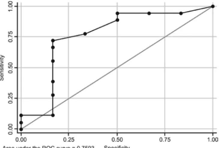
had been decannulated. Forty-six pa-tients had a satisfactory decannulation outcome, while 11 had decannula-tion failure, requiring the return to an artiicial airway within 2 weeks. he calculated Medical Research Coun-cil peripheral muscle strength score was signiicantly lower for the failure group than for the successful decannu-lation group (28.33 ± 15.31 vs. 41.11 ± 11.52; P = 0.04). Scores above or equal 26 had 94.4% sensitivity and 50.0% speciicity for the decannula-tion outcome, with an area under the ROC curve of 0.7593. In addition, white blood cell counts were higher in decannulation failure group patients (14,070 ± 3,073 vs. 10,520 ± 3,402 cells/μL; P = 0.00).
conclusion: his study has shown that peripheral muscle strength and blood leucocyte counts evaluated on the day of decannulation may inluen-ce the tracheostomy decannulation success rate.
Keywords: Tracheostomy; Decannulation; Weaning; Mechanical ventilation
Study developed at Hospital Agamenon Magalhães – HAM – Recife (PE), Brazil.
conlicts of interest: None
Submitted on August 5, 2010 Accepted on December 23, 2010
corresponding author:
Eduardo Eriko Tenório de França Hospital Agamenon Magalhães Estrada do Arraial - Casa Amarela, s/n Zip Code: 50000-000 - Recife (PE), Brazil.
space, less movement inside the trachea, patient comfort, and more effective deglutition.(4) However, recent studies have shown that prolonged tracheos-tomy may lead to late complications, such as tracheal stenosis, bleeding, fistulae, infections, hemorrhage
and bronchoaspiration.(2,4) Additionally, the
mor-tality of tracheostomized patients discharged from the intensive care unit (ICU) to the ward is higher. Therefore, decannulation is a fundamental step in the rehabilitation of critically ill patients.(2,4,5)
Recommendations confirm the benefit of MV weaning protocols for tracheostomized patients and the relevance of physiotherapists’ engagement in this process. However, few studies are available proposing decannulation criteria. The decannulation decision is still based on subjective evaluations rather than on standardized protocols.(1,6)
Usually, the following are considered criteria for decannulation: consciousness level, appropriate oxy-genation and mechanical ventilation, spontaneous breathing without MV, lack of airways obstruction, controlled secretion status, and appropriate deglu-tition. Other factors predictive of decannulation success include MV time, cough effectiveness (rep-resented by the peak expiratory flow) and the abil-ity of the respiratory muscle to provide satisfactory maximal expiratory pressure (PEmax).(2,7,8)
Therefore, considering the lack of parameters known to predict decannulation success, this study aimed to identify possible factors associated with decannulation success.
MetHodS
This was an observational retrospective evaluation study involving the analysis of medical records and including all decannulated patients from a general ICU of a high-complexity reference hospital, which is part of the public Recife – Pernambuco (Brazil’s) healthcare network (Hospital Agamenon Magalhães – HAM), who were admitted from March 1, 2007, to August 8, 2009. This study was approved by the institution’s Ethics Committee.
Routine decannulation criteria included the fol-lowing: clearance of the respiratory failure cause, clinical stability, Glasgow scale rating above 9, lack of tracheal or glottic stenosis, preserved deglutition, and
PEmax above 40 cmH2O, reflecting effective cough.
Personal information, underlying disease, co-morbidities, length of ICU stay, MV days before
decannulation, orotracheal tube days, tracheostomy days, spontaneous breathing days before decannula-tion, number of extubation failures and spontaneous breathing tests failures, antibiotics use, and decan-nulation success or failure were collected.
Data on respiratory muscle (maximal inspiratory pressure - PImax, PEmax and rapid shallow breathing index - RSBI) and peripheral muscle strength (Medical
Research Council [MRC] score),(6) white blood cell
counts and arterial gas on the decannulation day (pH, PaCO2, PaO2 and oxygenation rate – OR) were also collected.
The MRC peripheral muscle evaluation score was calculated for alert patients. This score rated the mus-cle strength between 0 (total palsy) and 5 (normal strength) for specific voluntary bilateral movements (Table 1). The total score ranged between 0 (com-plete tetraparesis) to 60 (normal muscle strength).(6)
table 1 – Medical research council (Mrc) Score
Evaluated movements Shoulder abduction Elbow lexion Wrist extension Hip lexion Knee extension Ankle dorsal lexion Muscle strength degrees
0 = No movement is observed
1 = Visible contraction, no segment movement
2 = Active movement upon resistance of gravity removed 3 = Active movement, against gravity
4 = Active movement against gravity and examiners’ resistance 5 = Normal strength
Consists of six bilaterally evaluated movements, and each movement muscle force was rated between 0 (total palsy) and 5 (normal muscle strength). Total scores ranged between 0 (complete tetraparesis) and 60 (normal muscle strength). Source: Adapted from De Jonghe et al. (2005).(6)
using GraphPad Prism version 4.0, Stata 10.1, and Microsoft Office Excel 2007.
reSultS
During the evaluation period, 1,541 distinct diagnosis patients were admitted to the general ICU of which 143 were tracheostomized; however, only 57 patients met the criteria and were decannulated during their general ICU stay, and 46 were successfully decannulated (81%) (Figure 1).
57 decannulated patients 143 TCT patients 1,541 patients admitted to the ICU
Failure (N = 11) Success (N = 46)
ICU – intensive care unit; TCT - tracheostomy.
Figure 1 - Included patients disposition.
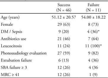
Fifty-seven patients were evaluated of which 37 were female. The diabetes mellitus (DM) prevalence, sepsis prevalence, use of antibiotics, leucocytosis sta-tus, phonoaudiology evaluation, extubation failure, spontaneous breathing attempt (SBA) failure and peripheral muscle strength evaluation data for these 57 patients are displayed in Table 2. The comparison of the decannulation success group patients with the failure group patients showed significantly higher DM and/or sepsis prevalences and more cases of leu-cocytosis in the decannulation failure group.
Table 3 compares the decannulation success and failure groups with respect to the length of ICU stay; ventilatory support time; artificial airway time; ven-tilatory support-free time; pH, PaCO2, PaO2 and OR profile; white blood cell count; PImax and PEmax values; RSBI; and MRC score, with significantly higher leucocyte counts and less peripheral strength in the decannulation failure group (P < 0.01 and P < 0.05, respectively).
Figure 2 displays the MRC versus the decannula-tion outcome ROC curve, with 94.4% sensitivity and 50.0% speciicity for MRC values greater than or equal to 26, with an area under the ROC curve of 0.7593.
table 2 – Sample characterization
Success (N = 46)
Failure (N = 11)
Age (years) 51.12 ± 20.57 54.00 ± 18.22
Female 29 (63) 8 (73)
DM / Sepsis 9 (20) 4 (36)*
Antibiotics use 21 (46) 7 (64)
Leucocitosis 11 (24) 11 (100)*
Phonoaudiology evaluation 27 (59) 9 (82)
Extubation failure 6 (13) 4 (36)
SBA failure ≥ 3 12 (26) 4 (36
MRC ≥ 41 12 (26) 1 (9)
DM – diabetes mellitus; SRA – spontaneous breathing attempt; MRC - Medical Research Council. Results expressed as mean ± standard deviation or number (%). * P < 0.01; Fisher’s exact test.
table 3 – decannulation success predictive factors
Success (N = 46)
Failure (N = 11)
p value*
TICU (days) 44.00 ± 25.64 43.36 ± 21.09 0.94
TMV (days) 35.79 ± 24.36 36.64 ± 21.80 0.82
TOTT (days) 14.50 ± 4.71 15.45 ± 2.77 0.52
TTCT (days) 27.11 ± 25.04 29.00 ± 22.60 0.27
TSB (days) 5.86 ± 4.90 5.54 ± 3.35 0.84
pH 7.44 ± 0.05 7.46 ± 0.05 0.43
PaCO2 (mmHg) 41.85 ± 10.46 34.88 ± 8.16 0.10
PaO2 (mmHg) 106.10 ± 28.84 98.28 ± 32.53 0.53
OR 371.60 ± 122.20 357.40 ± 92.72 0.77
WBC (cél/μL) 10520 ± 3402 14070 ± 3073 0.00
PImax (cmH2O) 61.45 ± 22.68 59.00 ± 16.09 0.75
PEmax (cmH2O) 77.21 ± 27.92 73.20 ± 27.64 0.69
RSBI (ipm/L) 68.80 ± 28.79 73.40 ± 38.94 0.84
MRC 41.11 ± 11.52 28.33 ± 15.31 0.04
Figure 2 – Medical research council (Mrc) score and decannulation outcome roc curve
dIScuSSIon
No decannulation decision-making recommenda-tions are currently available in the literature; howe-ver, some trials suggest different tracheal decannula-tion success predictive factors.(8)
In this study, the current parameters suggested in the literature were adopted; however, we observed a 19% decannulation failure rate. Similar to our results, Mendes et al. had a 20% failure rate when using PImax, PEmax, vital capacity and peak expiratory flow criteria; these two last variables were also analyzed
in our study.(8) Aiming to reduce this population
decannulation failure rate, this study analyzed other factors possibly correlated with tracheal decannulation success.
In our study, both the success and failure groups had no differences with respect to MV time; length of ICU stay; orotracheal tube, tracheostomy and spontaneous breathing times; and arterial gas evaluation on the decannulation day, showing that these variables were not effective tracheostomy decannulation success predictors. These criteria were not considered in previous studies; however, Ceriana et al. established that only patients spontaneously breathing for at least 5 days with stable arterial gasometry and PaCO2 below 60 mmHg would be decannulated, with a low 3% failure rate.(1) In our study, both the success and failure groups had more than 5 spontaneous breathing days, stable arterial gasometry, and PCO2 levels lower
than 60 mmHg.
The MV times were 35.79 and 36.54 days for success and failure patients, respectively. Studies such as that by Jubran et al. and Levine et al. have
shown that respiratory muscle strength is MV time-dependent, i.e., ventilator-related injuries impair the strength of the inspiratory muscles and render MV weaning difficult.(9-11)
Inspiratory muscle strength and appropriate ventilation work load indices, such as PImax and RSBI, are supposed to predict decannulation success. However, in our study, the mean values of these indices were similar for both groups, proving them to be ineffective decannulation success predictors.
Mendes et al. conducted a retrospective study to evaluate the MV and decannulation phases and also failed to identify any PImax differences between the groups, although all decannulated patients had
a PImax above 30 cmH2O.(8) Our study’s similarity
between the PImax and RSBI results may have been influenced by the routine muscle training completed by our tracheostomized patients.
Regarding white blood cell counts, Bach and Saporito reported that decannulation patients should have an antibiotic-free normal leucogram, but this is neither a consensus criterion nor has this criterion been used in other studies.(7) Our findings showed that antibiotic use was unable to predict decannulation success or failure. For leucogram readings, however, the results were significant, showing that 100% of the failure group patients had leucocytosis versus only 24% in the decannulation success group; additionally, the white blood cell count on the day of decannulation was significantly higher in the failure group. Although not directly reflecting an active infective process, the leucocyte count is indicative of ongoing and resolving infective and inflammatory processes and is used for disease phasing guidance.(12)
Another relevant finding in this study was the relationship between peripheral muscle strength, as evaluated using the MRC score, and decannulation success. Immobilization and skeletal muscle weakness are the most common and relevant ICU complications, especially in prolonged MV patients, such as tracheostomized patients. The literature reports that peripheral muscle strength loss is correlated with reduced respiratory muscle strength and MV weaning failure. It was also shown that an MRC score ≥ 41 could be predictive of MV weaning success; however, in our study, this value was not predictive of decannulation success.(13-16)
had 94.4% sensitivity and 50% specificity, represen-ting the best categorization of patients with respect to outcome (success or failure) in our sample. Given that this method is reliable, we could assure that a direct correlation between peripheral muscle strength and decannulation success was found in our findings.(16)
However, additional evaluation is warranted, with larger longitudinal trials and sample sizes, to evaluate the selected variables’ predictive value and to identify values above which the decannulation success rate is significant.
concluSIon
We may conclude that, for the studied population, peripheral muscle strength inluenced the decannulation success rate, as did the white blood cell count on the day of the procedure.
AcKnoWledgMentS
We would like to acknowledge the support of all of the Intensive Care Unit of the Hospital Agamenon Magalhães staff, especially the physiotherapists and intensive physiotherapy residents who supported us in the writing of this manuscript. This manuscript is published in memory of Prof. Jáder Carneiro Júnior, a great professional.
reSuMo
Introdução: A traqueostomia é provavelmente o procedi-mento cirúrgico mais comum realizado em pacientes críticos
objetivando facilitar o desmame do suporte ventilatório. Dire-trizes baseadas em evidências têm conirmado o benefício de protocolos de desmame da traqueostomia e a participação dos isioterapeutas neste processo, porém não existe consenso quan-to aos critérios para decanulação. Portanquan-to, o objetivo do estudo é avaliar a inluência da força muscular periférica e outros índi-ces sobre o suíndi-cesso na decanulação.
Métodos: Análise retrospectiva por meio de levantamento de prontuário de pacientes internados na unidade de terapia intensiva do Hospital Agamenon Magalhães no período de março de 2007 a agosto de 2009.
Método: Este é um estudo observacional, retrospectivo, dos prontuários dos pacientes internados na unidade de terapia intensiva clínico-cirúrgica do Hospital Agamenon Magalhães no período de março de 2007 a agosto de 2009. Foi avaliada a força muscular respiratória e periférica dos pacientes decanulados nesse período.
resultados: Foram avaliados 1.541 pacientes, dos quais, 143 realizaram a traqueostomia, mas apenas 57 pacientes preencheram os critérios de inclusão para serem decanulados, sendo que destes 46 evoluíram com sucesso e 11 com insucesso, considerado a necessidade de retorno a via aérea artiicial no período de duas semanas. A força muscular periférica obtida através do escore do Medical Research Council (MRC) foi signii-cativamente menor no grupo insucesso comparada ao sucesso (28,33 ± 15,31 vs 41,11 ± 11,52; p = 0,04). Valores de MRC ≥ 26 apresentaram uma sensibilidade de 94,4% e uma especii-cidade de 50,0% em relação ao desfecho da decanulação, com uma área sob a curva ROC de 0,7593. Já os leucócitos foram
maiores no grupo insucesso (14070 ± 3073 vs 10520 ± 3402
células/μL ; p = 0,00).
conclusão: O estudo mostrou que a força muscular peri-férica e a contagem dos leucócitos no dia da decanulação inlu-enciaram no sucesso de remoção do traqueóstomo.
descritores: Traqueostomia; Decanulação; Desmame; Ventilação mecânica
reFerÊncIAS
1. Ceriana P, Carlucci A, Navalesi P, Rampulla C, Delmastro M, Piaggi G, et al. Weaning from tracheotomy in long-term mechanically ventilated patients: feasibility of a decisional lowchart and clinical outcome. Intensive Care Med. 2003;29(5):845-8.
2. Stelfox HT, Crimi C, Berra L, Noto A, Schmidt U, Bigatello LM, Hess D. Determinants of tracheostomy decannulation: an international survey. Crit Care. 2008;12(1):R26. 3. Veelo DP, Schultz MJ, Phoa KY, Dongelmans DA,
Binnekade JM, Spronk PE. Management of tracheostomy: a survey of Dutch intensive care units. Respir
Care. 2008;53(12):1709-15.
4. Martinez GH, Fernandez R, Casado MS, Cuena R, Lopez-Reina P, Zamora S, Luzon E. Tracheostomy tube in place at intensive care unit discharge is associated with increased ward mortality. Respir Care. 2009;54(12):1644-52. 5. Hsu CL, Chen KY, Chang CH, Jerng JS, Yu CJ, Yang
PC. Timing of tracheostomy as a determinant of weaning success in critically ill patients: a retrospective study. Crit Care. 2005;9(1):R46-52.
6. De Jonghe B, Sharshar T, Lefaucheur JP, Outin H. Critical illness neuromyopathy. Clin Pulm Med. 2005;12(2):90-6. 7. Bach JR, Saporito LR. Criteria for extubation and
failure. A diferent approach to weaning. Chest. 1996;110(6):1566-71.
8. Mendes TAB, Cavalheiro LV, Arevalo RT, Soneght R. Estudo preliminar sobre a proposta de um luxograma de decanulação em traqueostomia com atuação interdisciplinar. Einstein (São Paulo). 2008;6(1):1-6. 9. Hefner JE. Tracheostomy decannulation: marathons and
inish lines. Crit Care. 2008;12(2):128.
10. Jubran A. Critical illness and mechanical ventilation: efects on the diaphragm. Respir Care. 2006;51(9):1054-61; discussion 1062-4.
11. Levine S, Nguyen T, Taylor N, Friscia ME, Budak MT, Rothenberg P, et al. Rapid disuse atrophy of diaphragm ibers in mechanically ventilated humans. N Engl J Med. 2008;358(13):1327-35.
12. Andriolo A. Guia de medicina laboratorial. Barueri: Manole; 2005.
13. Vassilakopoulos T. Ventilator-induced diaphragm dysfunction: the clinical relevance of animal models. Intensive Care Med. 2008;34(1):7-16.
14. Chiang LL, Wang LY, Wu CP, Wu HD, Wu YT. Efects of physical training on functional status in patients with prolonged mechanical ventilation. Phys her. 2006;86(9):1271-81. 15. De Jonghe B, Bastuji-Garin S, Durand MC, Malissin
I, Rodrigues P, Cerf C, Outin H, Sharshar T; Groupe de Rélexion et d’Etude des Neuromyopathies en Réanimation. Respiratory weakness is associated with limb weakness and delayed weaning in critical illness. Crit Care Med. 2007;35(9):2007-15.
16. Chambers MA, Moylan JS, Reid MB. Physical inactivity and muscle weakness in the critically ill. Crit Care Med. 2009;37(10 Suppl):S337-46. Review.