J of Evolution of Med and Dent Sci/ eISSN- 2278-4802, pISSN- 2278-4748/ Vol. 4/ Issue 41/ May 21, 2015 Page 7241
DELAYED PRESENTATION OF TENSION GASTROTHORAX COMPLICATING
ACUTE TRAUMATIC DIAPHRAGMATIC RUPTURE
Sahajananda H.1,K. T. Venkatesh Murthy2, Karthik3, Soumya Rohit4, Deepak Sivalingam5
HOW TO CITE THIS ARTICLE:
Sahajananda H, K. T. Venkatesh Murthy, Karthik, Soumya Rohit, Deepak Sivalingam. Delayed Presentation of Tension Gastrothorax Complicating Acute Traumatic Diaphragmatic Rupture . Journal of Evolution of Medical and Dental Sciences 2015; Vol. 4, Issue 41, May 21; Page: 7241-7248, DOI: 10.14260/jemds/2015/1051
ABSTRACT: We report a case who presented with respiratory distress after trauma that was treated for a left-sided haemopneumothorax. This was finally diagnosed as giant diaphragmatic hernia with tension gastrothorax. The diagnostic difficulties and complications of wrong diagnosis are discussed. KEYWORDS: Diaphragmatic tear, cardio respiratory embarrassment, Haemopneumothorax, tension gastro thorax.
INTRODUCTION: Blunt trauma to the chest or abdomen can result in diaphragmatic tear with prolapse of abdominal contents into the thoracic cavity.1 Motor vehicle accidents cause the vast majority of these mishaps, although falls and crushing injuries due to thoraco abdominal trauma may also be responsible. The incidence of diaphragmatic rupture after blunt thoracic and abdominal trauma is 0.8 percent to 5 percent.2 The diagnosis is often delayed due to absence of typical symptoms or other major injuries. An isolated blunt traumatic diaphragmatic rupture is rare. An early and precise diagnosis requires a high index of suspicion as missed diaphragmatic injury may result in strangulation of the herniated intra-abdominal contents in the thoracic cavity. Stomach, spleen, transverse colon and small bowel are the most common organs to become incarcerated in the pleural cavity, causing pulmonary compression and varying degrees of mediastinal shift.1,3 Progressive increase in the intrathorasic contents can present as tension thorax with severe cardiorespiratory compromise posing immense challenges.4 Anaesthetizing such patients is indeed a mammoth task. The diagnosis is often delayed due to absence of typical symptoms or other major injuries. We hereby present one such case of blunt traumatic diaphragmatic rupture that had massive viscero-thorax mimicking a haemopneumothorax.
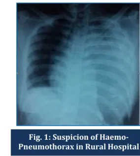
CASE REPORT: A 20 years old female with history of alleged road traffic accident who was a pillion rider, was hit by a tractor. She was taken to a private hospital where an intercostal chest drainage tube was placed after evaluation and chest radiograph, suspecting a haemo-pneumothorax (Fig. 1). After three days when she failed to improve, she was referred to the emergency department of our hospital with tube in-situ.
J of Evolution of Med and Dent Sci/ eISSN- 2278-4802, pISSN- 2278-4748/ Vol. 4/ Issue 41/ May 21, 2015 Page 7242 absence of breath sounds on the left side. Also noted were lacerations on abdomen & right lower limbs and swelling over the right thigh.
Two large bore IV cannulas were secured and crystalloid infusion started. Nasogastric tube was inserted and continuous suction applied and bladder was catheterized to monitor urine output. Two units of packed cells were arranged before shifting her to OT.
Her routine blood investigations were normal except a WBC count of 17600/dl. Arterial blood gas analysis showed pH-7.32, PO2-76mm of Hg, PCO2-45 mm of Hg, BE-4 ECG showed sinus tachycardia and ST-T wave changes depicting strain & mild ischemia in V4 V5 V6. Also there was a fracture of the right femur.
Pre-operative vitals were Pulse Rate - 143 bpm, Blood pressure-94/50mm of Hg, oxygen saturation–80-85% on 6L of O2, RR- 34pm with no air entry on auscultation, over the left lung fields.
In the operation theatre, monitors were applied (Non-invasive B. P., ECG, oxygen saturation & Et.Co2), and she was premedicated with inj. Glycopyrrolate 0.2mg, and inj. Fentanyl 75mcg. Rapid sequence induction and intubation with cuffed ETT 7.0mm were done using Inj. Etomidate, Suxamethonium and cricoid pressure.
Anesthesia was maintained with Oxygen 6 L/min, Sevoflurane 2%, and Intermittent doses of Atracurium. One lung ventilation of the right lung was instituted until the herniated contents were reduced back into the abdomen. Positive pressure ventilation of both lungs was done to inflate the collapsed lung.
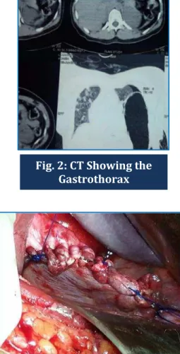
Laparotomy was done using a left paramedian incision. Abdominal organs stomach, jejunum, colon were in the left hemithorax with a 10-centimetre tear in the diaphragm (Fig. 3). The tear was repaired, and finally, IM nailing was done to fix fracture femur. 2 units of packed cells were transfused. The surgery lasted for over 3 hours. Her post op vitals were, PR- 124bpm, BP-128/70 mm Hg, sPO2 97-99 %, urine output of 220mL and on auscultation left upper lobe had mild crepts and ronchi with absent sounds in lower lung fields. Arterial blood gas analysis showed pH-7.34, PO2 -110 mm of Hg, PCO2-37 mm of Hg.
She was shifted to the SICU for further ventilator support and monitoring. She was electively ventilated for 72 hours on Pressure control mode with sedation and non-depolarizing muscle relaxants using Inj. fentanyl. 50mcg/hr, inj. Midazolam 2mg bolus, 1mg/hr. infusion, & inj. vecuronium 1mg/hr infusion. She was administered Inj. Hydrocortisone 100mg, inj. Dexona 8 mg, & inj. Deriphiline 150mg. slow I. V. were also added along with Salbutamol & Budecort nebulizations. Thrombo prophylaxis was administered for 3 days.
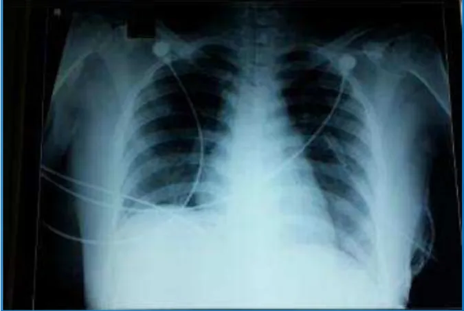
She was gradually weaned off the ventilator over the next 3 days. Lung opacities on the chest x-ray gradually disappeared with improved breath sounds mainly of left lung fields (Fig. 4, 5, 6). On the third day, ICD was removed & she was extubated. She was monitored for another 2 days in ICU and shifted to the ward.
DISCUSSION: Traumatic rupture of the diaphragm may occur due to blunt, penetrating or iatrogenic injuries and mostly involving the left hemi diaphragm.1 Blunt trauma occurs mostly due either to road traffic accident or falls from a height.1
Miller and Howie suggest that three factors are involved in the causation of traumatic diaphragmatic hernia:3
J of Evolution of Med and Dent Sci/ eISSN- 2278-4802, pISSN- 2278-4748/ Vol. 4/ Issue 41/ May 21, 2015 Page 7243 2. Distortion of the diaphragm by chest compression resulting in shearing forces which act to
produce a tear.
3. Congenital weakness of the diaphragm.
In our patient it was a road traffic accident leading to a direct anterior blow to the abdomen which leads to sudden transmission of force through the abdominal viscera (Acts as a hydrodynamic fluid wave leading to significantly increase intra-abdominal pressure and disruption.)2
With the protection afforded to the right leaf of the diaphragm by the liver, about 95% of these hernias occur through the left diaphragm. Stomach, spleen, transverse colon and small bowel are the most common organs to become incarcerated in the pleural cavity, causing pulmonary compression and varying degrees of mediastinal shift.2,4 The resultant cardiopulmonary embarr-assments may be further augmented by pulmonary contusion, pneumothorax or acute blood loss from associated injuries such as fractures and lacerations. Laceration of the spleen is particularly common. Rarely, the herniation may enter the pericardial sac alone, resulting in tamponade.4,5
Tension gastro thorax has previously been described. In the spontaneously ventilating patient the negative pressure generated in the thoracic cavity progressively sucks the stomach into the chest with each breath.6 Eventually, respiratory and hemodynamic compromise ensues, as with a classic tension pneumothorax.6,7 Tension gastro thorax is a rare life-threatening condition occurring when the stomach herniates through a defect in the diaphragm into the chest and becomes distended with air, leading to hemodynamic compromise.6,7,8 This is seen most commonly in the perinatal period in a patient with a congenital diaphragmatic hernia, or after acute major abdominal trauma or surgery in an otherwise healthy individual, or sometimes years later. In these settings, diagnosis requires a high level of suspicion, as this will often be mistaken for a tension pneumothorax, a far more common and equally life-threatening condition.8,9 A poorly differentiated diaphragm on chest radiograph, or visualization of a nasogastric tube in the stomach (in the chest), can help differentiate these conditions.10
Various methods have been used to treat the condition acutely. Nasogastric tubes can be placed to decompress the stomach-although placement may be difficult due to kinking at the level of the diaphragm.10 Needle decompression of the stomach has also been suggested but this may theoretically lead to contamination of the thoracic cavity. Positive pressure ventilation allows immediate re-expansion of the lung and forces intraperitoneal contents back into the abdomen. As the patient will require operative repair, ventilation is already indicated.9,10
Our patient also had a left sided disruption of diaphragm presenting to us with cardio respiratory embarrassment & upon laparotomy stomach, jejunum & colon being the contents intruding into the left hemithorax.
Diagnosis of diaphragmatic tears is indeed a clinical challenge. Cardio respiratory compromise prompts towards early detection and consequently leading to immediate surgical corrections.11 Early diagnosis and treatment abuts further complications like strangulation of the herniated contents which may lead significant morbidity & mortality. The clinical signs in the diagnosis of diaphragmatic hernia include diminished expansion of the chest wall, impaired resonance, adventitious sounds, cardiac displacement, circulatory collapse, cyanosis and dyspneoic and asymmetry of the hypochondria.
J of Evolution of Med and Dent Sci/ eISSN- 2278-4802, pISSN- 2278-4748/ Vol. 4/ Issue 41/ May 21, 2015 Page 7244 of the initial injury and starts acting as a barrier against herniation. Delayed detection becomes evident only when the intrathoracic pressure becomes negative leading to herniation.11
But, in our clinical scenario, due to misdiagnosis in the private hospital, our patient ended up with tension gastrothorax with significant mediastinal shift. Also, there was severe hemodynamic instability with respiratory compromise. The importance of early recognition and treatment of the condition is to be emphasized here: as it can lead to fatal complications like pulmonary contusions & cardiac tamponade.11
Clinical diagnosis can be confirmed radiologically. Initial chest X-ray is the best diagnostic aid in the evaluation of diaphragmatic rupture.12 A diaphragmatic hernia should be suspected if X-ray chest shows absence of fundic gas in its normal position, elevation of the hemi diaphragm and absence of a sharp hemi diaphragm or presence of a hemopneumothorax. On CT absence of continuity of diaphragm are the most sensitive sign & Collar sign being specific.13 The evaluation of the diaphragm by laparotomy remains the gold standard for diagnosis.12,13
Anesthetic management is very challenging in such cases & needs expertise & preparedness. Due to the pre-existing cardio respiratory embarrassment, one can expect extreme degrees of collapse at the time of induction of anesthesia.14 Further positive pressure ventilation leads to re-expansion of the collapsed lungs resulting in shift of mediastinal structures to the opposite side in turn leading to obstruction to the great veins from kinking often causing drastic falls in cardiac output. Such precipitating events in conjunction with hypoxia and acidosis can be responsible for these unexplained deaths.14 LOBB & BUTLIN recommended spontaneous ventilation after intubation with the patient awake until the hernia is reduced or if assisted ventilation is necessary, then the use of low tidal volumes preferable.15
During the anesthetic management of our patient, care was taken to adequately preload before induction and rapid sequence induction was sought for. Further one lung ventilation (right lung) was done with high frequency and low tidal volumes till the abdominal contents were removed from the hemithorax. Extreme vigilance was sought while re expanding the collapsed lung in order to prevent pneumothorax of normal lung & re expansion edema.
Complications like suture-line dehiscence, hemi diaphragmatic paralysis secondary to iatrogenic phrenic nerve injuries, respiratory insufficiency, empyemas and subphrenic abscess can lead to morbidity. Pulmonary embolism can be associated with such trauma but with aggressive prophylaxis this can be prevented.14,15
Post operatively our patient received ventilator support for 72 hours with good tracheal toileting, humidification along with lung recruitment maneuvers.
J of Evolution of Med and Dent Sci/ eISSN- 2278-4802, pISSN- 2278-4748/ Vol. 4/ Issue 41/ May 21, 2015 Page 7245
REFERENCES:
1. Petrone P, Leppaniemi A, Inaba K, Soreid e K, Asensio JA. Diaphragmatic injuries: challenges in the diagnosis and management. Trauma. 2007; 9: 227–36.
2. Gelman R, Mirvis SE, Gens D. Diaphragmatic rupture due to blunt trauma: sensitivity of plain chest radiographs. AJR Am J Roentgenol. 1991; 156: 51–7.
3. MILLER, J. D. & HowI~., P. W. Traumatic rupture of the diaphragm after blunt injury. Brit. J. Surg. 55: 423 (1968).
4. Farhan R, Mallicka MC, Rajeev S, etal. A review on delayed presentation of diaphragmatic rupture. World J Emerg Surg. 2009; 4: 32.
5. D’Cruz, P. Sugathan Compression of right atrium by traumatic diaphragmatic hernia Am Heart J, 133 (1997), pp. 380–383.
6. Tadler SC, Burton JH. Intrathoracic stomach presenting as acute tension gastrothorax. Am J Emerg Med 1999; 17: 370-1.
7. Slater RG. Tension gastrothorax complicating acute traumatic diaphragmatic rupture. J Emerg Med 1992; 10: 25-30.
8. Acute gastric distension: a lesson from the classics. Hospital Medicine Volume 62 Number 3. 9. Horst M, Sacher P, Molz G, et al. Tension gastrothorax. J Pediatr Surg. 2005; 40: 1500–1504. 3. 10.Sterns, l. P., jensen, n. K., sch~, mt, w. R., garamella, j. J., & lynch, m. F. Diaphragmatic disruption
in major thoracic trauma: a review of 16 cases. Canad. J. Surg. 12: 426 (1969).
11.Scrrw~rrOT, W. D. & GALE, J. W. Late recognition and treatment of traumatic diaphragmatic hernias. Arch. Surg. 94: 330 (1967).
12.Meyers BF, McCabe CJ. Traumatic diaphragmatic hernia. Occult marker of serious injury. Ann Surg. 1993; 218: 783–90.
13.Mirvis SE. Shanmuganagthan K. Imaging hemi-diaphragmatic injury. Eur Radiol. 2007; 17: 1411–21.
14.T. R. Lobb, G. R. Butlin: Anaesthesia and traumatic diaphragmatic hernia. Can Anaesth Soc J, 21 (1974), pp. 173–180.
15.Loehnin6, r. W., takaori, m., & safar, p. Circulatory collapse from anaesthesia for diaphragmatic hernia. Arch. Surg. 90: 109 (1965).
J of Evolution of Med and Dent Sci/ eISSN- 2278-4802, pISSN- 2278-4748/ Vol. 4/ Issue 41/ May 21, 2015 Page 7246
Fig. 3: Open Laparotomy and Closure of Rent Fig. 2: CT Showing the
Gastrothorax
J of Evolution of Med and Dent Sci/ eISSN- 2278-4802, pISSN- 2278-4748/ Vol. 4/ Issue 41/ May 21, 2015 Page 7247
Fig. 5: Portable Chest X-Ray on the 3rd Post-Operative day
J of Evolution of Med and Dent Sci/ eISSN- 2278-4802, pISSN- 2278-4748/ Vol. 4/ Issue 41/ May 21, 2015 Page 7248
AUTHORS:
1. Sahajananda H.
2. K. T. Venkatesh Murthy 3. Karthik
4. Soumya Rohit 5. Deepak Sivalingam
PARTICULARS OF CONTRIBUTORS:
1. Professor & HOD, Department of Anaesthesiology, Critical Care & Pain Medicine, Rajarajeswari Medical College & Hospital, Bangalore.
2. Professor, Department of
Anaesthesiology, Critical Care & Pain Medicine, Rajarajeswari Medical College & Hospital, Bangalore.
3. Associate Professor, Department of Anaesthesiology, Critical Care & Pain Medicine, Rajarajeswari Medical College & Hospital, Bangalore.
FINANCIAL OR OTHER
COMPETING INTERESTS: None
4. Associate Professor, Department of Anaesthesiology, Critical Care & Pain Medicine, Rajarajeswari Medical College & Hospital, Bangalore.
5. Post Graduate, Department of Anaesthesiology, Critical Care & Pain Medicine, Rajarajeswari Medical College & Hospital, Bangalore.
NAME ADDRESS EMAIL ID OF THE CORRESPONDING AUTHOR:
Sahajananda H, # 413, 7th Main,
Vijaya Bank Colony, Bannerghatta Road, Bangalore-76.
E-mail: [email protected]