Jemds.com
Case Report
Journal of Evolution of Medical and Dental Sciences/ eISSN- 2278-4802, pISSN- 2278-4748/ Vol. 4/ Issue 95/ Nov. 26, 2015 Page 16124
SPOROTRICHOSIS: A CASE REPORT
N. Sudheer1, A. Venkatakrishna2, M. Nagaswetha3, G. Manmohan4
1Assistant Professor, Department of Dermatology, Venerology & Leprology, Osmania Medical College and Hospital.
2Associate Professor, Department of Dermatology, Venerology & Leprology, Osmania Medical College and Hospital. 3Post Graduate, Department of Dermatology, Venerology & Leprology, Osmania Medical College and Hospital. 4Professor, Department of Dermatology, Venerology & Leprology, OsmaniaMedical College and Hospital.
ABSTRACT
Sporotrichosis is a rare disease in the Southern part of India. A sporadic case of sporotrichosis restricted to primary site of inoculation in a 11-years old student from a non–endemic region of Telangana has been described.
KEYWORDS
Sporotrichosis, Dimorphic.
HOW TO CITE THIS ARTICLE: N. Sudheer, A. Venkatakrishna, M. Nagaswetha, G. Manmohan. Sporotrichosis: A Case Report.
Journal of Evolution of Medical and Dental Sciences 2015; Vol. 4, Issue 95, November 26; Page: 16124-16125, DOI: 10.14260/jemds/2015/2361.
INTRODUCTION
Sporotrichosis is a subcutaneous/systemic fungal infection caused by dimorphic fungus sporothrix schenckii. The fungus occurs in natural environment, presumably in mould form but develops into a yeast like form.[1] S. schenckii is endemic
in the sub-Himalayan region.[2] Agriculturists, foresters,
gardeners, florists and nursery workers handling plants or plant material are particularly at risk. Traumatic inoculation is the typical mode for acquisition of skin infection in immunocompetent hosts. S. schenckii exhibits temperature dimorphism; it exists as a mold at room temperature (26°C) and as yeast in the host tissues (37°C). Both cutaneous and systemic forms of sporotrichosis exist. Three clinical types are described: (i) Fixed Cutaneous, (ii) Lympho Cutaneous, (iii) Disseminated.
CASE REPORT
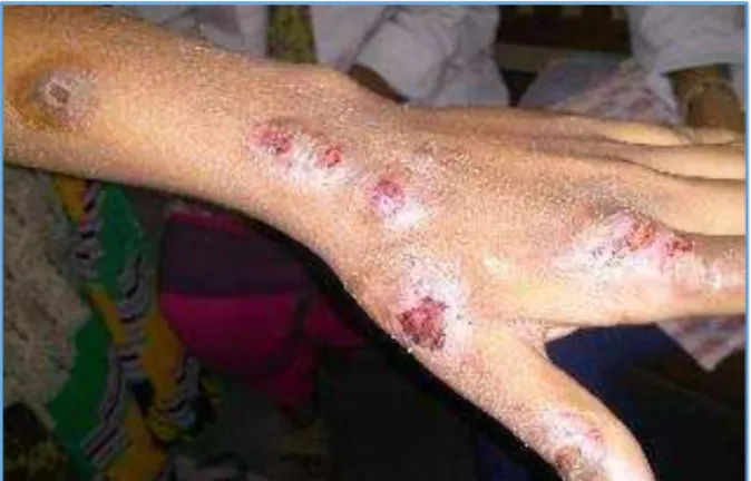
Eleven years boy presented with multiple, well defined, red crusted lesions over dorsum of left hand and arm since 3 years. Initially started as small papule over the dorsum of left hand and then progressed to present stage. H/o trauma present 3 years back, after which lesions appeared 2 months later. No H/o itching, pus discharge, pain or fever. H/o partial remission of lesions on taking medication and relapse within one month. There was no H/o cough, hemoptysis or weight loss.
ON EXAMINATION
Well defined erythematous crusted plaques on left forearm of 2x2 cms, about 3 to 4 in number. The surface was covered with dirty yellow crust. On removing the crust, there was slight serosanguineous discharge. No mucosal involvement or similar lesions elsewhere were seen.
Financial or Other, Competing Interest: None. Submission 03-11-2015, Peer Review 04-11-2015, Acceptance 17-11-2015, Published 26-11-2015. Corresponding Author:
Dr. N. Sudheer,
H. No. 2-2-24/BL/15/6, D.D Colony, Bagh Amberpet,
Hyderabad, Telangana.
E-mail: [email protected] DOI: 10.14260/jemds/2015/2361.
Thickened lymphatic cords were not palpable in the vicinity. Epitrochlear and axillary lymphnodes were enlarged. His general physical and systemic examination were normal. His routine laboratory parameters including the liver and renal function tests and x-ray chest were within normal limits. HBSAg, HCV and mantoux test were negative. Fungal culture was negative.
HPE- evidence of chronic inflammatory dermatosis. On KOH mount, fungal hyphae are seen. PAS and ZN staining were negative. Specimen for both histopathology and KOH mount was taken from crusted lesions (Deep biopsy). The patient was treated with oral antibiotics and tab. itraconazole 100mg once daily. The lesions started resolving 2 weeks after starting therapy. D/D considered and ruled out: cutaneous anthrax, atypical mycobacterial infection, cutaneous tuberculosis, cutaneous leishmaniasis, staphylococcal lymphangitis.
DISCUSSION
The lymphocutaneous variety of sporotrichosis presents a distinctive clinical picture with nodules and ulcers arranged linearly along the lymphatics with thickened lymphatic cords between the nodules, usually on exposed skin.[3],[4] The fixed
variety, where the pathogen remains localized, is less common.[5],[6] It may be a nodular, acneiform, ulcerated or
verrucous form of variable duration.[7] Although reports say
that histopathological examination of tissue stained with conventional H and E lacks sensitivity, a high index of suspicion of sporotrichosis should be exercised whenever a biopsy of a sporotrichoid lesion is received.
Until recently the standard therapy for
lymphocutaneous and cutaneous sporotrichosis was
saturated solution of potassium iodide concentration. Currently, oral itraconazole has become the drug of choice for lymphocutaneous sporotrichosis.[8],[9] The recommended
dosage is 100-200 mg daily, to be administered for 3-6 months. In our patient, the diagnosis of sporotrichosis was suspected clinically and started on tab. itraconazole 100mg daily.
Jemds.com
Case Report
Journal of Evolution of Medical and Dental Sciences/ eISSN- 2278-4802, pISSN- 2278-4748/ Vol. 4/ Issue 95/ Nov. 26, 2015 Page 16125
to various antibiotics for a duration of 3 years and prompt response to oral itraconzole within 2 weeks of treatment made us to report the case.
REFERENCES
1. Venugopal VP, Venugopal VT. Deep fungal infection. In:Valia RG, Valia AR, Siddappa K. editors, IADVL text book & Atlas of Dermatology 2nd edition. Indra Bhalani
publishers: Mumbai; 2001;p.258-84.
2. Agarwal S, Gopal K, Umesh, Kumar B. Sporotrichosis in Uttarakhand (India): a report of nine cases.
Int J Dermatol 2008;47:367-71.
3. Hay RJ, Moore M. Mycology. In: Champion RH, Burton SL, Burns DA, Breathnach SM, editors, Text book of dermatology. 6th edition oxford: Blackwell science;
1998;p.1277-376.
4. Itoh M, Okamoto S, Kanya S. Survey of 260 cases of sporotrichosis. Dermotologica 1986;172:203-13. 5. Hemashettar BM, Kuchbal DS, Hanchinameni Suman P.
Fixed cutaneous sporotrichosis from North Karnataka. Indian J Dermatol Venerol Leprol 1992;58:45-7. 6. Singh P. Sharma RC, Gupta ML. Localised cutaneous
sporotrichosis of face- a case report from India. Indian J Dermatol Venerol Leprol 1980;46:381.
7. Villaca-Neto CM, Rossetti RB, Fischman O, Paschoal LH. Localized cutaneous verrucous sporotrichosis of 26 years' duration. Mycoses 1988;31:353-5.
8. Ghosh A, Chakrabarti A, Sharma VK, Singh K, Singh A. Sporotrichosis in Himachal Pradesh (North India). Trans Royal Soc Trop Med Hyg 1999;93:41-45.
9. Kauffman CA, Bustamante B, Chapman SW, Pappas PG. Clinical Practice Guidelines for the Management of sporotrichosis: 2007 Update by the Infectious Diseases Society of America. Clin Infect Dis 2007;45:1255-65.
Fig. : Pre-Treatment
Fig. : Post Treatment