Percutaneous 17ß-estradiol
replacement therapy in hypertensive
postmenopausal women
1Unidade de Endocrinologia Ginecológica, Hospital de Clínicas de Porto Alegre, 2Departamento de Fisiologia, Universidade Federal do Rio Grande do Sul, and 3Instituto de Cardiologia do Rio Grande do Sul, Porto Alegre, RS, Brasil
M.C. Osório-Wender1,
D. Vitola3 and
P.M. Spritzer1,2
Abstract
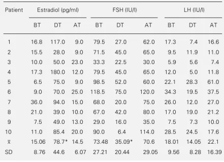
The present study evaluated the short-term effects of percutaneous 17ß-estradiol on blood pressure, metabolic profile and hormonal levels in postmenopausal women with systemic arterial hypertension. After a wash-out period of 15 days, 10 hypertensive patients were treated with guanabenz acetate to control blood pressure, followed by 17ß-estradiol in the form of hydroalcoholic gel administered for 21 of 28 days of each cycle, for 3 cycles. Patients were evaluated before, during and 2 months after estrogen administration. Systolic and diastolic blood pressure or heart rate did not present any significant change in any patient when compared to those periods with the antihypertensive drug only (pretreatment period and 60 days after estrogen therapy was discontinued). Plasma biological markers of hepatic estrogenic action (plasma renin activity, antithrombin III, triglycerides, total cholesterol and lipoproteins) also remained un-changed during the study. Hormone treatment was effective, as indi-cated by the relief of menopausal symptoms, a decrease in FSH levels (73.48 ± 27.21 to 35.09 ± 20.44 IU/l, P<0.05), and an increase in estradiol levels (15.06 ± 8.76 to 78.7 ± 44.6 pg/ml, P<0.05). There was no effect on LH (18.0 ± 9.5 to 14.05 ± 8.28 IU/l). Hormone levels returned to previous values after estrogen treatment was discontinued. The data indicate that short-term percutaneous 17ß-estradiol replace-ment therapy, at the dose used, seems to be a safe hormone therapy for hypertensive menopausal women. Nevertheless, a controlled, pro-spective, randomized clinical assay with a larger number of subjects is needed to definitely establish both the beneficial and harmful effects of hormone replacement therapy in hypertensive women.
Correspondence
P.M. Spritzer
Departamento de Fisiologia Universidade Federal do Rio Grande do Sul
Rua Sarmento Leite, 500 90050-170 Porto Alegre, RS Brasil
Fax: 55 (051) 226-7191
Presented at the XI Annual Meeting of the Federação de Sociedades de Biologia Experimental, Caxambu, MG, Brasil, August 21-24, 1996.
Research supported by FINEP (No. 66.91.0509.00) and CNPq (No. 520107/94-2).
Received April 19, 1996 Accepted July 15, 1997
Key words •Menopause
•Hormone replacement therapy
•Hypertension •17ß-Estradiol •LH
•FSH
Introduction
Three major benefits derive from hor-mone replacement therapy (HRT) in meno-pausal women: a better quality of life due to relief of vasomotor symptoms (1) and treat-ment of urogenital atrophy (2), prevention
and treatment of osteoporosis (3,4), and a decrease in mortality caused by cardiovascu-lar disease (5-8).
HRT using conjugated estrogens adminis-tered orally is related to the onset of hyper-tension in up to 5% of treated women (11). The renin-angiotensin-aldosterone system seems to be one of the mechanisms respon-sible for the association between higher blood pressure levels and the use of oral estrogen in some women (12). Previous studies evalu-ating blood pressure and HRT in normoten-sive women revealed that the use of a non-oral natural estrogen did not alter or even reduced blood pressure levels (13-15).
The aim of the present study was to evalu-ate the effectiveness of percutaneously ad-ministered 17ß-estradiol (17ß-E2), as well as
its metabolic and cardiovascular tolerance by postmenopausal women with blood pres-sure levels controlled by antihypertensive treatment.
Patients and Methods
Ten postmenopausal hypertensive women were selected among those seen at the Gyne-cological Endocrinology Unit of the Hospi-tal de Clínicas de Porto Alegre (HCPA). The criteria for selection were as follows: natural menopause established at least one year be-fore the beginning of the study or surgical menopause with estrogen and gonadotropin levels compatible with postmenopause; no use of drugs capable of affecting the hepatic metabolism or any treatment with hormones for six months before the study; normal gy-necological examination, including palpa-tion of the breasts, mammogram, cytology of the cervix, and histology of the endometrium; normal hepatic and renal function tests and no pathology other than hypertension; symp-toms of estrogen deficit, and hypertension classified as slight to moderate in the ab-sence of antihypertensive treatment (160/ 114 mmHg maximum, or higher blood pres-sure levels in the absence of hemorrhage, exudate, or papilledema upon fundoscopy). In order to determine the presence of hypertension, the various antihypertensive
treatments of the patients (diuretics, methyl-dopa or propranolol) were discontinued for a period of approximately 15 days (wash-out period) during which they underwent car-diologic evaluation. The 10 patients who fulfilled the criteria for inclusion were sub-mitted to complete clinical examination, fun-doscopy, electrocardiogram, and echocardi-ogram (uni- and bidimensional, using a Dop-pler-SSD 730 Aloka apparatus, according to the technique recommended by the Ameri-can Society of Echocardiography).
Guanabenz acetate was used as the anti-hypertensive treatment at progressively higher doses for three to four weeks until blood pressure levels were normalized. Thereafter, the patients were kept on a fixed dose of this drug until the end of the study.
A single researcher using the same ran-dom-zero sphygmomanometer measured blood pressure levels and pulse rate weekly. After a rest period of 10 min, blood pressure and pulse rate were determined three times at 5-min intervals, first in the supine and then in the standing position. Patient weight was checked every 15 days, and the variable used was the body mass index (BMI = weight/ height2).
The following serum concentrations were determined for all patients: estradiol, LH, FSH, plasma renin activity (PRA), aldoster-one, fasting glucose levels and glucose lev-els 2 h after the ingestion of 75 g glucose, total cholesterol, high, low, and very low density lipoproteins (HDL-c, LDL-c and VLDL-c, respectively), and antithrombin III (A-III). Blood samples were collected at three different times: before hormone treatment, at the end of the second cycle using percuta-neous 17ß-E2, and 2 months after hormone
the HCPA using the A-III chromogenic as-say (Baxter Healthcare Corporation, Deerfield, IL). Hormone levels were deter-mined at the Radioimmunoassay Laboratory of the HCPA using commercial radioimmu-noassay kits (Serono, E2-DPC, and
PRA-Baxter for LH, FSH and aldosterone, respec-tively).
After blood pressure was controlled with the antihypertensive drug and samples had been collected for laboratory tests, patients were given 17ß-E2 in the form of
hydroalco-holic gel (Besins-Iscovesco Laboratory, Paris, France). The patients were instructed to ap-ply 2.5 g of the gel (1.5 mg 17ß-E2) to the
skin of the abdomen, thighs and arms daily for 21 days, for 3 cycles of 28 days each. At the end of the second cycle blood samples were collected, and a second echocardio-graphic examination was performed during the third cycle. When the 3-month period of estrogen administration was completed, all patients with a uterus were submitted to hysteroscopy with a microcolpohysteroscope (model Hamou II), in addition to biopsy of the endometrium, and were treated with daily doses of 10 mg medroxyprogesterone ace-tate for 10 days.
The study design was a prospective study, and each patient was her own control. The data were analyzed by analysis of variance and the Student-Newman-Keuls test, with the level of significance set at P<0.05. The data are reported as means ± SD or median and range. The study was approved by the Ethics Committee of the Hospital de Clínicas and informed consent was obtained from each subject.
Results
The median age of the patients was 55 years (48-70 years), the median postmeno-pausal period was 7.5 years (2-23 years), and mean BMI was 28.8 kg/m2 (SD ± 4.37).
Eight of the 10 women had natural meno-pause and the other two had undergone
sur-gical menopause.
Figure 1 shows blood pressure levels dur-ing the study. With the antihypertensive treat-ment, blood pressure levels were controlled and were significantly lower than those dur-ing the period without treatment. The use of hormone therapy associated with the antihy-pertensive drug did not cause any significant change in blood pressure for any patient when compared to the periods with the anti-hypertensive drug only (pretreatment period and 60 days after estrogen therapy was dis-continued).
Menopausal symptoms decreased during estradiol treatment as evaluated weekly by the Kupperman index. No adverse skin ef-fect caused by the use of the gel was re-ported. Serum estradiol (E2)levels during
the pretreatment period were low (15.06 ± 8.76 pg/ml), as typically observed in post-menopausal women. During estrogen re-placement therapy, E2 rose to 78.7 ± 44.6 pg/
ml (P<0.05), a value compatible with that of the follicular phase of normal young fe-males, returning to postmenopausal levels two months after discontinuation of estro-gen therapy (14.5 ± 6.07 pg/ml) (Table 1).
Gonadotropin levels at the initial evalua-tion were 73.48 ± 27.21 IU/l for FSH and 18.01 ± 9.56 IU/l for LH. With the use of percutaneous 17ß-E2, these levels decreased
WASH OUT
BT
DT
AT
0 25 50 75 100 125 150 175 200
BP ( mmHg)
Systolic BP, supine Diastolic BP, supine
Systolic BP, standing Diastolic BP, standing
Table 2. No statistically significant changes were observed in BMI or carbohydrate me-tabolism during the study. The mean values ± SD for PRA, aldosterone, and A-III did not reveal any statistically significant change during any period studied, and values were normal at all stages (Table 2).
The different parameters of the lipid pro-file were altered in most patients before any treatment. Total cholesterol values were high for all 10 patients, triglycerides were above normal levels in 7 patients, LDL-c and VLDL-c were abnormally high in 8 patients, and the HDL-c level was normal in only 4 patients. The mean values of total cholesterol and of the three lipoproteins were high during the three periods studied. However, no signifi-cant differences were observed between the three periods (Table 3).
Plasma renin levels did not change throughout the study; fundoscopy was char-acteristic of a slight hypertensive retinopa-thy in 7 patients, and normal in the other 3; the electrocardiographic exams of 6 patients were normal; there were signs of an over-loaded left ventricle in 3 patients and of an overloaded left auricle in 1 patient (data not shown). Two echocardiograms were per-formed for each patient, with a mean interval of 4 months between exams (an initial exam and another one before discontinuation of estrogen therapy). The results always showed hypertrophy of the left ventricle and de-creased compliance, compatible with hyper-tension. The echocardiographic evaluation performed during treatment with percutane-ous 17ß-E2 did not show any changes in
parietal thickness or systolic function when compared to the initial exam (data not shown). At the beginning of the study, a biopsy of the endometrium obtained by suction ruled out the presence of any pathology in the 8 women with a uterus, revealing atrophic dometria in 6 patients and proliferative en-dometria in the other 2.
Hysteroscopy supplemented with an en-dometrial biopsy performed in the patients at
Table 1 - Hormonal profile of patients at the three periods studied (BT = before treatment, DT = during treatment, AT = two months after discontinuation of treat-ment).
Data are reported as individual values and as means ± SD for each period. *P<0.05 vs BT and AT (Student-Newman-Keuls test).
Patient Estradiol (pg/ml) FSH (IU/l) LH (IU/l)
BT DT AT BT DT AT BT DT AT
1 16.8 117.0 9.0 79.5 27.0 62.0 17.3 7.4 16.6
2 15.5 28.0 9.0 71.5 45.0 65.0 9.5 11.9 11.0
3 10.0 50.0 23.0 33.3 22.5 30.0 5.9 5.6 7.4
4 17.3 180.0 12.0 79.5 45.0 65.0 12.0 5.0 11.8
5 6.5 75.0 9.0 98.5 52.0 60.0 22.1 28.3 61.0
6 9.0 70.0 25.0 118.5 75.0 120.0 34.3 19.5 37.5
7 36.0 94.0 15.0 68.0 20.0 75.0 26.0 12.0 27.0
8 21.0 39.0 10.0 67.0 42.0 80.0 17.0 19.0 21.2
9 7.5 49.0 13.0 29.0 16.0 35.0 7.5 7.3 10.0
10 11.0 85.4 20.0 90.0 6.4 114.0 28.5 24.5 17.6
x
- 15.06 78.7* 14.5 73.48 35.09* 70.6 18.01 14.05 22.1
SD 8.76 44.6 6.07 27.21 20.44 29.05 9.56 8.28 16.39
Table 2 - Lack of effect of percutaneous 17ß-estradiol on body mass index (BMI), fasting glucose, plasma renin activity (PRA), aldosterone and plasma antithrombin III (A-III).
Data are reported as means ± SD for 10 women before treatment (BT), during treatment (DT) and two months after discontinuation of treatment (AT). Patients received 1.5 mg/day 17ß-estradiol percutaneously. There were no significant differ-ences between groups (Student-Newman-Keuls test).
BT DT AT
BMI (kg/m2) 28.82 ± 4.37 28.56 ± 4.36 28.28 ± 4.25
Fasting glucose (mg/dl) 102.5 ± 16.63 101.0 ± 16.58 102.4 ± 14.88
Glucose after 2 h (mg/dl) 123.9 ± 30.66 123.9 ± 37.02 118.6 ± 29.75
PRA (ng ml-1 h-1) 0.53 ± 0.39 0.58 ± 0.56 0.71 ± 0.54
Aldosterone (ng/dl) 10.2 ± 3.24 11.3 ± 5.1 11.5 ± 4.9
A-III (%) 96.5 ± 8.5 95.0 ± 8.74 103.4 ± 11.35
to 35.09 ± 20.44 (P<0.05) and 14.05 ± 8.28 IU/l, respectively. Two months after discon-tinuing the use of estradiol, gonadotropin levels increased again, with FSH being 70.6 ± 29.05 IU/l (P<0.05) and LH 22.1 ± 16.39 IU/l (Table 1).
the end of the estrogen administration period did not reveal any hyperplastic changes in any case. Endometrial histology after hor-mone therapy revealed proliferative endome-tria in 4 women and atrophic endomeendome-tria in the other 4. At the end of the study, the 8 patients with a uterus were instructed to take 10 mg medroxyprogesterone acetate every day for 10 days. Half of them had slight deprivation bleeding and the other half did not bleed.
Discussion
The effectiveness of percutaneous 17ß-E2 observed in the present study through the
relief of menopausal symptoms, the increase in serum estradiol levels and decrease in gonadotropin levels confirmed our prelimi-nary data (16) and the results obtained by other authors who used the same route of estradiol administration (17,18).
In women in the reproductive period, estrogen secreted by the ovary is first re-leased into the systemic circulation and then
reaches the liver, already at physiological concentrations. On the other hand, it is known that the high initial levels of estrogen in the liver following oral administration may cause considerable changes in hepatic metabolism. Many of the negative side effects of estrogen replacement therapy derive from changes in the synthesis of liver proteins that result from first-passage metabolism (19,20).
Recent studies using non-orally adminis-tered estrogens have shown a smaller inci-dence of these side effects, thus confirming the importance of avoiding supraphysiologi-cal hepatic concentrations of this steroid, particularly in patients who are hypertensive or at the risk of suffering thromboembolism (19,20). In the present study we observed that, during the hormone treatment, the plasma levels of liver proteins considered to be specific markers of estrogen action (PRA, A-III, lipids, and lipoproteins) remained stable, showing that the percutaneous ad-ministration of the drug did not alter the estrogen-dependent hepatic metabolism.
A-III is the main physiological inhibitor
Table 3 - Lipid profile of patients receiving 17ß-estradiol percutaneously.
Data are reported as individual values and as means ± SD. The Student-Newman-Kuels test showed no statistically significant difference between the mean values of the variables analyzed at periods BT (before treatment), DT (during treatment) and AT (two months after discontinuation of treatment).
Patient Total cholesterol (mg/dl) HDL-c (mg/dl) LDL-c (mg/dl) VLDL-c (mg/dl) Triglycerides (mg/dl)
BT DT AT BT DT AT BT DT AT BT DT AT BT DT AT
1 246 242 198 39 41 85 163 136 77 44 65 36 222 323 182
2 333 241 304 65 67 57 139 220 193 28 27 54 140 118 268
3 342 316 295 49 45 48 267 232 165 50 39 72 243 195 362
4 204 202 171 60 60 52 115 126 102 19 18 17 95 88 86
5 291 280 291 58 60 28 187 172 194 51 54 28 254 148 141
6 281 217 272 56 48 69 186 144 145 39 25 58 194 123 289
7 287 305 273 43 48 45 190 225 200 31 32 28 156 161 152
8 265 257 265 47 65 69 171 144 169 46 48 27 229 239 236
9 276 251 264 45 43 35 97 98 125 107 101 104 538 483 521
10 334 344 291 59 47 54 198 222 153 72 55 84 360 273 418
x
- 285.9 265.5 262.4 52.1 52.4 54.2 181.3 171.9 152.3 48.7 46.4 50.8 243.5 225.1 265.5
of coagulation and can be significantly re-duced by orally administered estrogen therapy (21). In the present study A-III levels remained unchanged, reinforcing the advan-tage of using this mode of drug administra-tion for those patients whose coagulaadministra-tion factors must not be altered.
Hypertension can occur or become exac-erbated in women who are on estrogen re-placement therapy, probably because of changes in the renin-angiotensin-aldoster-one system. Under physiological conditions, the concentration of the renin substrate rep-resents a constraint in the system. The oral administration of estrogen stimulates the hepatic synthesis of this protein, causing an increase in the levels of angiotensin I and the secretion of aldosterone. Therefore, a sig-nificant increase in renin substrate levels may lead to a state that, together with other predisposing factors, could enhance or start the development of hypertension in suscep-tible women (22). In the group of patients studied, with a record of hypertension prior to menopause, the use of percutaneous 17ß-E2 did not induce modifications either in the
levels of PRA and aldosterone or in the echocardiographic measurements. Moreover, blood pressure levels, controlled by the anti-hypertensive treatment, remained stable throughout the hormone therapy as well as after it was discontinued. Only few studies have been carried out to evaluate HRT in hypertensive menopausal women (23). Lip et al. (23) did not use a wash-out period in order to determine whether the patients actu-ally were hypertensive, or use the same anti-hypertensive drug for all patients, nor did they describe types, doses, and modes of administration of HRT.
Endometrial evaluation of our patients by hysteroscopy with a biopsy performed after the estrogen therapy revealed no hyper-plastic transformations. This confirmed our
expectations, since previous studies carried out to assess the risk of endometrial hyper-plasia due to the exclusive use of estrogen (with no opposing progestogen) have shown that an average period of 8 to 12 months of estrogen therapy was required to bring about a hyperplastic transformation (24). The 4 patients who presented proliferative endome-tria at the end of the study had deprivation vaginal bleeding after the use of medroxy-progesterone acetate. The patients who still had an atrophic endometrium even after es-trogen therapy did not present vaginal bleed-ing after usbleed-ing progestogen.
The data obtained in the present study indicate that percutaneous 17ß-E2, at the
dose used, was effective both in correcting menopausal symptoms and in increasing se-rum E2 while reducing the levels of
gonado-tropins during hormone therapy. The non-oral administration of the steroid, by avoid-ing the first hepatic passage of the molecule, prevented the stimulation of liver protein synthesis responsible for undesirable side effects. The present results indicate good cardiovascular tolerance for this modality of hormone therapy. Furthermore, the data sug-gest that short-term percutaneous 17ß-E2, at
the dose used, seems to be a safe hormone therapy for hypertensive menopausal women. Nevertheless, a controlled, prospective, ran-domized clinical assay with a larger number of subjects is needed to definitely establish both the beneficial and harmful effects of HRT in hypertensive women.
Acknowledgments
References
1. Steingold KA, Laufer L & Chetkowski RJ (1985). Treatment of hot flashes with transdermal estradiol administration. Jour-nal of Clinical Endocrinology and Metabo-lism, 61: 627-632.
2. Bathia NN, Bergman A & Karam MM (1989). Effects of estrogen on urethral function in women with urinary inconti-nence. American Journal of Obstetrics and Gynecology, 160: 176-181.
3. Lindsay R, Hart DM & Clark DM (1984). The minimum effective dose of estrogen for prevention of postmenopausal bone loss. Obstetrics and Gynecology, 63: 759-763.
4. Christiansen C & Riis BJ (1990). 17ß-Es-tradiol and continuous norethisterone: a unique treatment for established os-teoporosis in elderly women. Journal of Clinical Endocrinology and Metabolism, 71: 836-841.
5. Bush TL, Barret-Connor E & Cowan LD (1987). Cardiovascular mortality and con-traceptive use of estrogen in women: re-sults from the Lipid Research Clinics Pro-gram Follow-up Study. Circulation, 75: 1102-1119.
6. Stampfer MJ, Colditz GA, Willet WC, Manson JE, Rosner B, Speizer FE & Hennekens CH (1991). Postmenopausal estrogen therapy and cardiovascular dis-ease: ten year follow-up from the Nurse’s Health Study. New England Journal of Medicine, 325: 756-762.
7. Petitti DB, Perlman JA & Sidnei S (1987). Noncontraceptive estrogens and mortal-ity: long-term follow-up of women in the Walnut Creek study. Obstetrics and Gy-necology, 70: 289-293.
8. Henderson BE, Paganini-Hill A & Ross RK (1991). Decreased mortality in users of estrogen replacement therapy. Archives of Internal Medicine, 151: 75-78. 9. Kornhauser C, Malacara JM, Donato F &
Botello D (1994). High prevalence of arte-rial hypertension in women over 50 years of age in the city of Leon, Guanajuato. Revista de Investigacion Clinica, 46: 287-294.
10. Staessen J, Bulpitt CJ, Fagard R, Lijnen P & Amery A (1989). The influence of meno-pause on blood pressure. Journal of Hy-pertension, 1: 361-364.
11. Lobo RA (1987). Different types of estro-gens and progestoestro-gens. Obstetrics and Gynecology Clinicsof North America, 14: 143-167.
12. Ganten D, Takahashi S, Lindpainter K & Mullins J (1991). Genetic basis of hyper-tension. The renin-angiotensin paradigm. Hypertension, 18 (Suppl III): 109-114. 13. Hassager C, Strom V, Guyene TT &
Christiansen C (1987). The long-term ef-fect of oral and percutaneous estradiol on plasma renin substrate and blood pres-sure. Circulation, 76: 753-758.
14. Von Eiff AW, Plotz EJ, Beck KJ & Czernik A (1971). The effects of estrogens and progestins on blood pressure regulation of normotensive women. American Jour-nal of Obstetrics and Gynecology, 4: 31-47.
15. Osório MC, Vitola D & Spritzer PM (1992). Gel de estradiol 17ß no tratamento de pacientes pós-menopáusicas hipertensas. Arquivos Brasileiros de Endocrinologia e Metabolismo, 36: 40-43.
16. Wren BG, Brown LB & Routledge DA (1982). Differential clinical response to oestrogens after menopause. Medical Journal of Australia, 2: 329-332. 17. Philippe E, Faguer A, Charpin C, Loubière
R, Mascarel A, Leduc F, Dupin Ph, Hemet J, Vauzelle JL, Bigel P, Bapin M, Pagès A, Lenne Y, Laguillaumie B, De Lignières B & Gillet JY (1993). L’endomètre sous traitement hormonal substitutif de la menopause par estradiol percutané et progestérone micronisée à faible dose. Pathologica, 85: 465-487.
18. Gillet JV, Faguer B, Andre G, Magnin G, De Lignières B & Philippe E (1994). Re-cherche d’un traitement hormonal substitutif “sans règles” avec la proges-terone micronisée orale. Journal de Gynecologie, Obstetrique et Biologie de la Reproduction, 23: 407-412.
19. Holst J, Cajander S, Carlstrom K, Damber MG & von Shoultz B (1983). A compari-son of liver protein induction in postmeno-pausal women during oral and percutane-ous oestrogen replacement therapy. Brit-ish Journal of Obstetrics and Gynaecol-ogy, 90: 355-360.
20. De Lignières B, Basdevant A, Thomas G, Thalabard JC, Bodadr CM, Conard J, Guyenne TT, Mairon N, Corvel P, Guy-Grand B, Mauvais-Jarvis P & Sitruk-Ware R (1986). Biological effects of estradiol-17ß in post-menopausal women: oral ver-sus percutaneous administration. Journal of Clinical Endocrinology and Metabolism, 62: 536-541.
21. De Lignières B, Conard J, Samama M, Basdevant A & Guy-Grand B (1983). Dif-ferential AT III response to oral and parenteral administration of 17ß-estradiol. Thrombosis and Haemostasis, 49: 245. 22. Lobo RA (1990). Cardiovascular
implica-tions of estrogen replacement therapy. Obstetrics and Gynecology, 75 (Suppl 4): 18s-25s.
23. Lip GYH, Beevers M, Churchill D & Beevers DG (1994). Hormone replace-ment therapy and blood pressure in hy-pertensive women. Journal of Human Hy-pertension, 8: 491-494.