ISSN 1414-431X
www.bjournal.com.br
www.bjournal.com.br
Volume 45 (12) 1102-1340 December 2012
Braz J Med Biol Res, December 2012, Volume 45(12) 1248-1254
10.1590/S0100-879X2012007500169
doi:
Effects of short-term administration of estradiol on reperfusion
arrhythmias in rats of different ages
S.Q. Savergnini, A.M. Reis, R.A.S. Santos, P.E.B. Santos, A.J. Ferreira and A.P. Almeida
Institutional Sponsors
The Brazilian Journal of Medical and Biological Research is partially financed by
Faculdade de Medicina de Ribeirão Preto Campus
Ribeirão Preto
Explore High - Performance MS Orbitrap Technology In Proteomics & Metabolomics
analiticaweb.com.br S C I E N T I F I C
BIOMEDICAL SCIENCES
AND
Effects of short-term administration of
estradiol on reperfusion arrhythmias
in rats of different ages
S.Q. Savergnini
1, A.M. Reis
1, R.A.S. Santos
1, P.E.B. Santos
2†,
A.J. Ferreira
3and A.P. Almeida
11Departamento de Fisiologia e Biofísica, Instituto de Ciências Biológicas,
Universidade Federal de Minas Gerais, Belo Horizonte, MG, Brasil 2Departamento de Farmacologia, Instituto de Ciências Biológicas,
Universidade Federal de Minas Gerais, Belo Horizonte, MG, Brasil 3Departamento de Morfologia, Instituto de Ciências Biológicas,
Universidade Federal de Minas Gerais, Belo Horizonte, MG, Brasil
Abstract
Little is known about age-related differences in short-term effects of estradiol on ischemia-reperfusion (I/R) insults. The present study was designed to evaluate the effects of short-term treatment with estradiol on reperfusion arrhythmias in isolated hearts of 6-7-week-old and 12-14-month-old female rats. Wistar rats were sham-operated, ovariectomized and treated with vehicle or ovariectomized and treated with 17β-estradiol (E2; 5 µg·100 g-1·day-1) for 4 days. Hearts were perfused by the Langendorff
technique. Reperfusion arrhythmias, i.e., ventricular tachycardia and/or ventricular fibrillation, were induced by 15 min of left coronary artery ligation and 30 min of reperfusion. The duration and incidence of I/R arrhythmias were significantly higher in young rats compared to middle-aged rats (arrhythmia severity index: 9.4 ± 1.0 vs 3.0 ± 0.3 arbitrary units, respectively, P < 0.05). In addition, middle-aged rats showed lower heart rate, systolic tension and coronary flow. Four-day E2 treatment caused
an increase in uterine weight. Although E2 administration had no significant effect on the duration of I/R arrhythmias in
middle-aged rats, it induced a marked reduction in the rhythm disturbances of young rats accompanied by a decrease in heart rate of isolated hearts. Also, this reduction was associated with an increase in QT interval. No significant changes were observed in the QT interval of middle-aged E2-treated rats. These data demonstrate that short-term estradiol treatment protects against
I/R arrhythmias in hearts of young female rats. The anti-arrhythmogenic effect of estradiol might be related to a lengthening of the QT interval.
Key words: Cardioprotection; Estrogen; Ischemia/reperfusion; Langendorff technique; QT interval
Introduction
Premenopausal women are less susceptible to heart diseases and sudden cardiac death than men at a similar age (1). The incidence of coronary diseases in women increases substantially after menopause (2). Animal mod-els of heart diseases clearly support the cardioprotective effects of estrogen supplementation. Basic experiments have revealed that estrogen could attenuate ischemia- or reperfusion-induced ventricular arrhythmias, thereby sug-gesting that sex hormones may have protective effects against cardiac arrhythmias (3). Patten et al. (4) showed
that ovariectomized rats supplemented with 17β-estradiol
(E2) presented reduced cardiomyocyte apoptosis in vitro
and in vivo. Additionally, E2 had an antiarrhythmic activity
and reduced the L-type calcium current (ICaL) in female and
male rats; however, approximately 10-fold less hormone was required to produce these effects in female rats (5). The natural cardioprotection observed in female rats is
absent after ovariectomy (OVX) and E2 supplementation
of estrogen-deficient animals provides protection of myo -cardial function against ischemia/reperfusion (I/R) injury by
decreasing inflammation and apoptotic signaling (6).
Although the effects of chronic administration of
estro-Correspondence: A.J. Ferreira, Departamento de Morfologia, ICB, UFMG, Av. Antônio Carlos, 6627, 31270-901 Belo Horizonte, MG, Brasil. Fax: +55-31-3409-2810. E-mail: [email protected]
†In memoriam
Estradiol and reperfusion arrhythmias 1249
gen have been widely explored in the last few decades, especially in studies involving postmenopausal hormone replacement therapy (7-11), the short-term actions of estro-gen have not been fully evaluated. In spite of this, short-term estrogen therapy might be an important strategy to treat cardiovascular diseases. Indeed, short-term administration
of E2 to ovariectomized spontaneously hypertensive rats
(SHR) causes a significant reduction in blood pressure on
the fourth day of treatment (12), suggesting that the
ben-eficial effects of E2 on the cardiovascular system can be already observed after a few days of hormonal supplement. Thus, the aim of the present study was to investigate the effects of short-term estrogen administration on cardiac reperfusion arrhythmias in young and middle-aged rats since age-related differences might change the cardiovascular
responses to the administration of E2.
Material and Methods
Ethical approval
All experimental protocols were performed in accor-dance with the guidelines for the humane use of laboratory animals of our Institute and approved by the local authority, the Ethics Committee in Animal Experimentation (CETEA), protocol #017/07.
Animals
Female Wistar rats [6-7 weeks old, 200-250 g (young, N = 28) and 12-14 months old, 350-400 g (middle-aged, N = 29)] were obtained from the animal facility of the Instituto de Ciências Biológicas, Universidade Federal de Minas Gerais (CEBIO-UFMG). The animals were housed in a tempera-ture- and humidity-controlled room maintained on a 14:10-h light-dark cycle with free access to food and water.
Surgical ovariectomy
Rats underwent surgical ovariectomy by standard
pro-cedures. Briefly, each ovary was removed and the animals
were allowed to recover from the surgery for 18 days before
experimentation. Successful OVX was confirmed by a reduc -tion in the uterine weight, which was expressed by the ratio between uterine weight (mg) and body weight (g). Control animals (control group: young, N = 10, middle-aged, N = 12) were left with intact ovaries. OVX rats received daily
injec-tions of E2 (E2 group: 5 μg·100 g-1·day-1, SARSA, Hoechst
Marion Roussel, France; young, N = 11, middle-aged, N =
7) or vehicle (vehicle group: 0.1 mL corn oil·100 g-1·day-1;
young, N = 7, middle-aged, N = 10) for 4 days. We chose the
dose and duration of treatment with E2 (4 days) based on a
previous study by Belo et al. (12). In the cited study, the
au-thors showed that short-term E2 treatment (5 µg·100 g-1·day-1
for 4 days) reduced the arterial pressure of ovariectomized animals. Also, this effect was correlated with an increase in plasma atrial natriuretic peptide levels (12). Thus, these
results suggest that only 4 days of administration of E2 can
induce beneficial cardiovascular effects in female rats.
Isolated heart preparation
After a 4-day period of treatment, the rats were decapitated 10 to 15 min after intraperitoneal injection of 400 IU heparin. The thorax was immediately opened and the heart was carefully dissected and perfused with Krebs-Ringer solution
(118.4 mM NaCl, 4.7 mM KCl, 1.2 mM KH2PO4, 1.2 mM
MgSO4.7H2O, 2.5 mM CaCl2.2H2O, 11.7 mM glucose, and
26.5 mM NaHCO3) through a 1.0 ± 0.3-cm aortic stump. The
perfusion fluid was maintained at 37 ± 1°C with a constant
pressure of 65 mmHg and constant oxygenation (5% CO2-95%
O2). A force transducer was attached through a heart clip to
the apex of the ventricles to record the contractile force
(ten-sion, g) in a computer equipped with a data-acquisition system
(Biopac System, USA). A diastolic tension of 1.0 ± 0.2 gwas
applied to the hearts. Electrical activity was recorded with an electrocardiogram (Nihon Kohden, Japan) with the aid of 2 cotton wicks placed directly on the surface of the right atrium
and left ventricle. Coronary flow was measured by collecting
the perfusate over a period of 1 min at regular intervals. The
systolic tension and coronary flow parameters were normalized
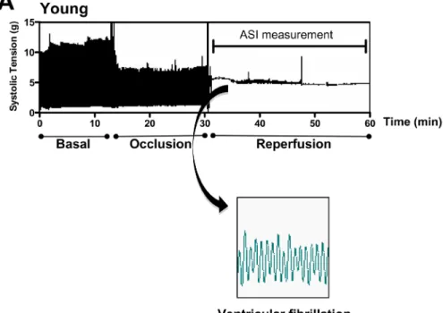
to the heart mass. Because a diastolic tension of 1.0 ± 0.2 g was applied to the hearts, diastolic tension was not normalized to heart mass. After the equilibration period (approximately 30 min), the left anterior descending coronary artery was com-pletely occluded by the method described by Lubbe et al. (13), beneath the left auricular appendage together with the adjacent veins. The ligature was released after 15 min and reperfusion was performed for an additional 30 min. Cardiac arrhythmias
were defined as the presence of ventricular tachycardia and/ or ventricular fibrillation after the ligature of the coronary artery
was released. To obtain a quantitative measurement, the ar-rhythmias were graded arbitrarily by their duration, with the duration of 30 min considered to be irreversible arrhythmias. Therefore, the occurrence of cardiac arrhythmias for 0 to 3 min was assigned a factor of 2; 3 to 6 min was assigned a factor of 4; 6 to 10 min was assigned a factor of 6; 10 to 15 min was assigned a factor of 8; 15 to 20 min was assigned a factor of 10; 20 to 25 min was assigned a factor of 11, and 25 to 30 min was assigned a factor of 12. A value of 0 to 12 was thus obtained in each experiment and was denoted as arrhythmia severity index (ASI) (14).
Statistical analysis
Data are reported as mean ± SEM. Statistical analysis
was performed by the Student t-test or one- or two-way
ANOVA followed by the Bonferroni test. P < 0.05 was
con-sidered to be significant (GraphPad Prism 4.0, USA).
Results
As depicted in Figure 1, ventricular tachycardia and/or
ventricular fibrillation were observed in young and
duration of I/R arrhythmias was significantly longer in
young rats compared to middle-aged rats (ASI: 9.4 ± 1.0
vs 3.0 ± 0.3 arbitrary units in young and middle-aged rats,
respectively; Figure 2A). In addition, 50% of the hearts from
Estradiol and reperfusion arrhythmias 1251
young rats presented irreversible arrhythmias while none of the hearts from middle-aged animals showed irrevers-ible arrhythmias during the reperfusion period. Thus, the incidence of irreversible arrhythmias was markedly higher in young rats compared to older rats (Figure 2B).
The success of the OVX procedure and of E2 treatment
was confirmed by the analysis of uterine weight. OVX caused a significant reduction of the uterine weight of both young and
middle-aged rats, which was reversed by 4-day E2
adminis-tration (Figure 3). No significant changes in body weight were
observed when comparing the young groups (control: 179.8
± 4.8 g; vehicle: 173.8 ± 4.9 g; E2: 172.0 ± 6.8 g) or when
comparing the middle-aged groups (control: 351.4 ± 15.8 g;
vehicle: 332.0 ± 13.3 g; E2: 356.4 ± 12.8 g).
We next determined if short-term administration of E2
might modify the duration and incidence of I/R arrhythmias
in young and middle-aged rats. Short-term E2 administration
induced a significant reduction in the rhythm disturbances
of young rats (ASI: 6.6 ± 1.7 in vehicle group vs 3.8 ± 0.8
in E2 group; Figure 4A). On the other hand, treatment with
E2 did not cause any significant effect on the duration of I/R arrhythmias in middle-aged rats (Figure 4B).
In terms of cardiac function, control and ovariectomized E2-treated middle-aged rats presented lower coronary flow
Figure 2. Effect of age on ischemia/reperfusion arrhythmias in female rats. Arrhythmias were produced by 15-min occlusion of the left anterior descending coronary artery in isolated rat hearts followed by reperfusion. A, Averaged arrhythmia severity index (ASI) and B, percentage of irreversible arrhythmias. N = 8-10 rats. *P < 0.05 vs young group (Student t-test).
Figure 3. Effects of ovariectomy (OVX) and hormonal treatment (estradiol) on uterine weight. Uterine weight is reported as the ratio between uterine weight (mg) and body weight (g). N = 4-6 rats. *P < 0.05 vs control of the same age, #P < 0.05 vs vehicle-treated group of the same age (two-way ANOVA followed by the Bonferroni test).
compared to young rats. In ovariectomized E2-untreated rats no significant differences were observed between young
and middle-aged animals (Figure 5A). The systolic tension
was lower in control and in ovariectomized E2-untreated and
-treated middle-aged animals compared to young rats.
How-ever, systolic tension was significantly
higher in vehicle-treated rats than in control middle-aged animals (Figure
5B). No significant differences in
diastolic tension were observed in any of the groups (Figure 5C). Heart rate was lower only in control middle-aged animals compared to young rats. In OVX-untreated and OVX + E2-treated rats, no significant differ -ences were observed between young and middle-aged animals. However,
the heart rate of young E2-treated
rats was significantly lower than that
of young control animals (Figure 5D). In addition, E2
adminis-tration induced a significant increase in QT and PR intervals in young rats compared to control. No significant changes in
QT or PR intervals were observed in middle-aged E2-treated
rats (Table 1).
Figure 5. Effects of estradiol on coronary flow (A), systolic tension (B), diastolic tension (C), and heart rate (D) of isolated perfused hearts from young and middle-aged female rats during the basal period. OVX = ovariectomy. N = 5-11 rats. *P < 0.05 (two-way ANOVA followed by the Bonferroni test).
Table 1. Effects of estradiol on PR and QT intervals of hearts isolated from young and middle-aged female rats during the basal period.
Young rats (6-7 weeks old) Middle-aged rats (12-14 months old)
PR (ms) QT (ms) PR (ms) QT (ms)
Control 33.15 ± 1.41 75.95 ± 2.37 37.76 ± 0.52 79.10 ± 3.55 OVX + Veh 34.74 ± 1.82 70.51 ± 4.56 36.53 ± 2.06 73.91 ± 2.91 OVX + E2 39.67 ± 1.87+ 87.65 ± 3.12+* 37.32 ± 2.20 77.05 ± 1.74
Estradiol and reperfusion arrhythmias 1253
Discussion
The results obtained in this study show that E2
treat-ment was less effective in attenuating cardiac reperfusion arrhythmias in middle-aged female rats compared to young female rats, thereby suggesting that the protective effects of estrogen on I/R insults are age-dependent. One possible
explanation for this observation is that aging influences the
cellular regulation of cardiac estrogen receptors (ERs).
The cardiac effects of E2 are mediated, at least in part,
by two distinct receptors: ER-alpha and ER-beta (15-17). Vornehm et al. (18) found that both ER-alpha and ER-beta
are involved in mediating E2-induced rapid cardioprotection
after I/R injury. Additionally, a membrane-bound G
protein-coupled estrogen receptor (GPER) with high affinity for estrogen has been identified in the heart (19,20). Activation
of GPER improves the functional recovery and reduces the infarct size in isolated rat hearts following I/R through a PI3K-dependent and gender-independent mechanism (21). Although we did not evaluate cardiac ER expression in the current study, Jazbutyte et al. (22) demonstrated
that ER-alpha expression in hearts was significantly lower
in sham-operated and in ovariectomized senescent (24 months old) SHR compared to young animals (3 months old). On the other hand, the expression of ER-beta was detected at comparable levels in hearts of young and senescent
rats. Taken together, these findings suggest that cardiac ER expression decreases with age and may influence the
responses to E2 treatment. Consequently, this leads to a
reduction in the cardioprotective effects of E2.
One may argue that 4-day E2 treatment could not be
sufficient to modulate the cardiovascular system. However,
as shown by Belo et al. (12), short-term E2 administration
resulted in intense reduction of SHR blood pressure with a concomitant increase in the synthesis and release of atrial natriuretic peptide, which is mainly synthesized, stored and secreted by cardiac atria (23). Also, many studies have demonstrated the effects of estrogen infusion on cardiac
activity in rats (5,24). We found that the efficiency of
short-term E2 treatment was demonstrated by the increase in
uterine weight observed in ovariectomized E2-treated
young and middle-aged rats. These latter data suggest that estrogen metabolism is similar in young and middle-aged rats, thereby indicating that aging did not interfere with the
metabolism of E2.
In keeping with previous reports, we observed that, in addition to the antiarrhythmogenic effect induced by
1. Wenger NK. Clinical characteristics of coronary heart dis-ease in women: emphasis on gender differences. Cardio-vasc Res 2002; 53: 558-567.
2. Rosano GM, Simon T, Mercuro G, Sans S,
Schenck-Gustaff-son K, StevenSchenck-Gustaff-son JC, et al. Hormone replacement therapy: where we stand in Europe. Eur Heart J 2001; 22: 439-441. 3. Wang Y, Wang Q, Zhao Y, Gong D, Wang D, Li C, et al.
Pro-tective effects of estrogen against reperfusion arrhythmias
References
estrogen treatment, young animals had an increased QT interval. Indeed, Burke et al. (25) have reported that the QT interval tends to be longer in women than in men. Also,
Philp et al. (5) have shown that E2 induces a blockade
of Ca2+ channels in female rats. Ca2+ channel blockers
possess antiarrhythmogenic actions in animal models of myocardial ischemia (26) and may contribute to the longer
QT interval observed in females (27). Additionally, the ICaL
transmural gradient together with lower IK and IK1 densities
in female than in male rabbit ventricles (28) could result in prolonged repolarization and greater transmural
disper-sion of repolarization (27). Thus, our findings suggest that
prolongation of the QT interval might be involved in the antiarrhythmogenic effects observed in ovariectomized young rats treated with estrogen.
Although there is evidence that E2 can contribute to
the reduction of arrhythmias as a consequence of vasore-laxation (29,30), in our study this possibility is an unlikely
mechanism since E2-treated rats presented no alterations
in coronary flow.
Also, it is important to note that, although we did not
measure the plasma E2 levels of the animals, Belo et al. (12)
demonstrated that treatment with E2 for 4 days induces a
sustained increase in plasma E2 levels, reaching 40-50 pg/
mL. These levels are higher than the plasma concentration
of E2 in female control rats, whose values range between
5 and 40 pg/mL during the estrous cycle (12,31). It is likely
that estrogen levels were higher in the OVX + E2-treated
group compared to both the OVX + vehicle and the intact control group. Thus, estrogen treatment producing blood concentrations exceeding endogenous levels might be required to reduce ASI in young animals.
The current study demonstrated that short-term E2
treat-ment protects against I/R arrhythmias in hearts of young females rats, but not of middle-aged rats, thereby
indicat-ing that the anti-arrhythmogenic effect of E2 is dependent
on age. This cardioprotective effect might be related to a lengthening of the QT interval. However, further experiments are needed to clearly identify the mechanisms underlying
the anti-arrhythmogenic effects of aging and of E2 in young
female rats.
Acknowledgments
following severe myocardial ischemia in rats. Circ J 2010; 74: 634-643.
4. Patten RD, Pourati I, Aronovitz MJ, Baur J, Celestin F, Chen X, et al. 17Beta-estradiol reduces cardiomyocyte apoptosis
in vivo and in vitro via activation of phospho-inositide-3 kinase/Akt signaling. Circ Res 2004; 95: 692-699.
5. Philp KL, Hussain M, Byrne NF, Diver MJ, Hart G, Coker SJ. Greater antiarrhythmic activity of acute 17beta-estradiol in female than male anaesthetized rats: correlation with Ca2+ channel blockade. Br J Pharmacol 2006; 149: 233-242. 6. Wang M, Tsai BM, Reiger KM, Brown JW, Meldrum DR.
17-Beta-estradiol decreases p38 MAPK-mediated myocar-dial inflammation and dysfunction following acute ischemia.
J Mol Cell Cardiol 2006; 40: 205-212.
7. Hulley S, Grady D, Bush T, Furberg C, Herrington D, Riggs B, et al. Randomized trial of estrogen plus proges-tin for secondary prevention of coronary heart disease in postmenopausal women. Heart and Estrogen/Progestin Replacement Study (HERS) Research Group. JAMA 1998; 280: 605-613.
8. Grady D, Herrington D, Bittner V, Blumenthal R, Davidson M, Hlatky M, et al. Cardiovascular disease outcomes during 6.8 years of hormone therapy: Heart and Estrogen/Progestin Replacement Study follow-up (HERS II). JAMA 2002; 288: 49-57.
9. Hulley S, Furberg C, Barrett-Connor E, Cauley J, Grady D, Haskell W, et al. Noncardiovascular disease outcomes during 6.8 years of hormone therapy: Heart and Estrogen/ Progestin Replacement Study follow-up (HERS II). JAMA
2002; 288: 58-66.
10. Rossouw JE, Anderson GL, Prentice RL, LaCroix AZ, Koop-erberg C, Stefanick ML, et al. Risks and benefits of estrogen plus progestin in healthy postmenopausal women: principal results from the Women’s Health Initiative randomized con-trolled trial. JAMA 2002; 288: 321-333.
11. Anderson GL, Limacher M, Assaf AR, Bassford T, Beresford SA, Black H, et al. Effects of conjugated equine estrogen in postmenopausal women with hysterectomy: the Women’s Health Initiative randomized controlled trial. JAMA 2004; 291: 1701-1712.
12. Belo NO, Silva-Barra J, Carnio EC, Antunes-Rodrigues J, Gutkowska J, Dos Reis AM. Involvement of atrial natriuretic peptide in blood pressure reduction induced by estradiol in spontaneously hypertensive rats. Regul Pept 2004; 117: 53-60.
13. Lubbe WF, Daries PS, Opie LH. Ventricular arrhythmias as-sociated with coronary artery occlusion and reperfusion in the isolated perfused rat heart: a model for assessment of antifibrillatory action of antiarrhythmic agents. Cardiovasc Res 1978; 12: 212-220.
14. Ferreira AJ, Santos RA, Almeida AP. Angiotensin-(1-7): cardioprotective effect in myocardial ischemia/reperfusion.
Hypertension 2001; 38: 665-668.
15. Grohe C, Kahlert S, Lobbert K, Stimpel M, Karas RH, Vetter H, et al. Cardiac myocytes and fibroblasts contain functional estrogen receptors. FEBS Lett 1997; 416: 107-112. 16. Taylor AH, Al-Azzawi F. Immunolocalisation of oestrogen
receptor beta in human tissues. J Mol Endocrinol 2000; 24: 145-155.
17. Mendelsohn ME, Karas RH. Molecular and cellular basis of cardiovascular gender differences. Science 2005; 308: 1583-1587.
18. Vornehm ND, Wang M, Abarbanell A, Herrmann J, Weil B, Tan J, et al. Acute postischemic treatment with estrogen receptor-alpha agonist or estrogen receptor-beta agonist im-proves myocardial recovery. Surgery 2009; 146: 145-154. 19. Filardo EJ, Quinn JA, Frackelton AR Jr, Bland KI. Estrogen
action via the G protein-coupled receptor, GPR30: stimula-tion of adenylyl cyclase and cAMP-mediated attenuastimula-tion of the epidermal growth factor receptor-to-MAPK signaling axis. Mol Endocrinol 2002; 16: 70-84.
20. Thomas P, Pang Y, Filardo EJ, Dong J. Identity of an estro-gen membrane receptor coupled to a G protein in human breast cancer cells. Endocrinology 2005; 146: 624-632. 21. Deschamps AM, Murphy E. Activation of a novel estrogen
receptor, GPER, is cardioprotective in male and female rats.
Am J Physiol Heart Circ Physiol 2009; 297: H1806-H1813. 22. Jazbutyte V, Hu K, Kruchten P, Bey E, Maier SK, Fritzemeier
KH, et al. Aging reduces the efficacy of estrogen substitution to attenuate cardiac hypertrophy in female spontaneously hypertensive rats. Hypertension 2006; 48: 579-586. 23. Lang RE, Tholken H, Ganten D, Luft FC, Ruskoaho H, Unger
T. Atrial natriuretic factor - a circulating hormone stimulated by volume loading. Nature 1985; 314: 264-266.
24. Li J, Xiao J, Liu Y, Zhang G, Zhang H, Liang D, et al. Mito-chondrial benzodiazepine receptors mediate cardioprotec-tion of estrogen against ischemic ventricular fibrillacardioprotec-tion.
Pharmacol Res 2009; 60: 61-67.
25. Burke JH, Ehlert FA, Kruse JT, Parker MA, Goldberger JJ, Kadish AH. Gender-specific differences in the QT interval and the effect of autonomic tone and menstrual cycle in healthy adults. Am J Cardiol 1997; 79: 178-181.
26. Curtis MJ, Walker MJ. The mechanism of action of calcium antagonists on arrhythmias in early myocardial ischaemia: studies with nifedipine and DHM9. Br J Pharmacol 1988; 94: 1275-1286.
27. Pham TV, Robinson RB, Danilo P Jr, Rosen MR. Effects of gonadal steroids on gender-related differences in transmural dispersion of L-type calcium current. Cardiovasc Res 2002; 53: 752-762.
28. Liu XK, Katchman A, Drici MD, Ebert SN, Ducic I, Morad M, et al. Gender difference in the cycle length-dependent QT and potassium currents in rabbits. J Pharmacol Exp Ther
1998; 285: 672-679.
29. Kitazawa T, Hamada E, Kitazawa K, Gaznabi AK. Non-genomic mechanism of 17 beta-oestradiol-induced inhibition of contraction in mammalian vascular smooth muscle. J Physiol 1997; 499 (Part 2): 497-511.
30. Santos RL, Abreu GR, Bissoli NS, Moyses MR. Endothelial mediators of 17 beta-estradiol-induced coronary vasodila-tion in the isolated rat heart. Braz J Med Biol Res 2004; 37: 569-575.