CHLOROTIC SPOTS ON
Clerodendrum
, A DISEASE
CAUSED BY A NUCLEAR TYPE OF
Brevipalpus
(ACARI:
TENUIPALPIDAE) TRANSMITTED VIRUS
Elliot Watanabe Kitajima1*; Karen Sumire Kubo2; Paulo de Tarso Oliveira Ferreira2; Berenice Kussumoto de Alcântara3; Alessandra Jesus Boari4; Renata Takassugi Gomes2; Juliana Freitas-Astua5,6; Jorge Alberto Marques Rezende1; Gilberto José de Morais1; Renato Barbosa Salaroli1
1
USP/ESALQ Depto. de Entomologia, Fitopatologia e Zoologia Agrícola, C.P. 09 13418900 Piracicaba, SP Brasil.
2
USP/ESALQ - Programa de Pós- Graduação em Fitopatologia. 3
USP/ESALQ - Graduanda em Engenharia Florestal. 4
Universidade Federal de Sergipe - Depto. de Agronomia, Av. Marechal Rondon, s/n - 49100-000 - São Cristóvão, SE - Brasil.
5
Embrapa Mandioca e Fruticultura Tropical - Rua Embrapa, s/n - 44380-000 - Cruz das Almas, BA - Brasil. 6
IAC/APTA - Centro Citros “Sylvio Moreira”, C.P. 4 - 13490-970 - Cordeirópolis, SP - Brasil. *Corresponding author <[email protected]>
ABSTRACT: Chlorotic spots have been observed in plants of Clerodendrum x speciosum growing in residential gardens and parks in Piracicaba, SP, Brazil. Thin sections of diseased tissues revealed characteristic cytopathic effects of the nuclear type of the Brevipalpus (Acari: Tenuipalpidae) mite-transmitted viruses (BTrV). Brevipalpus mites, identified as B. phoenicis, infesting symptomatic C. x speciosum plants transmitted the pathogen to healthy C. x speciosum and to C. thomsonae, Gomphrena globosa, Hibiscus cannabinus, H. coccineus, H. schizopetalus, Salvia leucantha, Spathiphyllum wallasi and Tetragonia expansa causing chlorotic spots on their leaves. Mechanical inoculation using leaf extracts from infected C. x speciosum resulted in chlorotic spots on inoculated C. x speciosum, Chenopodium quinoa, C. amaranticolor, G. globosa, H. cannabinus, H. coccineus and T. expansa leaves. C. amaranticolor and C. quinoa kept at 28 - 30°C became systemically infected. The same cytopathic effects caused by the nuclear type of BTrV were seen in tissues from all infected test plants by electron microscopy. The virus was purified from systemically infected leaves of C. amaranticolor and C. quinoa. A polyclonal antiserum obtained from an immunized rabbit presented a strong reaction with the homologous antigen in ELISA tests. The results suggest that this chlorotic spot disease of C. x speciosum is caused by a new species of the nuclear type of BTrV, tentatively named Clerodendrum chlorotic spot virus (ClCSV).
Key words: Brevipalpus phoenicis, Clerodendrum x speciosum, host range, transmission, purification
MANCHA CLORÓTICA DO
Clerodendrum
, UMA ENFERMIDADE
CAUSADA POR UM VÍRUS DO TIPO NUCLEAR, TRANSMITIDO PELO
ÁCARO
Brevipalpus phoenicis
(ACARI: TENUIPALPIDAE)
folhas com infecção sistêmica de C. amaranticolor e C. quinoa. Injeções de preparações purificadas em coelho geraram um anti-soro policlonal que reagiu especificamente com o antígeno homólogo em teste de ELISA. As evidências obtidas indicam que as manchas cloróticas do Clerodendrum estão associadas a um VTB do tipo nuclear, tentativamente denominado de vírus da mancha clorótica do Clerodendrum (Clerodendrum chlorotic spot virus- ClCSV).
Palavras-chave: Breviplapus phoenicis, Clerodendrum x speciosum, transmissão, gama de hospedeiras, purificação
fee ringspot (CoRSV) and Passion fruit green spot (PFGSV) (Kitajima et al., 2003; 2006b). The Brevipalpus genus includes about 300 species world-wide (Welbourn et al., 2003), but only three species (B. californicus (Banks), B. obovatus Donnadieu and B. phoenicis) are involved in plant virus transmission so far (Childers et al., 2003a). These three species may naturally infest up to 900 different plant species in 513 genera and 139 families (Childers et al., 2003b). De-spite the worldwide distribution in tropical and sub-tropical regions of these mites, infection of plant vi-ruses transmitted by Brevipalpus (BTrV) are restricted to the American continent. Natural infection by BTrVs include more than 40 plant species of 24 botanical families (Kitajima et al., 2003; 2006b). The only ex-ception is OFV which has been found worldwide in orchids (Kondo et al., 2003).
This study presents a detailed description of the transmission and host range of a new species of a nuclear type of BTrV isolated from the chlorotic spots on leaves of C. x speciosum, which is tentatively named Clerodendrum chlorotic spot virus (ClCSV). Results of cytopathology, purification and serology are also reported.
MATERIAL AND METHODS
Virus source - Clerodendrum x speciosum plants growing in Piracicaba, State of São Paulo, Brazil (22º43’S and 47º38’W) exhibiting chlorotic spots on the leaves and heavily infested with B. phoenicis were used as source of inoculum of the virus. Infection was confirmed by the presence of the nuclear type of BTrV by electron microscopy.
Mite transmission assays - adult mites, identified as B. phoenicis were collected with a fine needle from symptomatic C. x speciosum plants and transferred to 39 species of test-plants (Tables 1 and 2) grown in pots in a greenhouse. Three plants of each species were used in the transmission tests, placing ten adult mites on four leaves of each tested plant. C. x speciosum plants not infested by mites were used as control. The mites were kept on the plants for five days. After eliminating the mites with chemical sprays, they were kept for symptom development and, sub-sequently, analyzed by electron microscopy.
INTRODUCTION
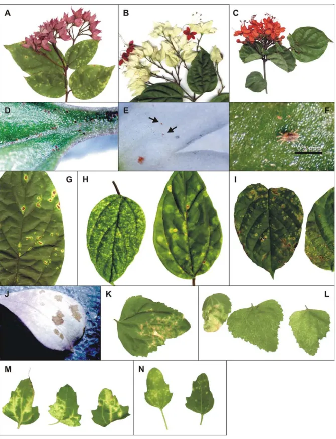
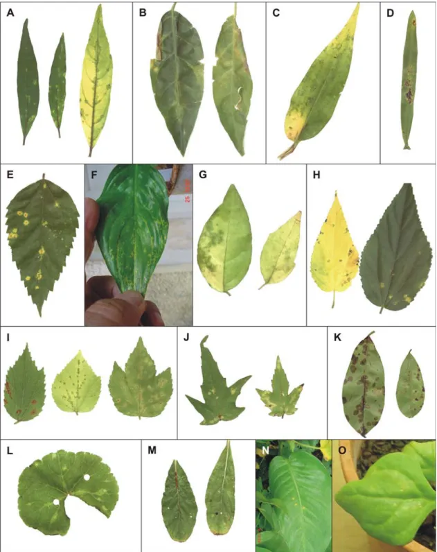
The genus Clerodendrum comprises about 400 species, most of which grow in warm temperate or tropical climates. The name is derived from the Greek words “Clerodendrum” for chance (klero) and tree (dendrum) and refers back to the original species name “fortunate”. Many publications refer to this genus as Clerodendron but Clerodendrum is the official spelling. Originally considered as a member of the family Verbenaceae, the genus Clerodendrum is now consid-ered as a member of the family Lamiaceae based on cla-distic analysis of the chloroplast DNA and internal tran-scriber spacer sequences (Steane et al., 2004). Clerodendrum plants appear as trees, shrubs and scrabbles and are commonly used as ornamentals (KVL, 2006). Clerodendrum thomsonae Baulf.(bleeding heart), C. x speciosum Tiej. et Bin.(glorybower, Java glory vine, heart vine, Pagoda flower) and C. splendens G. Don. (flaming glorybower)are among the most cultivated in home gardens and parks in Brazil and elsewhere, cov-ering fences and walls (Lorenzi & Souza, 2001) (Fig-ure 1, A-C). There are few reports of viral diseases af-fecting plants of this genus. A yellow mosaic of C. inerme, caused by a begomovirus was reported in In-dia (John et al., 2006) and a vein clearing of C. x speciosum, associated with an unidentified rhabdovirus, was found in Brazil (Schuta et al., 1997).
Plants of C. x speciosum exhibiting chlorotic and necrotic spots on their leaves (Figure 1, G) were found in a residential garden at Piracicaba, SP, Brazil, infested by mites identified as Brevipalpus phoenicis (Geijskes) (Figure 1 D, F). Electron microscopic examination of the tissue sections of these spots revealed cytopathic ef-fects of the nuclear type of the Brevipalpus mite trans-mitted viruses (BTrV) (Kitajima et al., 2003). Similar symptoms, always associated with Brevipalpus mite in-festation, were also found in C. thomsonae and C. splendens (Figure 1 H, I) in Piracicaba and other cities of the states of São Paulo, Santa Catarina, Amazonas and Distrito Federal. In C. thomsonae, brownish spots were also observed on flower petals (Figure 1, J) when infested by Brevipalpus (Figure 1, E).
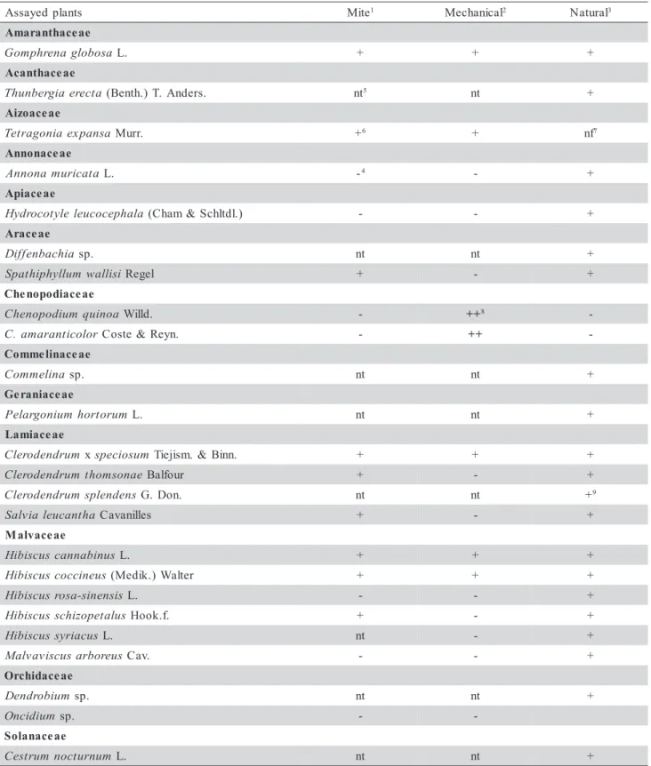
Table 1 - List of plants tested for susceptibility to Clerodendrum chlorotic spot virus (ClCSV) by mite, mechanical and natural transmission and respective symptoms.
1Brevipalpus phoenicis. 2 Extracts from local lesions of Chenopodium quinoa or C. amaranticolor used as inoculum. 3 Plants growing next to a group of Clerodendrum x speciosum plants naturally infected by Clerodendrum chlorotic spot virus (ClCSV) and infested by B. phoenicis. -4 no reaction. nt5- not tested. +6 Local chlorotic lesions. Infection by ClCSV demonstrated by electron microscopic examination of the lesions and mechanical inoculation of C. quinoa and C.amaranticolor. nf7- Plants not found near ClCSV - infected C.x splendens or C. thomsonae. ++8 Local chlorotic lesions followed by systemic infection (vein clearing and chlorotic spots) in
uninoculated leaves when plants were kept at 28 - 30oC for 2 weeks. 9Natural infection in an isolated plant.
s t n a l p d e y a s s
A Mite1 Mechanical2 Natural3 e a e c a h t n a r a m A a s o b o l g a n e r h p m o
G L. + + +
e a e c a h t n a c A a t c e r e a i g r e b n u h
T (Benth.)T. Anders. nt5 nt + e a e c a o z i A a s n a p x e a i n o g a r t e
T Murr. +6 + nf7
e a e c a n o n n A a t a c i r u m a n o n n
A L. -4 - +
e a e c a i p A a l a h p e c o c u e l e l y t o c o r d y
H (Cham&Schltdl.) - - +
e a e c a r A a i h c a b n e f f i
D sp. nt nt +
i s i l l a w m u l l y h p i h t a p
S Regel + - +
e a e c a i d o p o n e h C a o n i u q m u i d o p o n e h
C Willd. - ++8
-r o l o c i t n a r a m a .
C Coste&Reyn. - ++
-e a e c a n i l e m m o C a n i l e m m o
C sp. nt nt +
e a e c a i n a r e G m u r o t r o h m u i n o g r a l e
P L. nt nt +
e a e c a i m a L m u r d n e d o r e l
C xspeciosum Tiejism. &Binn. + + + e a n o s m o h t m u r d n e d o r e l
C Balfour + - +
s n e d n e l p s m u r d n e d o r e l
C G. Don. nt nt +9
a h t n a c u e l a i v l a
S Cavanilles + - +
e a e c a v l a M s u n i b a n n a c s u c s i b i
H L. + + +
s u e n i c c o c s u c s i b i
H (Medik.)Walter + + +
s i s n e n i s -a s o r s u c s i b i
H L. - - +
s u l a t e p o z i h c s s u c s i b i
H Hook.f. + - +
s u c a i r y s s u c s i b i
H L. nt - +
s u e r o b r a s u c s i v a v l a
M Cav. - - +
e a e c a d i h c r O m u i b o r d n e
D sp. nt nt +
m u i d i c n
O sp. -
-e a e c a n a l o S m u n r u t c o n m u r t s e
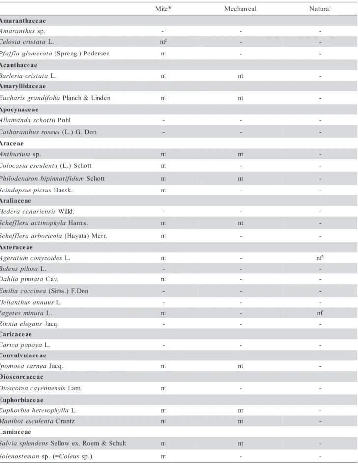
Table 2 - Other plant species tested negative for mite, mechanical and natural transmission of the Clerodendrum chlorotic spot virus. Continue... * e t i
M Mechanical Natural
e a e c a h t n a r a m A s u h t n a r a m
A sp. -1 -
-a t a t s i r c a i s o l e
C L. nt2 -
-a t a r e m o l g a i f f a f
P (Spreng.)Pedersen nt -
-e a e c a h t n a c A a t a t s i r c a i r e l r a
B L. nt nt
-e a e c a d i l l y r a m A a i l o f i d n a r g s i r a h c u
E Planch&Linden nt nt
-e a e c a n y c o p A i i t t o h c s a d n a m a l l
A Pohl - -
-s u e s o r s u h t n a r a h t a
C (L.)G. Don - -
-e a e c a r A m u i r u h t n
A sp. nt nt
-a t n e l u c s e a i s a c o l o
C (L.)Schott nt -
-m u d i f i t a n n i p i b n o r d n e d o l i h
P Schott nt nt
-s u t c i p s u s p a d n i c
S Hassk. nt -
-e a e c a i l a r A s i s n e i r a n a c a r e d e
H Willd. - -
-a l y h p o n i t c a a r e l f f e h c
S Harms. nt nt
-a l o c i r o b r a a r e l f f e h c
S (Hayata)Merr. nt -
-e a e c a r e t s A s e d i o z y n o c m u t a r e g
A L. nt - nf3
a s o l i p s n e d i
B L. - -
-a t a n n i p a i l h a
D Cav. nt -
-a e n i c c o c a i l i m
E (Sims.)F.Don - -
-s u u n n a s u h t n a i l e
H L. - -
-a t u n i m s e t e g a
T L. nt - nf
s n a g e l e a i n n i
Z Jacq. - -
-e a e c a c i r a C a y a p a p a c i r a
C L. - -
-e a e c a l u v l u v n o C a e n r a c a e o m o p
I Jacq. nt nt
-e a e c a e r o c s o i D s i s n e n n e y a c a e r o c s o i
D Lam. nt -
-e a e c a i b r o h p u E a l l y h p o r e t e h a i b r o h p u
E L. nt nt
-a t n e l u c s e t o h i n a
M Crantz nt nt
-e a e c a i m a L s n e d n e l p s a i v l a
S Sellowex. Roem&Schult nt nt -n o m e t s o n e l o
-Table 2 - Continuation. Continue... e a s o n i m u g e L n a j a c s u n a j a
C (L.) Millsp. nt - nf
s e m r o f i s n e a i l a v a n a
C DC nt - nf
a e s o r a i l a v a n a
C (Sw.)DC nt -
-a e c n u j a i r a l a t o r
C L. nt - nf
a c u e l o r h c o a i r a l a t o r
C G. Don nt - nf
b a l b a l s o h c i l o
D L. nt -
-x a m e n i c y l
G Merrill nt - nf
a m i r r e t a a n u c u
M (Piper&Tracy) Merr. nt -
-m u e r e n i c a n u c u
M L. nt - nf
s i r a g l u v s u l o e s a h
P L. nt -
-s i l a t n e d i c c o a n n e
S (L.) Link. nt -
-m u n a i g n i r e e d m u i b o l o z y t
S Bort. nt - nf
a t a l u c i u g n u a n g i
V (L.) Walp. - - nf
e a e c a i l i L m u i l l
A sp. nt nt
-s i l a n i m r e t e n i l y d r o
C (L.) Kunth nt nt
-a n a i r e d n a s a n e a c a r
D Hort. nt nt
-e a e c a v l a M a t n e l u c s e s u h c s o m l e b
A (L.) Moench - -
-m u t a i r t s n o l y t u b
A Dicks. ex-Lindl. nt nt
-e a e c a t r y M a r o l f i n u a i n e g u
E L. nt nt
-a r o l f i l u a c a i r a i c r y
M (DC.) Berg. nt nt
-e a e c a n i g a t c y N a e l l i v n i a g u o
B sp. nt nt
-a p a l a j s i l i b a r i
M L. - nt
-e a e c a r e l O m u d i c u l m u r t s u g i
L WTAiton nt nt
-e s n e n i s m u r t s u g i
L Lour. nt nt
-e a e c a r o l f i s s a P s i l u d e a r o l f i s s a
P Sims.f.flavicarpa Deg. - -
-e a e c a r e p i P m u r g i n r e p i
P L. nt nt
-e a e c a r o p s o t t i P a r i b o t m u r o p s o t t i
P (Thunb.) Ait. nt nt
-e a e c a n i g a b m u l P a t a l u c i r u a o g a b m u l
P Lam. nt nt
-e a e c a i b u R a c i b a r a a e f f o
C L. - -
-a e n i c c o c a r o x
I L. nt nt
-a l l y h p o r h t y r e a d n e a s s u
-Mechanical transmission assays - Extracts from chlorotic spots on C. x speciosum leaves were obtained by maceration in a 0.01M phosphate buffer, pH 7.0 containing activated charcoal (0.03% w/v) and nico-tine (2% v/v) (Chagas, 1978). In subsequent experi-ments, when transmission to Chenopodium quinoa Willd. and C. amaranticolor Coste & Reyn. was achieved (initial chlorotic spots and systemically in-fected leaves when plants were maintained at 28 - 30oC for two weeks), these plants were used as source of inoculum. Extracts were obtained using 0.01 M phos-phate buffer, pH 7.0, containing 0.1% sodium sulfite. Mechanical transmission tests were carried out with 65 different species of test-plants (Tables 1 and 2). Three to five test plants of each species were mechani-cally inoculated with the extract. Inoculation was made by rubbing the sap on the leaves dusted with carborundum.
Survey on possible natural infection by ClCSV -Other plant species (ornamentals, vegetables and fruit plants, weeds) are frequently present interspersed or next to symptomatic and Brevipalpus-infested C. x speciosum plants. These plants were surveyed for
symp-toms and the presence of the mites. Surveyed plants were located in the university campus of ESALQ and in three residential gardens of Piracicaba. A total of 87 species of plants growing under natural conditions (Tables 1 and 2) were surveyed for symptoms of chlo-rotic/necrotic lesions on the leaves. When leaves with suspected lesions were found, samples were collected and processed for electron microscopy analysis.
Electron microscopy - Small pieces of the chlorotic spots on the leaves (and in one case from a flower petal of C. thomsonae) were fixed in a modified Karnovsky solution (2% paraformaldehyde, 2.5% glu-taraldehyde in 0.05 M cacodylate buffer, pH 7.2) for 1 - 2 h, washed with buffer and post-fixed in 1% os-mium tetroxide in the same buffer for 1 h, dehydrated in a graded series of acetone, infiltrated and embed-ded in the low viscosity Spurr epoxy resin (Maunsbach & Afezelius, 1999). Blocks were sectioned in a Leica UT ultramicrotome equipped with a diamond knife. The sections were collected on copper grids, stained with 3% uranyl acetate and Reynold’s lead citrate, and examined in a Zeiss EM 900 transmission electron mi-croscope.
Table 2 - Continuation.
e a e c a t u R s i s n e n i s s u r t i
C (L.)Osbeck - -
-a y a r r u
M sp. nt nt
-e a e c a g a r f i x a S a l l y h p o r c a m a e g n a r d y
H (Thunb.)Serv. nt -
-e a e c a n a l o S a r o l f i n u a i s l e f n u r
B (Pohl)D. Don - -
-m u u n n a m u c i s p a
C L. - -
-s n e l o e v a u s a r u t a
D (Humb.&Bonpl.) . l s e r P & . t h c r e
B - -
-m u i n o m a r t s a r u t a
D L. - -
-m u t n e l u c s e n o c i s r e p o c y
L Mill. - -
-a n a i m a h t n e b a n a i t o c i
N Domin. nt - nf
i i d n a l e v e l c a n a i t o c i
N Gray nt -
-a s o n i t u l g a n a i t o c i
N L. nt -
-m u c a b a t a n a i t o c i
N L. - -
-o l i g m u n a l o
S Raddi nt -
-a n e g n o l e m m u n a l o
S L. - -
-m u i l o f e a l o i v m u n a l o
S Schott. - -
-e a e c a c i t r U i e r e i d a c a e l i
P Gagnep.&Guillaumin nt nt
-e a e c a n e b r e V s n e p e r a t n a r u
D L. nt nt
Virus purification - Fresh leaves of C. amaranticolor and C. quinoa exhibiting symptoms of systemic infec-tion were used for virus purificainfec-tion, based on the pro-tocol used for CoRSV purification (Boari et al., 2004), which is the procedure used to purify OFV (Chang et al., 1976) with small modifications. Infected leaf tissues frozen in liquid nitrogen were ground in 0.1 M phosphate buffer, pH 7.0, containing 0.01 M so-dium diethyl ditiocarbamide, 0.1% ascorbic acid and 5% Triton X-100. After clarification by low speed centrifugation, the suspension was centrifuged on top of a 20% sucrose cushion (150 min /40,000 g). The pellet was resuspended in 0.1 M phosphate buffer, pH 7.0, and submitted to a cycle of differential cen-trifugation and then centrifuged in a sucrose gradi-ent (10 - 40%) for 90 min at 40,000 g. One ml frac-tion were collected and analyzed by spectrophotom-etry, and the fraction with the peak of nucleic acid absorbance was separated and centrifuged for 150 min at 40,000 g. The pellet was resuspended and stored for antiserum production. A sample was ex-amined in the electron microscope by negative stain-ing with 1% uranyl acetate.
Antiserum production and ELISA - Four aliquots of 250 µL of a purified preparation (ca. 20 µg mL-1),
mixed with an equal volume of incomplete Freund´s adjuvant, were injected intramuscularly in a rabbit at weekly intervals. One week after the last injection, the serum was collected and stored at -20°C (Van Regenmortel, 1982). This antiserum was tested in Plate trapped antigen - Enzyme linked immunosorbent as-say (PTA-ELISA) (Mowat & Dawson, 1987) against purified virus and extracts from infected Clerodendrum. Antisera against OFV (Kondo et al., 1995) and CoRSV (Boari et al., 2004), and the respec-tive antigens were also included in the test to analyze possible serological relationships. Appropriate healthy controls were included in homologous and heterolo-gous reactions.
RESULTS
Symptoms - Symptoms caused by natural or experi-mental (mite or mechanical inoculation) transmission of ClCSV remained restricted to the inoculated leaves of susceptible hosts. The symptoms consisted usually of chlorotic spots (Figure 1 H, I, K, N; Figure 2 A, B, C, F, I, J, L, N, O), occasionally with a necrotic center (Figure 1 G, 2 E, H). Some hosts developed brownish (Figure 2 I, K) or necrotic spots (Figure 2 D, M). In some few cases, the chlorotic spots became greenish when the leaves became senescent (Figure 2 A, C, G, H, I). Necrotic stem lesions were only
no-ticed in H. cannabinus. In one instance, the white co-rolla of C. thomsonae growing in a residential garden presented brownish spots (Figure 1 J).
Mite transmission tests - Mites (Figure 1 D, E, F) infesting C. x speciosum, C. splendens and C. thomsonae exhibiting chlorotic lesions were identified as B. phoenicis based on external morphological char-acteristics (Welbourn et al., 2003). When mites col-lected from symptomatic C. x speciosum plants were transferred to healthy ones, chlorotic spots became vis-ible four to five weeks later. Electron microscopic ex-amination confirmed the presence of cytopathic effects similar to those found in the source plant. After this initial experiment, similar mite transmission tests were performed on plants of 39 species (Tables 1 and 2). Chlorotic spots developed only in nine of the tested plant species (Clerodendrum thomsonae, C. x speciosum, Gomphrena globosa, Hibiscus cannabinus, H. coccineus, H. schizopetalus, Salvia leucantha, Spathiphyllum wallisi and Tetragonia expansa). Elec-tron microscopy confirmed the presence of cytopathic effects characteristic of the nuclear type of BTrV in these spots. Attempts to rear B. phoenicis collected from field Clerodendrum plants on C. x speciosum or C. thomsonae under laboratory conditions were unsuc-cessful so far.
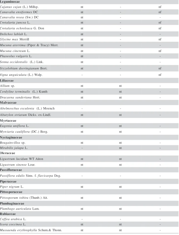
Mechanical transmission - Transmission of ClCSV was successfully achieved by mechanical means us-ing C. x speciosum as source of inoculum only when buffer containing nicotine and activated charcoal was used. In the initial trials, C. amaranticolor and C. quinoa demonstrated to be good indicator plants among those tested. Once infected, these plants became good sources of inoculum for mechanical transmission us-ing routine phosphate buffer containus-ing sodium sulfite. As already demonstrated with OFV and CoRSV, both C. quinoa and C. amaranticolor became systemically infected when kept at 28 - 30oC for 10 - 14 days af-ter inoculation. Systemically infected leaves of C. amaranticolor and/or C. quinoa were used as source of inoculum in later experiments. A total of 65 differ-ent plant species (Tables 1 and 2) were tested for me-chanical transmission of ClCSV, but only six were sus-ceptible (C. amaranticolor, C. quinoa, G. globosa, H. cannabinus, H. coccineus and T. expansa) besides C. x speciosum. Transmission was confirmed by back-inoculation to C. amaranticolor and C. quinoa.
search, therefore, was carried out on such plants grow-ing interspersed or next to ClCSV-infected C. x speciosum for those showing localized symptoms on their leaves. A total of 87 different plant species were analyzed (Tables 1 and 2). In 19 of them (Annona muricata, Cestrum nocturnum, C. thomsonae, Commelina sp., Dendrobium sp., Difenbachia sp., G. globosa, H. cannabinus, H. coccineus, H. rosa-sinensis, H. schizopetalus, H. syriacus, Hydrocotyle leucocephala, Malvaviscus arboreus, Oncidium, Pelargonium hortorum, S. leucantha, S. wallisi and Thunbergia erecta) chlorotic or ringshaped spots were noticed. Few isolated C. splendens plants with chlorotic spots on their leaves were found in residential gardens. These spots were sampled and examined by electron microscopy. In all of them the characteristic cytopathic effects of the nuclear type of BTrV were observed.
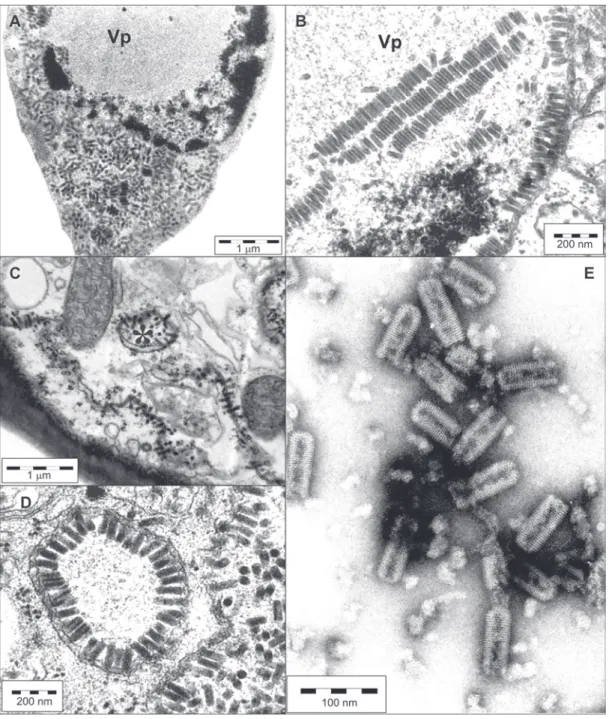
Electron microscopy - Cell alterations characterized by an electron lucent inclusion (viroplasm) in the nucleus and the presence of short rod shaped particles (ca. 40 nm × 100 - 110 nm) in the nucleus or
cyto-plasm were observed in the tissues of all symptom-atic plants, obtained by natural or experimental (mite or mechanical) transmission of the virus (Figure 3 A-D). In the nucleus, these rodlike particles could ap-pear scattered within viroplasm or nucleoplasm and sometimes arranged side-by-side forming lamellar ag-gregates (Figure 3 B). Also, they commonly appear ei-ther in the nucleus or in the cytoplasm arranged per-pendicularly onto membranous system of the nuclear envelope or endoplasmic reticulum (Figure 3 C). This arrangement is reminiscent of early phases of the bud-ding process during morphogenesis of most of the membrane bound viruses, which ends up with the complete envelopment of the nucleocapsid by the mem-brane. However, with the nuclear type of BTrV (Kitajima et al., 2003) this budding process appears to be incomplete in most cases. In some instances the endoplasmic reticulum cisternae form a tubular array with the particles arranged in a radial configuration in-side, producing a figure referred to as “spoke wheel” (Figure 3 C, D). These cell changes are characteristic of the so-called nuclear type of BTrV, and served as evidence that these plants are infected by ClCSV.
Virus purification - The protocol used for purifica-tion of OFV and CoRSV was successfully used to pu-rify ClCSV from extracts of systemically infected leaves of C. amaranticolor and C. quinoa. Examina-tion of the fracExamina-tion which had a UV absorpExamina-tion for nucleic acid contained a large amount of short bullet shaped particles 40 nm wide and 100 - 110 nm long, with cross striation of 5 nm, essentially similar to those observed in purified preparations of OFV and CoRSV
(Figure 3 E). Mechanical inoculation of these prepa-rations caused local lesions in C. quinoa. Transmis-sion electron microscopy of these leTransmis-sions indicated the presence of typical cytopathic effects caused by a nuclear type of BTrV, thus confirming that the puri-fied preparations were infectious.
PTA-ELISA - The purified preparations of ClCSV strongly reacted with the produced homologous anti-serum in PTA-ELISA. Antisera against OFV and CoRSV only reacted weakly with purified ClCSV (data not shown), suggesting that it may differ from OFV and CoRSV, but may share some common antigens or epitopes.
DISCUSSION
The chlorotic spots occurring naturally in at least three Clerodendrum species (C. x speciosum, C. thomsonae, C. splendens) are caused by a nuclear type of BTrV, which is named Clerodendrum chlorotic spot virus (ClCSV). The virus is naturally transmitted by mites and by mechanical inoculation to several other plant species. Nineteen of 87 plants growing near ClCSV-infected C. x speciosum (Table 1) showed lo-calized infection with cytopathic effects characteris-tic of the nuclear type of BTrV. They were possibly infected by B. phoenicis that acquired the ClCSV from infected C. x speciosum plants. Few isolated C. splendens plants were also found to be infected by ClCSV. Experimental mite transmission tests resulted in the infection of some of these plants (G. globosa, H. cannabinus, H. coccineus, S. leucantha, S. wallasi). Infection of H. cannabinus, H. coccineus, S. leucantha and Dendrobium sp., could be demonstrated by me-chanical inoculation on C. amaranticolor and C. quinoa. With 14 species of these plants, mechanical recovery of the virus was unsuccessful possibly due to inhibitors present in the extracts. Further studies are required to confirm infection of these plants by im-munological or molecular tools to detect ClCSV.
Two orchid species (Dendrobium sp. and On-cidium sp.) growing amidst ClCSV infected C. x speciosum presented leaf lesions undistinguishable from those caused by OFV. However, extracts from the le-sions did not react with OFV antiserum, and no am-plification was obtained by RT-PCR using primers spe-cific for OFV (data not shown). It is likely that these orchid species were infected by an isolate of ClCSV but further tests are required to confirm this hypoth-esis.
C. amaranticolor and C. quinoa with ClCSV resulted in local chlorotic spots and if the plants are maintained at high temperatures (28 - 30ºC), systemic infection occurs in the form of chlorotic spots or vein clear-ing. The systemic infection in hosts as C. amaranticolor and C. quinoa may be a common
fea-ture for most of the nuclear type of BTrV and de-serves attention to understand why temperature may affect the mechanisms that tend to restric the infec-tion. In other susceptible plant species, however, sys-temic infection was not yet observed under similar conditions.
Based on symptoms and electron microscopy examination of the infected tissues, the causal agent of some reported diseases like Malvaviscus ringspot (Kitajima et al., 2003), Soursop (Annona muricata) ringspot (Bitancourt, 1955; Kitajima et al., 2003) and Hibiscus chlorotic spot (Kitajima & Rodrigues, 2001) may be caused by ClCSV. Similar symptoms were ob-served for these plants growing near to ClCSV-infected C. x speciosum as above cited, and presumed to be naturally inoculated by the mite vector. This possibil-ity will be evaluated using immunological and molecular techniques.
Many hosts, which appeared not to be in-fected by ClCSV in this study like Citrus sinensis (Rodrigues et al., 2003a), Pittosporum tobira (Rodrigues et al., 2003b), Piper nigrum (Yamashita et al., 2004), Allamanda schottii, Hedera canariensis, Bidens pilosa and Mussaenda erythrophylla (Rodrigues et al., 2005) were reported to be infected by the nuclear type of BTrV. Now that immunologi-cal and molecular tools are available for three of the nuclear types of BTrV, respectively ClCSV, CoRSV and OFV, it will be possible to assess whether or not other diseases for wich cytopathology indicates the presence of the nuclear type of BTrV represent in-fection by isolates of one of these viruses or a dif-ferent virus.
So far, except for OFV, known BTrV are re-stricted to the American continent (Kitajima et al., 2003; 2006a) although Brevipalpus species that serve as vector are dispersed worldwide in tropical and sub-tropical regions (Childers et al., 2003b). The world-wide occurrence of OFV is explainable by the intense exchange of living plants which certainly resulted in the introduction of the virus and the vector, even in temperate regions. The absence of BTrVs in other parts of the world, however, is circumstantial and based on observations made only in parts of South Africa, Australia and few countries in South-east Asia (J.C.V. Rodrigues and E.W. Kitajima, un-published results) and a wider survey is required to confirm these observations. ClCSV has been found naturally infecting Clerodendrum species in the states of São Paulo, Santa Catarina, Amazonas and Distrito Federal indicating a wide dispersion in Brazil and is likely to be present in other parts of the American continent.
Examination of thin sections of the tissues from the lesions of experimental or naturally ClCSV-infected plants appeared similar to those reported for other nuclear types of BTrV as OFV and CoRSV (Kitajima et al., 2003; Kondo et al., 2003; Chagas et al., 2003). Typical electron lucent viroplasm in the nucleus and short rod shaped particles were seen mostly in
mesophyl parenchyma cells, and rarely in vascular parenchyma. The rod-like particles were demonstrated to be the virions by immunogold labeling in OFV-in-fected cells (Kitajima et al., 2001) and by analogy, those present in ClCSV-infected cells may represent ClCSV particles, though experimental demonstration is still required. These particles were commonly naked, without surrounding membrane, and quite often were perpendicularly arranged to the nuclear envelope or endoplasmic reticulum membranes, as shown for other nuclear type of BTrV, a process suggesting that the envelopment of the particles is not completed after an initial start. In very rare instances, membrane bounded particles could individually be seen within en-doplasmic reticulum elements, possibly after a success-ful budding process. A possible explanation might be that glycoprotein of most nuclear types of BTrV have some defect by which the budding process can not be completed and results in the accumulation of naked nucleocapsids. Similar observations were de-scribed for Tomato spotted wilt virus (Resende et al., 1991).
The same pattern of cell alterations observed in plant tissues was observed also in gland cells of B. phoenicis collected from ClCSV-infected plants (Kitajima et al., 2006a). This fact strongly suggests that the virus multiplies in the mite, thus B. phoenicis/ClCSV relationship is of the circulative/propagative type. This is in agreement with the conclusions reached in the transmission of OFV by B. californicus (Kondo et al., 2003).
informa-tion about ClCSV genome emerges, a better under-standing of the phylogenetic relationship among these viruses is expected. Also, the development of molecu-lar and immunological tools to detect ClCSV will help to confirm the cases of its natural transmission as well as the establishment of its natural host range, which is much wider than that of CoRSV and OFV.
ACKNOWLEDGMENTS
To FAPESP (2000/11805-0) and CNPq (41.0192/03-1). The authors are grateful to Dr. Kanchi Gandi from the International Plant Name In-dex for updating information regarding genus Clerodendrum.
REFERENCES
BITANCOURT, A.A. Estudos sobre a leprose dos citros. I-Distribuição geográfica e sintomatologia. II. Transmissão natural às folhas. III.Transmissão natural às frutas. IV. Experiências de tratamento. Arquivos do Instituto Biológico, v.22, p.161-231, 1955.
BOARI, A.J.; FIGUEIRA, A.R.; NEDER, D.G.; INFIESTA, L.R.; NOGUEIRA, N.L.; ROSSI, M.L.; KITAJIMA, E.W. Efeito da temperatura na infecção sistêmica de Chenopodium quinoa pelo Coffee ringspot virus (CoRSV). Fitopatologia Brasileira, v.28, p.246-247, 2003. Suplemento.
BOARI, A.J.; FREITAS-ASTUA, J.; FERREIRA, P.T.O.; NEDER, D.G.; NOGUEIRA, N.L.; ROSSI, M.L.; KITAJIMA, E.W. Purification and serology of the coffee ringspot virus. Summa Phytopathologica, v.30, p.453-458, 2004.
CHAGAS, C.M. Mancha anular do cafeeiro. São Paulo: USP/IB, 1978. 92p. (Tese – Doutorado).
CHAGAS, C.M.; KITAJMA, E.W.; RODRIGUES, J.C.V. Coffee ringspot virus vectored by Brevipalpus phoenicis (Acari: Tenuipalpidae) in coffee. Experimental and Applied Acarology, v.30, p.203-213, 2003.
CHANG, M.U.; ARAI, K.; DOI, Y.; YORA, K. Morphology and intracellular appearance of orchid fleck virus. Annals of the Phytopathological Society of Japan, v.42, p.156-167, 1976.
CHILDERS, C.C.; FRENCH, J.V.; RODRIGUES, J.C.V. Brevipalpus californicus, B. obovatus, B. phoenicis, and B. lewisi (Acari: Tenuipalpidae): a review of their biology; feeding injury and economic importance. Experimental and Applied Acarology, v.30, p.5-28, 2003a.
CHILDERS, C.C.; RODRIGUES, J.C.V.; WELBOURN, W.C. Host plants of Brevipalpus californicus, B. obovatus, and B. phoenicis (Acari: Tenuipalpidae) and their potential involvement in the spread of viral diseases vectored by these mites. Experimental and Applied Acarology, v.30, p.29-105, 2003b.
JOHN, P.; SIVALINGAM, P.N.; KUMAR, N.; MISHRA, A.; AHYLAWAT, Y.S.; MALATHI, V.G. A new begomovirus associated with yellow mosaic disease of Clerodendron inerme. Plant Pathology, v.55, p.291, 2006.
KITAJIMA, E.W.; RODRIGUES, J.C.V. Mancha verde e mancha clorótica de Hibiscus são causadas por tipos diferentes de vírus transmitidos por Brevipalpus. Summa Phytopathologica, v.27, p.105, 2001.
KITAJIMA, E.W.; KONDO, H.; MACKENZIE, A.; REZENDE, J.A.M.; GIORIA, R.; GIBBS, A.; TAMADA, T. Comparative cytopathology and immunocytochemistry of Japanese, Australian and Brazilian isolates of Orchid fleck virus. Journal of General Plant Pathology, v.67, p.231-237, 2001.
KITAJIMA, E.W.; CHAGAS, C.M.; RODRIGUES, J.C.V. Brevipalpus-transmitted plant virus and virus-like diseases: cytopathology and some recent cases. Experimental and Applied Acarology, v.30, p.135-160, 2003.
KITAJIMA, E.W.; MORAES, G.J.; CALEGÁRIO, R.F.; SALAROLI, R.B. Dados preliminares sobre detecção electrono-microscópica de vírus transmitidos por Brevipalpus (Acari: Tenuipalpidae) nos tecidos do ácaro vetor. In: SIMPÓSIO BRASILEIRO DE ACAROLOGIA, 1., Viçosa, 2006. Resumos. Viçosa, 2006a. p.248.
KITAJIMA, E.W.; MORAES, G.J.; RODRIGUES, J.C.V.; FREITAS-ASTUA, J. Plantas infectadas naturalmente por vírus transmitidos por ácaros Brevipalpus (Acari: Tenuipalpidae). In: SIMPÓSIO BRASILEIRO DE ACAROLOGIA, 1., Viçosa, 2006. Resumos. Viçosa, 2006b. p.251.
KONDO, H.; MATSUMOTO, J.; MAEDA, T.; INOUYE, N. Host range and some properties of orchid fleck virus isolated from oriental Cymbidium in Japan. Bulletin Research Institute for Bioresource, Okayama University, v.3, p.151-161, 1995.
KONDO, H.; MAEDA, T.; TAMADA, T. Orchid fleck virus: Brevipalpus californicus mite transmission, biological properties and genome structure. Experimental and Applied Acarology, v.30, p.215-223, 2003.
KONDO, H.; MAEDA, T.; SHIRAKO, Y.; TAMADA, T. Orchid fleck virus is a rhabdovirus with an unusual bipartite genome. Journal of General Virology, v.87, p.2413-2421, 2006.
KVL. September: Clerodendron trichotomum. Available at: http:/ /en.sl.kvl.dk/Faciliter/Arboretet/MaanedensPlante/2006/ September.aspx. 2006: Accessed at: 05 feb. 2007.
LORENZI, H.; SOUZA, H.M. Plantas ornamentais do Brasil. Arbustivas, herbáceas e trepadeiras. 3.ed. Nova Odessa: Instituto Plantarum, 2001. 1088p.
MAUNSBACH, A.B.; AFZELIUS, B. Biomedical electron microscopy. Illustrated methods and interpretations. San Diego: Academic Press, 1999. 548p.
MOWAT, W.P.; DAWSON, S. Detection and identification of plant viruses by ELISA using crude sap extracts and unfractioned sera. Journal of Virological Methods, v.15, p.233-247, 1987.
RESENDE, R.O.; DE HAAN, P.; ÁVILA, A.C.; KITAJIMA, E.W.; KORMELINK, R.; GOLDBACH, R.; PETERS, D. Generation of envelope and defective interfering RNA mutants of tomato spotted wilt virus by mechanical passage. Journal of General Virology, v.73, p.2375-2383, 1991.
RODRIGUES, J.C.V.; KITAJIMA, E.W.; CHILDERS, C.C.; CHAGAS, C.M. Citrus leprosis virus vectored by Brevipalpus phoenicis (Acari: Tenuipalpidae) on citrus in Brazil.
Experimental and Applied Acarology, v.30, p.161-179, 2003a.
RODRIGUES, J.C.V.; ACHOR, D.S.; CHILDERS, C.C.; KITAJIMA, E.W. Three new host plants of the Brevipalpus borne virus found in United States. In: INTERNATIONAL CONGRESS OF ACAROLOGY, 11., Brisbane, 2003. Program and abstract book. Brisbane, 2003b.
RODRIGUES, J.C.V.; ANTONY, L.M.M.A.; KITAJIMA, E.W. Levantamento de vírus transmitidos por Brevipalpus (Acari: Tenuipalpidae) na região de Manaus e Urucu, AM. Summa Phytopathologica, v.31, p.26, 2005. Suplemento.
SCHUTA, L.R.; LIMA, M.L.R.C.; COSTA LIMA NETO, V. Transmissão do vírus do clareamento das nervuras de Clerodendron speciosum (Verbenaceae). Summa Phytopathologica, v.23, p.54, 1997.
STEANE, D.A.; DE KOK, R.P.J.; OLMSTEAD, R.G. Phylogenetic relationships between Clerodendrum (Lamiaceae) and other Ajugoid genera inferred from nuclear and chloroplast DNA sequence data. Molecular Phylogenetics and Evolution,
VAN REGENMORTEL, M.H. Serology and immunochemistry of plant viruses. London: Academic Press, 1982. 302p. WELBOURN, W.C.; OCHOA, R.; KANE, E.C.; ERBE, E.F.
Morphological observations on Brevipalpus phoenicis (Acari: Tenuipalpidae) including comparisons with B. californicus and B. obovatus. Experimental and Applied Acarology, v.30, p.107-133, 2003.
Received March 13, 2007 Accepted August 14, 2007