Cardioprotection against experimental myocardial
ischemic injury using cornin
Y. Xu, Y. Xu, H. Luan, Y. Jiang, X. Tian and S. Zhang
School of Pharmaceutical Sciences, Binzhou Medical University, Yantai, China
Abstract
Phosphorylated-cyclic adenosine monophosphate response element-binding protein (Phospho-CREB) has an important role in the pathogenesis of myocardial ischemia. We isolated the iridoid glycoside cornin from the fruit ofVerbena officinalisL, investigated its effects against myocardial ischemia and reperfusion (I/R) injuryin vivo, and elucidated its potential mechanismin vitro. Effects of cornin on cell viability, as well as expression of phospho-CREB and phospho-Akt in hypoxic H9c2 cellsin vitro, and myocardial I/R injury in vivo, were investigated. Cornin attenuated hypoxia-induced cytotoxicity significantly in H9c2 cells in a concentration-dependent manner. Treatment of H9c2 cells with cornin (10 mM) blocked the reduction of expression of phospho-CREB and phospho-Akt in a hypoxic condition. Treatment of rats with cornin (30 mg/kg, iv) protected them from myocardial I/R injury as indicated by a decrease in infarct volume, improvement in hemodynamics, and reduction of severity of myocardial damage. Cornin treatment also attenuated the reduction of expression of phospho-CREB and phospho-Akt in ischemic myocardial tissue. These data suggest that cornin exerts protective effects due to an increase in expression of phospho-CREB and phospho-Akt.
Key words: Cornin; Myocardial ischemia and reperfusion; CREB; Akt; Hypoxia
Introduction
Primary myocardial ischemia-reperfusion (I/R) therapies such as percutaneous coronary intervention and thrombo-lysis are the standard of care for acute coronary syndromes. Prompt restoration of bloodflow to the ischemic myocardium limits infarct size and the risk of death. Paradoxically, the return of blood flow can also result in additional cardiac damage and complications such as reperfusion injury. Effective therapies to reduce or prevent reperfusion injury have proved elusive. Despite improved understanding of the pathophysiology of reperfusion injury and encouraging preclinical trials using multiple agents, most clinical trials focusing on prevention of reperfusion injury have been dis-appointing (1,2). Despite these problems, adjunctive thera-pies to limit reperfusion injury remain an active area of investigation.
Cyclic adenosine monophosphate response element-binding protein (CREB) is a cellular transcription factor that binds to certain DNA sequences called cyclic adeno-sine monophosphate response elements and influences transcription of downstream genes. Phosphorylated-CREB (Phospho-CREB) is an activated form of CREB involved in myocardial protection against I/R-related injury (3,4). Increased levels of phospho-CREB in myocardial tissue could attenuate I/R-related injury (5). Protein kinase B, also known as Akt, is a serine/threonine-specific protein
kinase that plays a key part in multiple cellular pro-cesses: glucose metabolism, apoptosis and transcrip-tion. Akt promotes cell survival by stimulating expression of cellular genes via the CREB nuclear transduction pathway (6).
Cornin is an iridoid glycoside isolated from the fruit of Verbena officinalisL. It has protective actions against cerebral ischemia injury (7) and induces angiogenesis in vitro(8). We wished to investigate the effects of cornin in a rat model of myocardial I/R as well as its potential cardioprotective mechanism in cultured H9c2 cells and intact rats.
Material and Methods
Material
Cornin (purity499.0%, CAS number, 548-37-8; mole-cular formula, C17H24O10; molemole-cular weight, 388.37) was dissolved in sterile physiologic (0.9%) saline to make a stock solution. Dilutions were prepared according to different administration doses. Troponin T (cTnT) enzyme-linked immunosorbent assay kits were purchased from Maisha Biology (China). Polyclonal rabbit anti-mouse phospho-CREB and phospho-Akt antibodies were purchased from Biosynthesis Biotechnology (China).
Correspondence: S. Zhang:<[email protected]>
Animals
All experimental designs and procedures were conducted in accordance with the Animal Care Guide-lines of the Animal Experimental Committee of Binzhou Medical University (China; authorization number, BYLY 2015-74).
Thirty adult male Sprague-Dawley rats (270–300 g) were housed individually under constant temperature (22 ±2°C) and humidity with a 12-h light/dark cycle. They had
free access to rodent food and water.
Cell culture
H9c2 cells (clonal line derived from embryonic rat hearts) were purchased from American Type Culture Collection (USA). Cells were cultured in Dulbecco’s modified Eagle’s medium (DMEM) containing D-glucose (4.5 g/L), 20% fetal bovine serum (FBS), 10,000 U/L penicillin, and 10 mg/L streptomycin using standard methods in an incubator with an atmosphere of 5% CO2 at 37°C. The medium was changed every 2 days. Upon reaching confluence, cells were subcultured by detach-ment with 0.25% trypsin-EDTA solution (Sigma-Aldrich, USA), re-seeded onto new plates at a ratio of 1:5, and incubated in DMEM containing 2% FBS. Cells were maintained at 37°C in a humidified incubator in an atmosphere of 5% CO2/95% air.
Hypoxia modelin vitro
To mimic hypoxia injuryin vitro, cells were incubated in a hypoxic solution for 6 h. The hypoxic solution (9) contained 0.9 mM NaH2PO4, 6.0 mM NaHCO3, 1.0 mM CaCl2, 1.2 mM MgSO4, 40 mM natrum lacticum, 20 mM HEPES, 98.5 mM NaCl, 10.0 mM KCl (pH adjusted to 6.8) and was bubbled with N2for 30 min before application. The partial pressure of oxygen of the hypoxic solution was adjusted top4.0 kPa. Hypoxia was elicited by placing the plates of cultured cardiomyocytes in a hypoxic incubator (Kendro, Germany) while oxygen was adjusted to 1.0% and CO2to 5.0%. Before hypoxia, cells were pretreated with cornin (1, 3, 10, and 30mM) for 24 h. Normal culture (DMEM containing 2% FBS under an atmosphere of 20% oxygen and 5% CO2) served as a negative control group, and the hypoxic culture solution served as the hypoxia group.
Cell viability assays
Cell viability was determined by the 3-(4,5-dimethylthi-azol-2-yl)-2,5-diphenyltetrazolium bromide (MTT) assay. Cells were seeded at 8103 cells/well in 96-well cell culture plates. After exposure to hypoxia, 20 mL of MTT solution (5 mg/mL) was added into each well and thefinal concen-tration made up to 0.5 mg/mL. Plates were incubated for an additional 2 h and the absorbance at 490 nm measured in a microplate reader. Percent viability was defined as the relative absorbance of treated cells compared with untreated control cells.
Western blotting of hypoxic H9c2 cell
Before hypoxia, cells were pretreated with cornin (1, 3, 10, and 30mM) for 24 h. Then, they were incubated in hypoxic solution for 6 h, washed twice with ice-cold phosphate-buffered saline and lysed in NP40 lysis buffer (50 mM Tris, pH 7.4, 250 mM NaCl, 5 mM EDTA, 50 mM NaF, 1 mM Na3VO4, 1% NP-40 and 0.02% NaN3; Biosource, USA) supplemented with 1 mM phenylmethanesulfonylfluoride and 1 protease inhibitor cocktail (Sigma-Aldrich). Equal amounts of cell protein (40 mg) were separated by sodium dodecyl sulfate-polyacrylamide gel electrophoresis (SDS-PAGE) and ana-lyzed by Western blotting using specific antibodies against phospho-CREB, phospho-Akt and b-actin (loading control). Absorbance of the bands was quantified with Gel Doc 2000 (Bio-Rad, USA). Data were normalized against those of the corresponding bands of proliferating cell nuclear antigen. Results were reported as fold-increase over control.
Induction of myocardial I/R injury
Myocardial I/R procedures were induced according to a procedure described previously (10). Briefly, rats were anesthetized with ketamine 100 mg/kg (im) and xylazine 10 mg/kg (im) and ventilated with room air using a rodent respirator. The chest was opened by middle thoracotomy. After pericardiotomy, a 4-0 black silk ligature was placed under the left anterior descending artery (LAD). The ends of the tie were threaded through a small vinyl tube to form a‘‘snare’’for reversible occlusion of the LAD. After 30 min of ischemia, the myocardium was re-perfused by loosen-ing the snare for 24 h.
A pilot study was conducted with four doses of cornin (7.5, 15, 30, and 60 mg/kg) to determine dose dependency in acute I/R-treated rats. Cornin post-treatment (15, 30, and 60 mg/kg) significantly (Po0.05) lowered elevated levels of creatine kinase-MB (CK-MB) and cardiac troponin (cTnT) in the serum of acute I/R-induced rats after 240 min. Hence, cornin at 30 mg/kg was chosen for the present study.
Ninety rats were divided into three groups: i) non-myocardial I/R (the silk suture crossed without ligation and I/R was not incurred); ii) I/R rats received saline only; iii) I/R rats received cornin (30 mg/kg,iv).
Rats in each group were divided into three subgroups of 10 and received drug treatment (iv) at the indicated dose after reperfusion for 5 min. Cornin was dissolved in sterile saline to make stock solutions and appropriate dilutions according to the doses required. The first subgroup of animals was used for evaluation of hemody-namics, infarct size, as well as serum levels of CK-MB and cTnT. The second subgroup was used for histopathologic and Western-blotting analyses. The third subgroup was used for evaluation of hemodynamics at day-14.
Evaluation of hemodynamics
left ventricular cavity via the right common carotid artery. Pressure was transduced and amplified by a pressure transducer. Left ventricular systolic pressure (LVSP) and maximal rate of rise of left ventricular pressure (±dp/dtmax) were recorded and programmed using a biotic signal collection and processing system (Biopic, USA).
Determination of serum levels of CK-MB and cTnT Blood samples were collected 24 h after I/R. Serum levels of CK-MB and cTnT were measured using enzyme-linked immunosorbent assay kits.
Analyses of myocardial infarction
Acute myocardial infarction was determined according to a method described previously (11). The non-ischemic area, area at risk, and infarct area of each tissue slice were separated, weighed, and calculated as a percentage of corresponding area multiplied by slice weight.
Western blotting of myocardial tissue
Heart samples (area at risk) were taken 24 h after I/R and suspended in a buffer containing 10 mM Tris, pH 7.5,1.5 mM MgCl2, 10 mM KCl, and 0.1% Triton X-100, and lysed by homogenization. Nuclei were recovered by microcentrifugation at 2000 g for 5 min at 4°C. Super-natants were collected and stored at –80°C for Western blotting. Nuclear proteins were extracted at 4°C by resuspending the nuclei pellet gently in buffer containing 20 mM Tris, pH 7.5, 20% glycerol, 1.5 mM MgCl2, 420 mM NaCl, 0.2 mM EDTA, and 0.1% Triton X-100, followed by 1-h incubation with occasional vortex-mixing at 4°C. After microcentrifugation at 5250 g for 15 min at 4°C, super-natants were collected. Protein concentrations of extracts were measured by bicinchoninic acid assay. Equal amounts of cell protein (50mg) were separated by SDS-PAGE and analyzed by Western blotting using specific antibodies against phospho-CREB, phospho-Akt and
b-actin. Absorbance of bands was quantified with Gel Doc 2000 (Bio-Rad). Data were normalized against those of correspondingb-actin bands. Results were reported as fold-increase over the sham group.
Histopathologic examination of myocardial tissue 24 h after I/R
Hearts were fixed in 10% formalin and embedded in paraffin. Sections were stained with hematoxylin & eosin afterfixation. Pathological scores were determined by an investigator blinded to the experimental design. Morpho-logical criteria were used to assess histopathoMorpho-logical damage: 0, no damage; 1 (mild), interstitial edema and focal necrosis; 2 (moderate), diffuse myocardial cell swelling and necrosis; 3 (severe), necrosis with con-traction bands, neutrophil infiltration, and compression of capillaries; 4 (very severe), widespread necrosis with contraction bands, neutrophil infiltration, capillary com-pression and hemorrhage.
Statistical analyses
Histopathological scores between groups were com-pared using the sum of ranks test. Quantitative data from experiments are reported as means±SD. Significance was determined by one-way analysis of ANOVA followed by Dunnett’s test. Po0.05 was considered significant.
Results
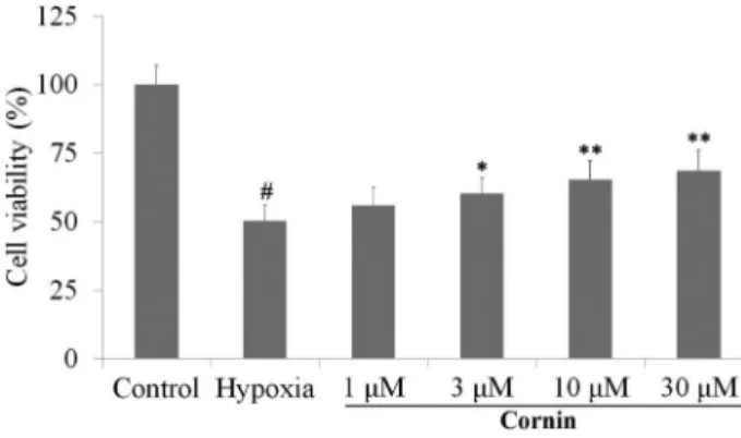
Cornin attenuated hypoxia-induced cytotoxicity Results of the cell viability assay are shown in Figure 1. After exposure to hypoxia for 6 h, only 50.3±5.7% viable cells remained as compared with control cells. Cornin (1, 3, 10, and 30 mM) prevented cells from incurring hypoxia-induced damage in a concentration-dependent manner, and restored cell survival to 56.0±6.4, 60.3±5.8, 64.4±7.0, and 68.7±7.3%, respectively (Figure 1).
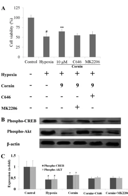
To clarify the mechanism of action of cornin on hypoxia-induced cytotoxicity, a selective inhibitor of CREB (C646, 1mM) or Akt (MK2206, 1mM) was used. We found that pretreatment of H9c2 cells with cornin plus C646 (1 mM) or MK2206 (1mM) for 120 min did not decrease hypoxia-induced cellular damage (Figure 2A).
Cornin attenuated the reduction in expression of phospho-CREB and phospho-Akt during hypoxia
We investigated the effect of cornin on hypoxia-induced reduction of expression of phospho-CREB and phospho-Akt in H9c2 cells. Pretreatment of H9c2 cells with cornin blocked the reduction in expression of hypoxia-induced phospho-CREB and phospho-Akt (Figure 2B,C). To clarify the mecha-nism of action of cornin on hypoxia-induced reduction in expression of phospho-CREB and phospho-Akt in H9c2 cells, a selective inhibitor of CREB (C646, 1 mM) or Akt (MK2206, 1mM) was used. We found that pretreatment of
H9c2 cells with cornin plus C646 (1 mM) or MK2206 (1 mM) for 120 min did not attenuate the reduction of expression of hypoxia-induced CREB or phospho-Akt (Figure 2B,C).
Cornin reduced myocardial infarct volume and ameliorated myocardial function
We examined the effect of cornin on infarct size. Infarct size was reduced significantly in the cornin 30 mg/kg group compared with the vehicle-treated group (Table 1). These data strongly suggested that cornin could attenuate myocardial I/R injury.
The effect of cornin on LVSP and±dp/dtmax of the left ventricle was evaluated 24 h and 14 days after myocardial I/R. The Heart Index was evaluated 14 days after myocardial I/R. Compared with vehicle-treated animals, rats treated with
cornin had significantly improved LVSP, +dp/dtmax and dp/dtmax 24 h and 14 days after myocardial I/R, and a lower Heart Index 14 days after myocardial I/R (Tables 1 and 2). These data suggested that cornin treatment provided immediate and long-term benefits for recovery of myocardial function after I/R.
Cornin decreased serum levels of CK-MB and cTnT Serum levels of CK-MB and cTnT were elevated significantly in vehicle-treated rats subjected to I/R injury. However, treatment with cornin (30 mg/kg) reduced serum levels of CK-MB and cTnT markedly (Table 3).
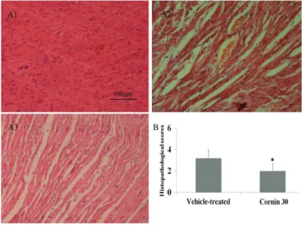
Twenty-four hours after I/R, the pathological features of the infarct area of vehicle-treated rats became apparent: widespread tissue necrosis, contraction bands, capillary compression, and abundant signs of hemorrhage in myocardial tissue. Upon treatment with cornin, these histological features were largely absent, and hearts appeared normal, or only minor architectural changes (interstitial edema, localized necrotic areas) were noted (Figure 3).
Cornin attenuated the reduction in expression of phospho-CREB and phospho-Akt
Western blotting was employed to measure expres-sion of phospho-CREB and phospho-Akt in ischemic myocardial tissue. Low expression of phospho-CREB and phospho-Akt in myocardial tissues was detected after I/R,
but high expression was noted in the hearts of sham-operated rats (Figure 4). Cornin treatment attenuated the reduction of expression of CREB and phospho-Akt markedly.
Discussion
We demonstrated that hypoxia for 6 h significantly decreased cell viability (as evidenced by the MTT assay) in the culture medium. However, pretreatment with cornin (3, 10 and 30mM) decreased cytotoxicity considerably in a concentration-dependent manner.
We observed significant improvement of myocardial function in rats treated with cornin during myocardial I/R challenge as reflected by a reduction in infarct size and histopathological scores. Simultaneously, cornin stimu-lated cardiodynamics directly and inhibited necrosis of myocardial cells. Hence, cornin treatment provided immediate benefits for recovery of myocardial function after I/R.
Increased levels of CK-MB can be detected 3–6 h after the onset of chest pain in individuals who have suffered cardiac arrest. Serum levels of CK-MB peak at 12–24 h and return to normal within 48–72 h. cTnT is a biomarker during myocardial damage (12), the level of which increases to a peak 12 h to 24 h during acute myocardial infarction (13). Determination of serum levels of cTnT is used in the diagnosis of ischemic heart diseases. Lowering the serum level of cTnT can lessen myocardial damage (14). Our results suggest that cornin Table 1.Effects of cornin (30 mg/kg) on left ventricular systolic pressure (LVSP), maximal rate of rise of
left ventricular pressure (±dp/dtmax) and infarct volume after ischemia and reperfusion (I/R).
Group LVSP (mmHg) +dp/dtmax dp/dtmax Infarct volume (%)
Sham 107±8 10987±1387 9325±941
– Vehicle-treated 74±9# 5863±677# 5795±871# 35.2±8.2#
Cornin 90±12* 7363±786* 7067±611* 22.5±5.4*
Data are reported as means±SD for n=10 per group. At 24 h after I/R, LVSP,±dp/dtmax and infarct volume were determined.#Po0.01 compared to the sham group; * Po0.01 compared to the vehicle-treated group (one-way ANOVA followed by Dunnett’s test).
Table 2.Effects of cornin (30 mg/kg) on left ventricular systolic pressure (LVSP), maximal rate of rise of left ventricular pressure (±dp/dtmax) and the Heart Index 14 days after ischemia and reperfusion (I/R).
Group LVSP (mmHg) +dp/dtmax dp/dtmax Heart index (100xg/g)
Sham 107±10 10536±1041 8912±827 0.312±0.029
Vehicle-treated 70±9# 5542±722# 5265±794# 0.374±0.027#
Cornin 89±11* 7052±931** 6999±804** 0.340±0.028*
Data are reported as means±SD, n=10 per group. At 14 days after I/R, LVSP and±dp/dtmax were determined. #Po0.01 compared to the sham group; * Po0.05, **Po0.01 compared to the vehicle-treated group (one-way ANOVA followed by Dunnett’s test).
Table 3.Effects of cornin (30 mg/kg) on serum levels of cTnT and CK-MB after ischemia and reperfusion (I/R).
Group cTnT (ng/mL) CK-MB (U/L)
Sham 0.20±0.03 85±9
Vehicle-treated 1.47±0.35# 422±55#
Cornin 0.85±0.22* 246±46*
(iv) significantly reduced serum levels of cTnT and CK-MB, and that it could lessen the severity of myocardial damage. Phospho-CREB has a key role in myocardial protec-tion against I/R-related injury. CREB is activated by phosphorylation at Ser-133 by protein kinase A (15,16),
which can also be mediated by Akt (17). CREB is a substrate for various cellular kinases (including Akt) (6). Attenuation of the reduction of expression of phospho-CREB in myocardial tissue can reduce the size of the myocardial infarct (18). Increasing expression of Akt in Figure 3.Effects of cornin on histopathological changes 24 h after ischemia and reperfusion (I/R).A, Effects of cornin on pathological injury. Representative light-microscopic appearance of rat myocardial morphology (hematoxylin staining; original magnification200) for sham (A1), vehicle-treated (A2), and cornin 30 mg/kg (A3) groups.B, Effects of cornin on histopathological changes. Data are reported as means±SD, n=10 per group. At 24 h after I/R, myocardial histopathological scores were determined. *Po0.01 compared to the vehicle-treated group (sum of ranks test).
myocardial tissue can also reduce the size of the myocardial infarct, and Akt-dependent activation is dependent upon phospho-CREB (19). Our results showed that expression of phospho-CREB and phospho-Akt was reduced in vitro and in vivo. Reduced expression of phospho-Akt was dependent upon reduced expression of phospho-CREB in vitro. Hence, cornin could reduce myocardial injury during hypoxia by CREB-dependent Akt signaling.
In summary, we demonstrated that cornin can protect myocardial function in rats during myocardial I/R injury. Cornin decreased infarct volume, improved hemody-namics, and alleviated myocardial damage. These effects of cornin were correlated with an increase in expression of
phospho-CREB and phospho-Akt in ischemic myocardial tissue. The main mechanism of action of cornin appeared to be modulation of CREB-dependent Akt signaling. Thesefindings suggest the therapeutic potential of cornin against myocardial I/R injury.
Acknowledgments
This research was supported by funds from the Science and Technology Plan of Colleges and Universities in Shandong Province (number J13LM07), as well as funds from the Binzhou Medical University for Scientific Research (BY2011KYQD05), and the Natural Science Foundation of China (31170321).
References
1. Bolli R, Becker L, Gross G, Mentzer R Jr, Balshaw D, Lathrop DA. Myocardial protection at a crossroads: the need for translation into clinical therapy.Circ Res2004; 95: 125–134, doi: 10.1161/01.RES.0000137171.97172.d7. 2. Cannon RO III. Mechanisms, management and future
directions for reperfusion injury after acute myocardial infarction.Nat Clin Pract Cardiovasc Med2005; 2: 88–94, doi: 10.1038/ncpcardio0096.
3. Marais E, Genade S, Lochner A. CREB activation and ischaemic preconditioning. Cardiovasc Drugs Ther 2008; 22: 3–17, doi: 10.1007/s10557-007-6078-3.
4. Nagy N, Shiroto K, Malik G, Huang CK, Gaestel M, Abdellatif M, et al. Ischemic preconditioning involves dual cardio-protective axes with p38MAPK as upstream target.J Mol Cell Cardiol2007; 42: 981–990, doi: 10.1016/j.yjmcc.2007. 02.010.
5. Li C, Tian J, Li G, Jiang W, Xing Y, Hou J, et al. Asperosaponin VI protects cardiac myocytes from hypoxia-induced apoptosis via activation of the PI3K/Akt and CREB pathways. Eur J Pharmacol 2010; 649: 100–107, doi: 10.1016/j.ejphar.2010.08.060.
6. Du K, Montminy M. CREB is a regulatory target for the protein kinase Akt/PKB.J Biol Chem1998; 273: 32377–32379, doi: 10.1074/jbc.273.49.32377.
7. Jiang WL, Zhang SP, Zhu HB, Tian JW. Cornin ameliorates cerebral infarction in rats by antioxidant action and stabili-zation of mitochondrial function.Phytother Res 2010; 24: 547–552.
8. Kang Z, Jiang W, Luan H, Zhao F, Zhang S. Cornin induces angiogenesis through PI3K-Akt-eNOS-VEGF signaling path-way.Food Chem Toxicol 2013; 58: 340–346, doi: 10.1016/ j.fct.2013.05.017.
9. Zhang N, Pei F, Wei H, Zhang T, Yang C, Ma G, et al. Isorhamnetin protects rat ventricular myocytes from ische-mia and reperfusion injury. Exp Toxicol Pathol 2011; 63: 33–38, doi: 10.1016/j.etp.2009.09.005.
10. Jiang WL, Fu FH, Xu BM, Tian JW, Zhu HB, Jian H. Cardioprotection with forsythoside B in rat myocardial ischemia-reperfusion injury: relation to inflammation response.
Phytomedicine2010; 17: 635–639, doi: 10.1016/j.phymed. 2009.10.017.
11. Zhu J, Qiu Y, Wang Q, Zhu Y, Hu S, Zheng L, et al. Low dose cyclophosphamide rescues myocardial function from ischemia-reperfusion in rats.Eur J Cardiothorac Surg2008; 34: 661–666, doi: 10.1016/j.ejcts.2008.05.035.
12. Wu AH, Lane PL. Metaanalysis in clinical chemistry: validation of cardiac troponin T as a marker for ischemic heart diseases.Clin Chem1995; 41: 1228–1233.
13. Mair J, Artner-Dworzak E, Lechleitner P, Smidt J, Wagner I, Dienstl F, et al. Cardiac troponin T in diagnosis of acute myocardial infarction.Clin Chem1991; 37: 845–852. 14. Jiang WL, Zhang SP, Zhu HB, Hou J. Cardioprotection of
Asperosaponin X on experimental myocardial ischemia injury. Int J Cardiol 2012; 155: 430–436, doi: 10.1016/j. ijcard.2011.06.010.
15. Shaywitz AJ, Greenberg ME. CREB: a stimulus-induced transcription factor activated by a diverse array of extra-cellular signals.Annu Rev Biochem1999; 68: 821–861, doi: 10.1146/annurev.biochem.68.1.821.
16. Kwak HJ, Park KM, Choi HE, Chung KS, Lim HJ, Park HY. PDE4 inhibitor, roflumilast protects cardiomyocytes against NO-induced apoptosis via activation of PKA and Epac dual pathways. Cell Signal 2008; 20: 803–814, doi: 10.1016/ j.cellsig.2007.12.011.
17. Das S, Tosaki A, Bagchi D, Maulik N, Das DK. Resveratrol-mediated activation of cAMP response element-binding protein through adenosine A3 receptor by Akt-dependent and -independent pathways.J Pharmacol Exp Ther2005; 314: 762–769, doi: 10.1124/jpet.105.084285.
18. Ye Y, Long B, Qian J, Perez-Polo JR, Birnbaum Y. Dipyridamole with low-dose aspirin augments the infarct size-limiting effects of simvastatin.Cardiovasc Drugs Ther
2010; 24: 391–399, doi: 10.1007/s10557-010-6252-x. 19. Caravatta L, Sancilio S, di Giacomo V, Rana R, Cataldi A, Di