Synthesis and anti-myocarditis activity in a
multifunctional lanthanide microporous metal-organic
framework with 1D helical chain building units
Chenglv Hong
1, Xinlang Zhou
2, Weijian Huang
1, Peiren Shan
1and Fengquan Dong
3 1Department of Cardiology, The First Affiliated Hospital of Wenzhou Medical University, Wenzhou, Zhejiang, China 2Department of Cardiology, Wenzhou City Hospital of Traditional Chinese Medicine and Western Medicine Combined, Wenzhou, Zhejiang, China 3Department of Cardiology, Shenzhen University General Hospital, Shenzhen, Guangdong, China
Abstract
A new microporous lanthanide metal-organic framework, {[Yb(BTB)(H2O) (DEF)2}n(1, DEF=N,N-Diethylformamide), with 1D nano-sized channels has been constructed by bridging helical chain secondary building units with 1,3,5-benzenetrisbenzoic acid (H3BTB) ligand. Structural characterization suggests that this complex crystallizes in the hexagonal space group P6122 and possesses 1D triangular channels with coordinated water molecules pointing to the channel center. In addition, anti-myocarditis properties of compound1were evaluatedin vivo. The results showed that compound1can improve hemodynamic parameters of, and it may be a good therapeutic option for heart failure in the future.
Key words: Metal-organic framework; Anti-myocarditis;In vivo
Introduction
Myocarditis, also known as inflammatory cardiomyo-pathy, is the inflammation of the heart muscle. Symptoms can include shortness of breath, chest pain, decreased ability to exercise, and an irregular heartbeat (1,2). The duration of problems can vary from hours to months. Com-plications may include heart failure due to dilated cardio-myopathy or cardiac arrest (3,4).
Metal–organic frameworks (MOFs) that are constructed
by coordination of metal centers with multiorganic con-nectors represent an emerging class of inorganic-organic hybrid crystalline materials (5,6). Their structural tenability, well-defined single crystal architectures, functionalized pore environment and modifiable building blocks make them useful in many potential applications including biological activity, catalysis, and luminescent sensing materials (7–9).
The organic ligand plays an important role in the construc-tion of porous MOFs because it not only guides the forma-tion of the secondary building units, but also determines the pore shapes and pore surroundings of the obtained products (10,11). MOFs prepared with ligands of high symmetry have been well studied because of synthetic and crystallographic considerations. As the elongated ligand of H3BTC, 1,3,5-benzenetrisbenzoic acid (H3BTB,
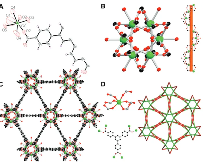
Figure 1) has been widely used in the construction of
porous MOFs (12,13). However, compared with the transi-tion BTB frameworks reported, the lanthanon metal-BTB frameworks are less studied (14).
Here, we present the synthesis and the structural analysis of a highly porous Yb-organic network {Yb(BTB)(H2O)]
(DEF)2}n(1, DEF=N,N-diethylformamide). This MOF is
com-posed of novel 1D helical chain building units and BTB3–
ligand, which represents the first example of Ln-MOFs based on 1D helical chain building units. In addition,in vivo
anti-myocarditis activity of compound1was investigated.
Material and Methods
Apparatus and materials
All the starting materials and reagents used in this work were obtained commercially and used without further purification. Element analyses (C, H, and N) were deter-mined with an elemental Vairo EL III analyzer (Bruker, Germany). Powder X-ray diffraction data were collected using PANalytical X’Pert Pro powder diffractometer (Bruker)
with Cu-Karadiation and 5°p2yp50°. Thermogravimetric
experiments were performed using a TGA/NETZSCH STA449C instrument heated from 30 to 800°C (heating rate of 10°C/min, nitrogen stream; Bruker). Single crystal
Correspondence: Fengquan Dong:<[email protected]>
Received August 17, 2017 | Accepted November 10, 2017
X-ray diffraction was carried out by an Oxford Xcalibur E diffractometer (Bruker).
Synthesis and characterization of {[Yb(BTB)(H2O)]
(DEF)2}n(1)
A mixture of Yb(NO3)26H2O (0.1 mmol, 0.031 g) and
H3BTB (35 mg, 0.062 mmol) was added to a solution of
DEF (4mL) and H2O (1 mL). The mixture was sealed in a
Pyrex tube, and heated at 140°C for 3 days. After cooling to room temperature, the colorless polyhedral-shaped
crystals formed werefiltered, washed with DEF, and then dried in air. Analytical data for compound 1 (C37H39N2
O9Yb): C, 53.23; H, 4.44; N, 3.29%. Calculated: C, 53.62;
H, 4.74; N, 3.38%.
Crystal structure determination
Suitable single crystal of compound 1 was carefully selected under optical microscope and glued on thin glass
fibers. The intensity data of1was collected on an Oxford Xcalibur E diffractometer. The empirical absorption cor-rections were applied to the data using the SADABS system. This structure was solved by direct method and refined by full-matrix least-squares method on F2 using the SHELXS–97 program (15). All non-hydrogen atoms of 1 were refined anistropically, and all the hydrogen atoms attached to carbon atoms were fixed at their ideal posi-tions. Pertinent crystal data and structural refinement results for compound1are summarized in Table 1.
In vivoanti-myocarditis activity
C57BL6/j mice were involved in our experiment. A total of 48 eight-week-old male mice were divided into four groups: control+PBS (G1, n=12), control+1(G2, n=12), CVB3+PBS (G3, n=12), CVB3+1 (G4, n=12). G3 and G4 were infected by intraperitoneal (ip) injection of 1105 Figure 1.Schematic representation of the H3BTB ligand used in
this research
Table 1.Crystal data and structure refinements for compound1.
Formula weight 624.43
Temperature/K 293 (2)
Crystal system hexagonal
Space group P6122
a/Å 18.0081 (15)
b/Å 18.0081 (15)
c/Å 21.8141 (13)
a/° 90
b/° 90
g/° 120
Volume/Å3 6126.4 (11)
Z 6
rcalcg/cm3 1.016
m/mm 1 2.316
Radiation MoKa(l=0.71073)
2Yrange for data collection/° 6.422 to 52.726
Reflections collected 16173
Independent reflections 4186 [Rint=0.0485, Rsigma=0.0462] Data/restraints/parameters 4186/111/162
Goodness-of-fit on F2 1.018
Final R indexes [I4=2s(I)] R1=0.0316,oR2=0.0684 Final R indexes [all data] R1=0.0440,oR2=0.0734 Largest diff. peak/hole / e Å 3 0.42/
–0.78
Flack parameter –0.026(10)
CCDC 1573543
plaque forming units (pfu) Coxsackie virus B3 (CVB3) per mouse, while G1 and G2 received ip injection of phosphate-buffered saline (PBS) on the same day. Compound1was orally applied at 50 mg/kg on the next day of infection (G2 and G4), while G1 and G3 were orally administrated the same dose of PBS. Animals were housed with a normal diet, 12 h light/dark cycle, 30–70% humidity, and 20–25°C. All mice were sacrificed
on day 7 post-CVB3 infection. We used a conductance catheter (DDS-307, Chang-Ai, China) to collect hemody-namic data (pressure and volume) before sacrificing the animals.
Statistical analysis was performed using Prism 6 (Bruker). One-way analysis of variance (ANOVA) was used for statistical analysis of the data with correction for multiple comparisons via the Tukey’s range test. Data are reported as means±SD. Differences were regarded to be signifi
-cant if the two-sided P-value was lower than 0.05.
Results and Discussion
Molecular structure
The solvothermal reaction of Yb(NO3)36H2O and
H3BTB in a mixed solvent of DEF and H2O provided
complex1 as colorless crystals. Single-crystal X-ray dif-fraction reveals that 1 crystallizes in a highly symmetric and chiral hexagonal space group P6122 and the 3-D
coordination network is constructed through the connec-tion of infinite 1-D helical chain building units and the BTB3–ligands. The asymmetric coordination unit consists
of one Yb ion situated on a symmetry site with one half occupancy, half BTB3–ligand and one coordinated water
molecule. As shown in Figure 2A, the Yb(III) ion is seven-coordinated by six carboxylic acid O atoms from six dif-ferent BTB3–ligands and one coordinated water molecule,
resulting in a pentagonal bipyramid geometry. The Yb-O bond distances are in the range of 2.212 (3) to 2.609 (5) Å.
Figure 2.A, view of the asymmetric unit in compound1with 30% thermal ellipsoid level;B, view of 1D helical chain building units in1;
Each Yb atom is connected with the neighboring ones though three carboxylic groups. Such a connection mode leads to the formation of a 1D right-handed chain along a 61axis, which represent a rare case of Ln-based helical
chain building units according to the Cambridge Crystal-lographic Data Centre database (Figure 2B). The pitch of the helical chain is 21.832 (3) Å. Furthermore, such 1D helical chain building units are further linked by BTB ligand through its three carboxylate groups to afford a 3D non-interpenetrating framework with 1D triangular channels with coordinated water molecule pointing to the channel center (Figure 2C). Based on the crystallographic data and considering the van der Waals radii of atoms, the pore size for the triangular channel is 5.4 Å. To understand the network of1more clearly, we use the software TOPOS to simplify its framework. Each Yb(III) ion is connected with six O atoms from six different BTB– ligands and each
BTB–ligand binds with six different Yb(III) ions. Thus, both
of the Yb(III) ions and the BTB–ligand could be viewed
as 6-connected nodes. In this case, the whole framework of 1 can be simplified to a 2-nodal (6,6)-connected net-work with the point symbol of (410.65)(47.68), which has
not been observed in MOF chemistry (Figure 2D). The effective free volume of1without guest water molecules is 58.3% of the crystal volume (3569 Å3of the 6126 Å3unit
cell volume), calculated with PLATON software.
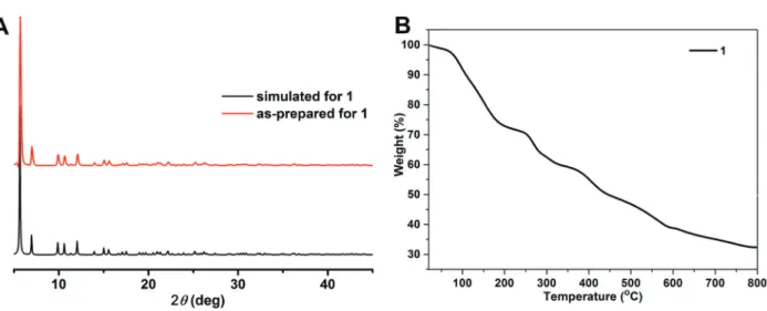
Powder X-ray diffraction analysis (PXRD) and thermal analysis
PXRD experiment was carried out to verify the phase purity of the as-synthesized samples. As shown in Figure 3A, the diffraction peak of compound1is in good agreement with that of the simulated one based on the single crystal diffraction data, indicating the pure phase of the obtained samples. From the thermogravimetric curve of compound1, we found that the first weight loss of 27.1% occurs from 25 to 210°C, which corresponds to the release of one coordinated water molecule and two lattice DEF mole-cules (Calcd: 26.5%). Then, the dissolved sample was stable up to 230°C, after which the framework began to collapse (Figure 3B).
In vivoanti-myocarditis activity
To evaluate the impact of compound 1 in CVB3-induced myocarditis, body weight, heart rate (HR), maximum left ventricle pressure (Pmax), maximum left ventricle pressure rise rate (dP/dtmax), and ejection fraction (EF) were analyzed in the present study. There was a significant difference in body weight between G3 and G1. Furthermore, there was a significant difference between G4 and G3 (Table 2). In comparison to G1 and G2, there was a sharp decrease in G3 animals in HR, Pmax, dP/dtmax and EF. Oppositely, G4 had a downward
Figure 3.A, powder X-ray diffraction analysis patterns for compound1.B, TGA curve for1.
Table 2.Body weight of mice at day 0 and day 7.
Control+PBS Control+1 CVB3+PBS CVB3+1
Day 0 BW (g) 24.83±0.66 24.88±0.69 24.88±0.64 24.79±0.59 Day 7 BW (g) 26.65±0.49 26.37±0.74 20.41±0.73*# 22.41±1.15*#+
Data are reported as means±SD. BW: body weight. CVB3: Coxsackie virus B3; PBS: phosphate buffered saline. *Po0.01vscontrol+PBS,#Po0.01vscontrol+1,+Po0.01vsCVB3
trend that compared to G1 and G2, but there was no significant difference between them (Table 3).
As known, human myocarditis can result in chest dis-comfort, palpitation, shortness of breath, dizziness, decreased activity, and poor appetite. CVB3-mice are a good myo-carditis model that we can easily see reduced activity, and get body weight data through weighing; the decreased appetite indicates that the myocarditis model works. From our in vivo experiment, we found that the CVB3 group significantly lost body weight, but it seemed to reverse after application of compound 1. The CVB3 group had a significant decrease of HR, Pmax, dP/dtmax, and EF, which are essential factors of heart failure, especially
systolic heart failure. In our investigation, compound 1
was effective in hemodynamics, indicating it could be a candidate for anti-myocarditis therapy.
In conclusion, we demonstrated the successful con-struction of a novel Yb-based MOF with 1D helical chain building units built up from 1,3,5-H3BTB ligand. Structural
characterization suggests that this complex crystallizes in the hexagonal space group P6122 and possesses 1D
triangular channels with coordinated water molecules point-ing to the channel center. In addition, the results showed that compound1can improve hemodynamic parameters, and may be a good therapeutic compound for heart failure in the future.
References
1. Ghatnur SM, Parvatam G, Balaraman M. Culture conditions for production of biomass, adenosine, and cordycepin from cordyceps sinensis CS1197: optimization by desirability func-tion method.Pharmacogn Mag2015; 11 (Suppl 3): S448– S456, doi: 10.4103/0973-1296.168946.
2. Root-Bernstein R, Fairweather D. Unresolved issues in theories of autoimmune disease using myocarditis as a framework.J Theor Biol2015; 375: 101–123, doi: 10.1016/ j.jtbi.2014.11.022.
3. Li-Sha G, Yi-He C, Na-Dan Z, Teng Z, Yue-Chun L. Effects of carvedilol treatment on cardiac cAMP response element binding protein expression and phosphorylation in acute coxsackievirus B3-induced myocarditis. BMC Cardiovasc
Disord2013; 13: 100, doi: 10.1186/1471-2261-13-100.
4. Massilamany C, Gangaplara A, Reddy J. Intricacies of cardiac damage in coxsackievirus B3 infection: Implications for therapy.Int J Cardiol2014; 177: 330–339, doi: 10.1016/ j.ijcard.2014.09.136.
5. Krejci J, Mlejnek D, Sochorova D, Nemec P. Inflammatory cardiomyopathy: a current view on the pathophysiology, diagnosis, and treatment. Biomed Res Int 2016; 2016: 4087632, doi: 10.1155/2016/4087632.
6. Li-Sha G, Jing-Lin Z, Guang-Yi C, Li L, De-Pu Z, Yue-Chun L. Dose-dependent protective effect of nicotine in a murine model of viral myocarditis induced by coxsackievirus B3.
Sci Rep2015; 5: 15895, doi: 10.1038/srep15895.
7. Nyland JF, Fairweather D, Shirley DL, Davis SE, Rose NR, Silbergeld EK. Low-dose inorganic mercury increases severity and frequency of chronic coxsackievirus-induced autoimmune
myocarditis in mice.Toxicol Sci2012; 125: 134–143, doi: 10.1093/toxsci/kfr264.
8. Junghans U, Kobalz M, Erhart O, Preibler H, Lincke J, Möllmer J, Krautscheid H, Gläser R. A Series of robust copper-based triazolyl isophthalate MOFs: impact of linker functionalization on gas sorption and catalytic activity.Materials 2017; 10: 338, doi: 10.3390/ma10040338.
9. Haydar MAL, Abid HR, Sunderland B, Wang S. Metal organic frameworks as a drug delivery system for fl urbi-profen. Drug Des Devel Ther2017; 11: 2685–2695, doi: 10.2147/DDDT.S145716.
10. Han Q, Qi B, Ren W, He C, Niu J, Duan C. Polyoxo-metalate-based homochiral metal-organic frameworks for tandem asymmetric transformation of cyclic carbonates from olefins.Nat Commun2015; 6: 10007, doi: 10.1038/ ncomms10007.
11. Kim JY, Jin M, Lee KJ, Cheon JY, Joo SH, Kim JM, et al. In situ-generated metal oxide catalyst during CO oxidation reaction transformed from redox-active metal-organic frame-work-supported palladium nanoparticles. Nanoscale Res Lett 2012; 7: 461, doi: 10.1186/1556-276X-7-461.
12. Wang Z, Wang J, Li M, Sun K, Liu C. Three-dimensional printed acrylonitrile butadiene styrene framework coated with Cu-BTC metal-organic frameworks for the removal of methylene blue.Sci Rep2014; 4: 5939, doi: 10.1038/ srep05939.
13. Mitra J, Guerrero EN, Hegde PM, Wang H, Boldogh I, Rao KS, et al. New perspectives on oxidized genome damage and repair inhibition by pro-oxidant metals in neurological Table 3.Hemodynamic data of mice.
Control+PBS Control+1 CVB3+PBS CVB3+1
HR (bpm) 531.48±40.32 529.98±39.46 386.77±124.59*# 475.79±79.13+
Pmax (mmHg) 113.96±21.64 109.66±12.91 90.25±22.37*# 105.34±11.43
dP/dtmax (mmHg/s) 9742.27±1766.32 9445.64±1920.23 6108.48±2592.93*# 8000.47±1378.99
EF (%) 78.60±2.65 77.76±5.20 69.66±7.74*# 73.64±4.59
Data are reported as means±SD. HR: heart rate; Pmax: maximum left ventricle pressure; dP/dtmax: maximum rate of rise of left ventricle pressure; EF: ejection fraction; CVB3: coxsackie virus B3; PBS: phosphate buffered saline. *Po0.05vscontrol+PBS,#Po0.05vscontrol+ compound1,+Po0.01vsCVB3
diseases. Biomolecules 2014; 4: 678–703, doi: 10.3390/ biom4030678.
14. Zhu Y, Zhu M, Xia L, Wu Y, Hua H, Xie J. Lanthanide metal-organic frameworks with six-coordinated ln(iii) ions and free functional organic sites for adsorptions and extensive
catalytic activities.Sci Rep2016; 6: 29728, doi: 10.1038/ srep29728.
15. Sheldrick GM. SHELXL-97, program for crystal structure
solution and refinement. University of Göttingen: Göttingen,