campestris
pv.
viticola
by rep-PCR Fingerprinting
Loiselene C. Trindade1*, Mirtes F. Lima2 & Marisa A. S. V. Ferreira1
1Departamento de Fitopatologia, Universidade de Brasília, CEP 70910-900, Brasília, DF, fax (61) 272-1793, e-mail:
marisavf@unb.br;2EMBRAPA- Semi Árido, Cx. Postal 23, CEP 56300-970, Petrolina, PE
(Accepted for publication on 13/12/2004)
Corresponding author: Marisa A.S.Velloso Ferreira
ABSTRACT
Bacterial canker of grapevine (Vitis vinifera), caused by Xanthomonas campestris pv. viticolawas first detected in Brazil in 1998, affecting grapevines in the São Francisco river basin, state of Pernambuco. The disease was also reported in Juazeiro, Bahia and later in Piauí and Ceará. Due to its limited geographical distribution and relatively recent detection in Brazil, very little is known about the pathogen’s biology and diversity. Repetitive DNA based-PCR (rep-PCR) profiles were generated from purified bacterial DNA of 40 field strains of X. campestris pv. viticola, collected between 1998 and 2001 in the states of Pernambuco, Bahia and Piauí. Combined analysis of the PCR patterns obtained with primers REP, ERIC and BOX, showed a high degree of similarity among Brazilian strains and the Indian type strain NCPPB 2475. Similar genomic patterns with several diagnostic bands, present in all strains, could be detected. Fingerprints were distinct from those of strains representing other pathovars and from a yellow non-pathogenic isolate from grape leaves. The polymorphism observed among the Brazilian strains allowed their separation into five subgroups, although with no correlation with cultivar of origin, geographic location or year collected.
Additional keywords: diversity, grapevine bacterial canker, Vitis vinifera, repetitive DNA. RESUMO
Caracterização molecular de isolados brasileiros de Xanthomonas campestris pv. viticolaatravés de rep-PCR O cancro bacteriano da videira (Vitis vinifera), causado por Xanthomonas campestris pv. viticola, foi primeiramente detectado no Brasil em 1998, em parreirais no município de Petrolina, Vale do São Francisco, Pernambuco. A doença foi também observada em Juazeiro, Bahia, e mais tarde nos estados de Piauí e Ceará. Devido à sua limitada distribuição geográfica e relativamente recente detecção no Brasil, pouco se conhece sobre a biologia e diversidade do patógeno. Padrões genômicos de rep-PCR foram gerados a partir do DNA purificado de 40 isolados de X. campestris pv. viticola, coletados entre 1998 e 2001, em diferentes localidades dos estados de Pernambuco, Bahia e Piauí. A análise combinada dos padrões obtidos com os “primers” REP, ERIC e BOX, mostraram alta similaridade entre os isolados brasileiros e o isolado tipo NCPPB 2475, originário da Índia. Os isolados brasileiros apresentaram padrões similares e algumas bandas diagnósticas, presentes em todos os isolados. Os padrões foram distintos dos isolados de outros patovares e de um isolado não-patogênico obtido de folhas de videira. O polimorfismo observado entre os isolados brasileiros permitiu a diferenciação de cinco subgrupos, sem nenhuma relação com cultivar de origem, local ou época de coleta.
Palavras-chave adicionais: diversidade, cancro bacteriano, Vitis vinifera, DNA repetitivo.
*Parte da Dissertação de Mestrado do primeiro autor. Universidade de Brasília (2002).
INTRODUCTION
In 1998, a new disease was reported affecting ‘Red Globe’ grapevines (Vitis vinifera L.) in Pernambuco and Bahia states. A gram-negative bacterium was isolated from all diseased plants and Koch’s postulates were completed. The causal agent was identified through biochemical and pathogenicity tests as
Xanthomonas campestris pv. viticola Nayudu (Dye) (Malavolta Jr. et al., 1999a; Lima et al., 1999). Later, the disease was also detected in the states of Piauí (Malavolta et al., 1999b) and Ceará (Freire & Oliveira, 2001).
plants could be detected from eight to 14 days after artificial inoculation depending on the method of inoculation used (Malavolta Jr. et al., 1999a; Lima et al.,1999).
Xanthomonas campestris pv. viticola was first described by Nayudu (1972) in India as Pseudomonas viticola Nayudu sp. nov., and its occurrence is apparently very restricted. Besides India, there are also reports of a “grapevine bacterial canker disease (GVBCD)” in Ukraine (Kochenko, 1993). Since the pathovar viticola has not been included in the reclassification work of Vauterin et al. (1995), its specific status may still be controversial. As with the pathovar mangiferaeindicae of the genus Xanthomonas, all strains could be referred to as members of the pathovar viticola. We chose here to use the name proposed by Dye (1978), considering that it has been used in all references to this disease found to date and until more information on their species classification is available.
Several molecular methods have been used to assess diversity and to detect plant pathogenic bacteria (Louws et al., 1999). The presence of repetitive DNA sequences is common in the prokaryotic and eukaryotic genomes. In bacteria, the observed occurrence of three families of repetitive sequences, REP, ERIC and BOX elements (Stern et al., 1984; Sharples & Lloyd, 1990; Koeuth et al., 1995) led to the development of repetitive DNA PCR-based genomic fingerprinting protocols (referred as rep-PCR), in which primers were designed to amplify intervening DNA between two adjacent repetitive sequences (Louwset. al., 1999). The amplified fragments, then separated by electrophoresis on agarose gels, generate specific fingerprints (DeBruijn, 1992). Repetitive-PCR has been successfully used for identification and differentiation of strains and the assessment of genetic diversity of phytopathogenic bacteria such as Xanthomonas spp. and
Pseudomonas spp. (Louws et al., 1994, 1995; Opgenorth et al., 1996; Weingart & Volksch, 1997; Restrepo et al., 2000),
Clavibacter michiganensis (Smith) Davis et al. (Louwset al., 1998), and Ralstonia solanacearum (Smith) Yabuuchi et al. (Horita & Tsuchiya, 2001).
Due to its relatively recent detection in Brazil, little is known about the biology and diversity of X. campestris pv.
viticola. The objective of this study was to compare several field strains of X. campestris pv. viticola, collected from 1998 to 2001, using rep-PCR fingerprinting. The molecular characterization of these isolates could provide information not only on their diversity but also on their origin. Specific fingerprints may also be helpful in the identification process leading to a more rapid and effective diagnosis of grapevine bacterial canker.
MATERIALS AND METHODS
Bacterial strains: source and maintenance
Forty grapevine bacterial canker-derived strains previously identified as X. campestris pv. viticola were used in this study (Table 1). Reference strain NCPPB 2475 (originated from India), another 18 Xanthomonassp. strains from various
host plants and one Ralstonia solanacearum strain were included for comparison purposes. One unidentified isolate, commonly isolated from grape leaves, was also included. This isolate grew on asparagine rich-medium, formed yellow, fast-growing and gram-negative colonies and did not induce a hypersensitive reaction on tobacco leaves. All isolates were cultivated on Kado’s 523 medium (Kado & Heskett, 1970). For long-term maintenance, isolates were preserved on sterile distilled water at room temperature and frozen in 30% glycerol at –80 ºC.
Biochemical and pathogenic characterization
Initially all grapevine-derived isolates were identified and characterized according to conventional biochemical and physiological tests (Bradbury, 1986; Lelliott & Stead, 1987; Schaadet al., 2001), except those obtained from other culture collections (IBSBF 1385, 1386 and 1369, and NCPPB 2475). The tests conducted were as described by Lima et al. (1999), except that the hypersensitivity test on tomato (cv. Santa Cruz), and growth on triphenyl tetrazolium chloride-containing medium (TTC) tests were included. All isolates were tested for pathogenicity by inoculation of petioles and leaves of grapevine cv. Catalunha after superficial wounding with a sterile needle.
DNA isolation
Total bacterial DNA was extracted using a modified version of the CTAB method from Ausubel et al. (1992). Single colonies grown on Kado’s 523 medium were transferred to 1.5 ml of 523 liquid medium and cultures were grown at 28 ºC, with shaking at 150 rpm. After 48 h the cultures were centrifuged at 4,600 g (8,000 rpm) for 5 min, the supernatant was removed and the pellets were resuspended in 520 µl of TE buffer (10 mM Tris-HCl, 1 mM EDTA, pH 8.0). Fifteen microliters of 20% SDS and 3 µl of Proteinase K (20 mg/ml) were added. The mixture was incubated for 1 hour at 37 ºC, then 100 µl of 5 M NaCl and 80µl of a 10% CTAB solution in 0.7 M NaCl were added and mixed. The suspension was incubated for 10 min at 65 ºC and kept on ice for 15 min. An equal volume of chloroform:isoamyl alcohol (24:1) was added, followed by incubation on ice for 5 min and centrifugation at 7,200 g (10,000 rpm) for 20 min. The aqueous phase was transferred to a new tube, 0.6 volumes of isopropanol were added and DNA was precipitated at –20 ºC for 16 h. DNA was collected by centrifugation at 7,200 g (10,000 rpm) for 10 min, washed with 500 µl of 70% ethanol, air-dried at room temperature for approximately 3 h and finally resuspended in 50 µl of TE buffer. The DNA was quantified on 0.8% agarose gels by comparison with DNA samples (phage λ DNA-Hind
III fragments) of known concentration, diluted to working aliquots of 50 ng/µl and stored at –20 ºC until needed.
Repetitive elements-based PCR (rep-PCR)
Amplification reactions were conducted with three primer sets: (1) REP1R-I (5’ IIICGICGICATCIGGC 3’) and REP2-I (5’ ICGITTATCIGGCCTAC 3’); (2) ERIC1R (5’
ATGTAAGCTCCTGGGGATTCA 3’) and ERIC2 (5’ AAGTAA
TABLE 1 - Species and strains used in this study
aNational Collection of Plant Pathogenic Bacteria, York, UK.
bColeção de Bactérias Fitopatogênicas da Universidade de Brasília, Brasília, D.F. c Instituto Biológico Setor de Bacteriologia Fitopatológica, Campinas, S.P.
Strain Host Location and year
collected Xanthomonas campestrispv.viticola Vitis vinifera
NCPPBa2475 cv. Anabi-e-Shahi India, 1972
UnBb1183,1185,1186,1187, 1188,1189, 1190,1193, 1194,1195, IBSBFc1369 cv. Red Globe Petrolina, PE, 1998
1191,1192 cv. Itália Petrolina, PE, 1998
IBSBF 1385 cv. Itália Teresina, PI, 1998
IBSBF 1386 cv. Ribier Teresina, PI, 1998
UnB 1201,1202,1203,1206,1207,1208, 1209 cv. Itália Petrolina, PE, 2000
UnB 1204 cv. Red Globe Juazeiro, BA, 2000
UnB 1216,1218 cv. Red Globe Petrolina, PE, 2000
UnB 1205 cv. Itália Sobradinho, BA, 2000
UnB1210 cv. IAC 572 (rootstock) Petrolina, PE, 2000
UnB 1211 cv. Catalunha Petrolina, PE, 2000
UnB 1212 cv. Itália Petrolina, PE, 2001
UnB1213 cv. Red Globe Petrolina, PE, 2001
UnB 1214,1219,1220,1221 cv. Catalunha Petrolina, PE, 2001
UnB 1222 cv. Perlette Petrolina, PE, 2001
UnB 1223, 1224 cv. Red Globe Juazeiro, BA, 2001
UnB 1225 cv. Festival Petrolina, PE, 2001
UnB 1226 cv. Thompson Seedless Petrolina, PE, 2001
UnB 1227 cv. Itália Petrolina, PE, 2001
X. axonopodis pv.glycines
UnB 306 soybean Planaltina, DF,1983
X. axonopodis pv.manihotis
UnB 1126 cassava Jataí, GO, 1996
UnB 1114 cassava Mirador, PR,1996
UnB 1111 cassava Paranavaí, PR,1996
UnB 1129 cassava Rolândia, PR, 1996
X. axonopodispv.passiflorae
UnB 642 passionfruit Planaltina, DF, 1986
X. axonopodispv.ricini
UnB 607 (white colonies) castor Ibiapina,CE,1985
X. campestrispv.campestris
UnB 159 cabbage Vargem Bonita, DF, 1981
UnB 606 cabbage Guaramiranga, CE,1985
X. campestrispv.carotae
UnB 107 carrot Vargem Bonita, DF, 1981
X. campestrispv.mangiferaeindicae
UnB 769 (yellow) mango Planaltina, DF, 1990
UnB 764 ( white) mango Planaltina, DF, 1990
IBSBF 1230 ( white) mango Brasil,1996
IBSBF1508 ( white) cashew Brasil,1999
X. campestris pv.vesicatoria
UnB 604 tomato Ibiapina, CE,1985
UnB 605 tomato CE, 1985
UnB 1174 pepper DF, 1998
X. melonis
UnB1080 cucumber Vargem Bonita, DF, 1995
Ralstonia solanacearum
UnB 486 potato Lagoa Seca, PB, 1984
unidentified non-pathogenic isolate (NI) Vitis viniferacv. Catalunha Petrolina, PE, 2001
CTCCGGCAAGGCGACGCTGAC3’), corresponding to REP, ERIC and BOX elements, respectively (Louws et al., 1999). Primers were synthesized by Life Technologies do Brasil Ltda. Each reaction consisted of: 1.2 µl of 10X Taq buffer (500 mM KCl, 100 mM TRIS-HCl); 1 mg/ml BSA; 1.5 mM MgCl2; 0.2 mM each dNTP (GIBCO, BRL); 2 µM of each primer, 1.25 U of Taq
DNA polymerase (GIBCO, BRL) and 50 ng of bacterial DNA, in
a final volume of 12 µl.
min, followed by 30 cycles of: 94 °C/1 min; 1 min at 52 °C (ERIC) or 53 ºC (BOX) for primer annealing, and 8 min at 65 °C for primer extension. After the extension step, the temperature was held at 65 °C for 15 min for all three primer sets. For each bacterial strain, reactions were repeated at least twice.
The PCR products were separated by electrophoresis on 1.5% agarose gels. Total volume of the reaction (12 µl) was analyzed. Electrophoresis was carried out at 80 V for 4 h in 0.5 X Tris-borate EDTA buffer. Gels were stained with ethidium bromide (0.5 µg/ml), and DNA migration patterns were analyzed visually. Sizes of fragments were determined by comparison with DNA molecular weight markers, 1 Kb and 100 bp DNA ladders (GIBCO, BRL).
Genomic fingerprint comparisons among bacterial strains were performed by measurement of band sizes. The presence or absence of each band was converted into binary data, scored as 1 for presence, and 0 for absence. Only the reproducible bands ranging from 200 bp to 3 kb were scored for each strain. Data generated with the three sets of primers were combined and analyzed using software MVSP 3.1 (Kovach, 1999). Genetic relationships among isolates were determined by Jaccard’s similarity coefficients followed by UPGMA (unweighted pair-group method using arithmetic averages) clustering analysis. The reliability of the dendrograms was assessed by bootstrap analysis (Swofford & Olsen, 1990) with the software program WinBoot (Yap & Nelson, 1996) with 1,000 repeated samplings.
RESULTS
All 37 field strains dated from 1998-2001, isolated from
V. vinifera plants with bacterial canker symptoms showed biochemical properties of the genus Xanthomonas(Schaadet al., 2001) and, apart from colony color, they could not be differentiated from a X. campestris pv. campestris (Pammel) Dowson strain (UnB 159) through biochemical and physiological tests. The pathogenicity assay on grape leaves and petioles along with the biochemical characteristics, confirmed the identity of all strains as the pathogen described by Nayudu (1972).
The DNA amplification from 41 X. campestris pv.
viticola strains with primers REP, ERIC and BOX (rep-PCR) showed multiple fragments varying from 190 bp to 3054 bp. Initially, several tests with REP primers were carried out in order to assess the reproducibility of the method with purified DNA and whole cells collected directly from agar or liquid cultures grown for 24-48 h. The cells were resuspended in sterile water and 1 µl was used in the amplification reaction. The REP-PCR banding patterns were reproducible only when purified DNA was used. Only the major and more intense bands of 1 kb and 600 bp were visualized when using whole cells in the amplification reactions. With purified DNA, the same electrophoretic patterns were produced from independent amplification reactions and also from two independent DNA extractions of the same strain (data not shown).
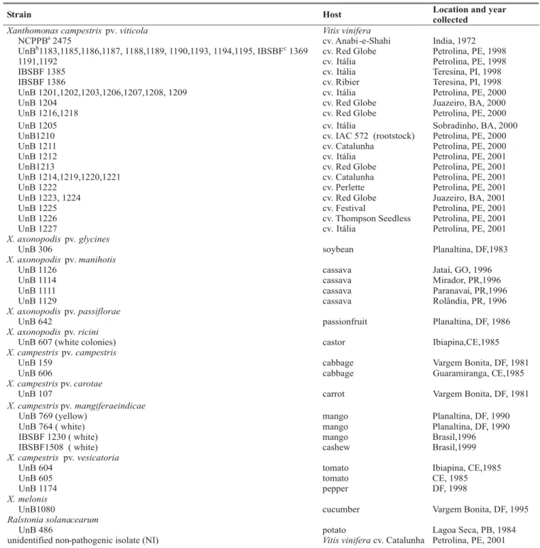
With ERIC primers, 19 bands varying from 190-3054 bp
were visualized and scored (Figure 1). The majority of strains and the type strain NCPPB 2475 showed a similar pattern overall (Figure 1A), with several diagnostic bands. Polymorphic bands were also observed, and nine patterns could be differentiated. The genomic patterns of X. campestris pv. viticola were visually distinct from a non-pathogenic isolate (NI) and other xanthomonads (Figure 1B).
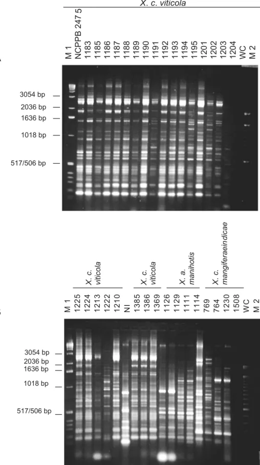
With primer BOX A1R a total of 12 bands varying from 210 bp to 2,036 bp were scored. An 800 bp-fragment was observed in the amplification patterns of 38 X. campestris pv.
viticolastrains. However, strains UnB 1212, 1222 and 1227 did not present that band. Visual analysis of the BOX fingerprinting patterns revealed five distinct patterns among the grape strains. Polymorphic bands were mostly detected in the region of 600-1000 bp. A high degree of similarity between Brazilian strains and the Indian strain was also observed (Figure 2).
The REP primers produced 16 scoreable bands ranging from 250 to 2900 bp. Seven distinct patterns were observed amongX. campestris pv. viticolastrains, with a 1,000 bp-band present in all strains. Polymorphism was observed between bands of 500 and 1000 bp.
The PCR products of all 41 pathovar viticola strains, regardless of the geographic location, host cultivar or year collected, showed an overall homogeneous pattern sharing multiple bands (Figures 1 and 2). For all three primers used, the non-pathogenic yellow strain (NI) showed a distinct banding pattern from the Xanthomonas spp. strains.
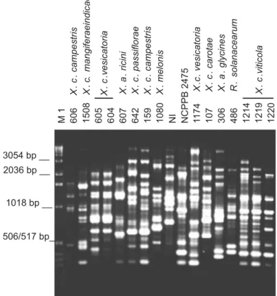
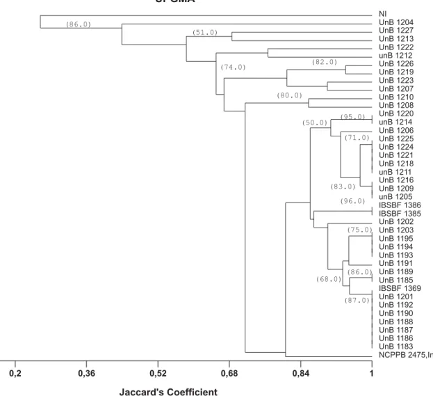
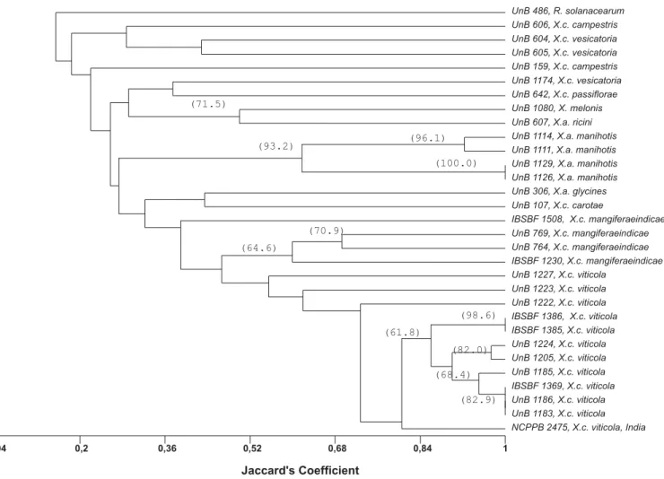
Considering only the grapevine-derived strains and the combined data from the three primer sets, two distinct groups could be differentiated at 26% similarity: group 1, formed by the non-pathogenic strain (NI) only, and group 2, comprising all pathovar viticolastrains (Figure 3). At 68% similarity, five subgroups were detected among the pathogenic strains. Thirty-two strains, representing 78% of the strains, formed one single group, which included the type strain (NCPPB 2475), 27 strains from Pernambuco, the two strains from Piauí (IBSBF 1385 and 1386) and two strains from Bahia (UnB 1224 and 1205). Reference strain NCPPB 2475 showed above 80% similarity to 29 grapevine strains (~73% of the tested strains). Dendrograms obtained with each individual set of primers showed an overall similar pattern of separation when compared to the combined analysis (data not shown). Of the three sets of primers tested, BOX was the most conserved, identifying a smaller number of subgroups among the strains. Pathovarviticola strains representing all geographic locations and different rep-PCR groups (Figure 3) were selected and compared with other Xanthomonas sp. strains, associated with different hosts and one R. solanacerum strain. The combined analysis of the patterns generated by BOX, ERIC and REP-PCR showed the separation into two groups at 15% similarity (Figure 4). The first group was composed only of
Ralstonia solanacearum (UnB 486), the second and largest group included all xanthomonads and the 12 selected X.
campestris pv. viticola strains. Three mango (Mangifera indica L.) strains of X. campestris pv. mangiferaeindicae
M1
X.c.viticola
1018 bp 1636 bp 2036 bp
517/506 bp 3054 bp
FIG. 1 - Genomic fingerprints of Xanthomonas spp. generated from ERIC-PCR. (A) field strains of X. campestris
pv. viticola collected in Brazil compared to type strain NCPPB 2475. (B) field strains of X. campestris pv. viticola
collected in Brazil compared to other Xanthomonas spp. from different host species. NI: unidentified strain; WC: water control; M: molecular size standards 1 Kb DNA ladder (M1) and 100 bp-DNA ladder (M2). PCR products were separated by electrophoresis on 1.5% agarose gels.
A
B
M1 1225
12
24
12
13
12
22
12
10
NI 13
85
13
86
13
69
11
26
11
29
11
11
11
14
76
9
76
4
12
30
15
08
WC M2
X
.
c
.
vi
ti
co
la
X
.
c
.
vi
ti
co
la
X
.
a
.
m
anihot
is
X
.
c
.
m
angif
er
aein
dic
a
e
2036 bp
1018 bp 1636 bp
FIG. 2 - Genomic fingerprints of Xanthomonas spp. from various host species and four strains of X. campestris pv. viticola generated from BOX-PCR. Lane M1: molecular size standard ,1 Kb- DNA ladder; NI: unidentified strain. PCR products were separated by electrophoresis on 1.5% agarose gel.
M1 606
X
.
c
.
campestris
1508 605 60
4
607
X
.
a
.
ricini
642
X
.
c
.
p
assiflorae
159
X
.
c
.
campestris
1080
X
.
melonis
NI NCPPB
2475
1
174
X
.
c
.
vesicatoria
107
X
.
c
.
carot
ae
306
X
.
a
.
glycines
486
R
.
solanacearum
1214 1219 1220
1018 bp 3054 bp __
506/517 bp_ 2036 bp __
__
X
.
c
.
mangiferaeindicae
X
.
c
.
vesicatoria
X
.
c
.
viticola
769 (yellow) and IBSBF 1230 (white), showed the highest similarity to X. campestris pv. viticola. Among the X. campestris
pv. mangiferaeindicae strains, the strain IBSBF 1508 (white) from cashew was the most divergent. All other xanthomonads were grouped separately with similarity levels below 36% in relation to the grapevine strains.
DISCUSSION
Reproducible genomic fingerprints were produced for all field strains of X. campestris pv. viticola, collected in three different years (1998, 2000 and 2001), different locations and isolated from different grape cultivars. Although most bands were present in all tested strains, there were differences in the intensity of some amplified fragments as well as in the occurrence of several polymorphic bands. The rep-PCR fingerprints clearly differentiated the grapevine strains from theR. solanacearum strain as well as from the unidentified yellow strain and members of other pathovars, all of which produced unique profiles (Figures 1 and 2). In the genus
Xanthomonas, rep-PCR has revealed both inter and intrapathovar variability (Louws et al., 1994). For example, the studies of Louws et al. (1995) with Xanthomonas campestris
pv. vesicatoria(Doidge) Dye., confirmed the separation of this pathovar into two genetically distinct groups, thus confirming the taxonomic studies of Vauterin et al. (1995). On
the other hand, with primers REP and ERIC, a very high degree of homogeneity was found within a population of Xanthomonas fragariae Kennedy & King, pathogenic on strawberries (Fragaria x ananassa Duch.), collected during a four-year-period in California (Opgenorth et al., 1996). Louws et al. (1994) suggested that intrapathovar diversity in Xanthomonas spp. could be grouped into two categories: (i) pathovars from which isolates had almost identical fingerprints or had overall unique profiles but shared multiple bands, and (ii) pathovars from which isolates could be divided into groups and did not share common banding patterns. The Brazilian strains of the pathovar
viticola characterized in this study were found to belong to the first category.
The ERIC-PCR revealed more polymorphic bands among the strains than the other two sets of primers. Other studies have also shown differences in the ability of primers to reveal diversity in Xanthomonasspp. The REP element, for example, was the most conserved and the less efficient in revealing polymorphism in X. campestris pv. vesicatoria group B strains (Louws et al.,1995). Within Xanthomonas axonopodis pv. manihotis(Bondar) Vauterin et al., both REP and ERIC primers showed a high level of polymorphism among strains originating from different edaphoclimatic zones in Colombia (Restrepo et al., 2000).
three-FIG. 3 - Phenogram based on Jaccard’s similarity coefficients among 40 Xanthomonas campestris pv. viticola strains collected in Brazil along with the type strain (NCPPB 2475) from India and a non-pathogenic yellow strain from grape (Vitis spp.) leaves (NI). Cluster analysis was performed using the UPGMA method. Values greater than 50% in 1,000 bootstrap samples are placed on the branches.
UPGMA
Jaccard's Coefficient
NCPPB 2475,India UnB 1183 UnB 1186 UnB 1187 UnB 1188 UnB 1190 UnB 1192 UnB 1201 IBSBF 1369 UnB 1185 UnB 1189 UnB 1191 UnB 1193 UnB 1194 UnB 1195 UnB 1203 UnB 1202 IBSBF 1385 IBSBF 1386 unB 1205 UnB 1209 UnB 1216 unB 1211 UnB 1218 UnB 1221 UnB 1224 UnB 1225 UnB 1206 unB 1214 UnB 1220 UnB 1208 UnB 1210 UnB 1207 UnB 1223 UnB 1219 UnB 1226 unB 1212 UnB 1222 UnB 1213 UnB 1227 UnB 1204 NI
0,04 0,2 0,36 0,52 0,68 0,84 1
(86.0)
(96.0)
(75.0)
(86.0) (68.0)
(87.0) (50.0)
(71.0) (95.0)
(83.0)
(74.0) (82.0)
(80.0) (51.0)
year period, showed similarity values above 80% in relation to the Indian strain. Considering their biochemical and physiological properties, the Brazilian and Indian strains have shown the same characteristics (Nayudu, 1972; Lima et al., 1999; Araújo, 2001) except in the milk proteolysis test, which was negative for the strains from India studied by Chand & Kishun (1990). The high genetic similarity between Brazilian isolates and the Indian type strain supports the hypothesis that this bacterium may have originated in India, and that it was disseminated and introduced through contaminated planting material (Robbs & Rodrigues Neto, 1999). It is also possible that multiple introductions have occurred over time and/or that the bacterium may have been present in the country for a longer period, remaining undetected until the favorable climatic conditions of the summer of 1998 (Lima et al., 1999). The development of irrigation projects in the São Francisco basin, including Bahia and Pernambuco started in the 1970’s (Araújo, 2001). Introduction and transit of planting material
from other regions occurred and may have been responsible for the introduction of the bacterium. However, the lack of favorable environmental conditions may have prevented bacterial spread as the occurrence of an epidemic was not observed until 1998. Another hypothesis is that the bacteria may have been introduced by other potential hosts like mango and neem (Azadirachta indica A. Juss.). It is not known if the bacterium is naturally associated with these hosts in the area. Occurrence of symptomatic mango or neem plants has not been reported.
Xanthomonas campestris pv. viticola and X. campestris
UPGMA
Jaccard's Coefficient
NCPPB 2475, X.c. viticola, India UnB 1183, X.c. viticola UnB 1186, X.c. viticola IBSBF 1369, X.c. viticola UnB 1185, X.c. viticola UnB 1205, X.c. viticola UnB 1224, X.c. viticola IBSBF 1385, X.c. viticola IBSBF 1386, X.c. viticola UnB 1222, X.c. viticola UnB 1223, X.c. viticola UnB 1227, X.c. viticola
IBSBF 1230, X.c. mangiferaeindicae UnB 764, X.c. mangiferaeindicae UnB 769, X.c. mangiferaeindicae IBSBF 1508, X.c. mangiferaeindicae UnB 107, X.c. carotae
UnB 306, X.a. glycines UnB 1126, X.a. manihotis UnB 1129, X.a. manihotis UnB 1111, X.a. manihotis UnB 1114, X.a. manihotis UnB 607, X.a. ricini UnB 1080, X. melonis UnB 642, X.c. passiflorae UnB 1174, X.c. vesicatoria UnB 159, X.c. campestris UnB 605, X.c. vesicatoria UnB 604, X.c. vesicatoria UnB 606, X.c. campestris UnB 486, R. solanacearum
0,04 0,2 0,36 0,52 0,68 0,84 1
(98.6)
(82.9) (82.0)
(68.4) (61.8)
(70.9)
(64.6) (71.5)
(93.2)
(100.0) (96.1)
FIG. 4 - Phenogram based on Jaccard’s similarity coefficients showing relationships between the Brazilian strains of Xanthomonas campestris
pv. viticola, the type strain from India (NCPPB 2475) and other bacterial species and genera. Cluster analysis was performed using the UPGMA method. Values greater than 50% in 1,000 bootstrap samples are placed on the branches.
should be considered that the use of neem for protecting vineyards may also represent a source of genetic variation since this species can be naturally infected thus providing more inoculum for potential disease outbreaks (Nayudu, 1972; Nascimentoet al., 2001).
The rep-PCR fingerprinting allowed us to identify X.
campestrispv. viticola strains by the presence of several diagnostic bands present in all genomic fingerprints. Those bands were not present in the fingerprinting patterns of other pathovars,R. solanacearum, or the yellow strain associated with grape leaves. Intrapathovar variation was also detected and its significance should be further investigated with additional molecular markers. The lack of correlation between rep-PCR groupings and geographic origin of strains is an indication that this bacterium has spread among different vineyards in the Petrolina-Juazeiro area. Correlation between rep-PCR groupings and cultivar of origin or year of isolation were not evident either. The existing diversity in the pathovar
viticola should be considered when adopting control measures based on selection of resistant material. Our study indicates
LITERATURE CITED
ARAÚJO, J.S.P. Perfil epidemiológico e subsídios para controle de
Xanthomonas campestris pv. viticola (Nayudu) Dye, agente do cancro bacteriano da videira (Vitis vinifera L.) no Brasil. (Tese de Doutorado). Rio de Janeiro. Universidade Federal Rural do Rio de Janeiro. 2001.
AUSUBEL, F.M., BRENT, R., KINGSTON, R.E., MOORE, D.D., SEIDMAN, J.G., SMITH, J.A. & STRUHL, K. (Eds). Short that rep-PCR fingerprinting can be considered an important tool for identifying and monitoring the diversity of this pathogen in the affected areas.
ACKOWLEDGEMENTS
protocols in molecular biology. 2nded. London. Jonh Wiley & Sons
Inc. 1992.
BRADBURY, J.F. Guide to plant pathogenic bacteria Kew. C.A.B. International. 1986.
CHAND, R. & KISHUM, R. Outbreak of grapevine bacterial canker disease in India. Vitis 29:183-188. 1990.
DeBRUIJIN, F.J. Use of repetitive (Repetitive Extragenic Palindromic and Enterobacterial Repetitive Intergenic Consensus) sequences and polymerase chain reaction to fingerprint the genomes of Rhizobium meliloti isolates and other soil bacteria. Applied and Environmental Microbiology 58:2180-2187. 1992.
DYE, D.W. Genus IX Xanthomonas Dowson 1939. In: Young, J.M., Dye, D.W., Bradbury, J.F., Panagopoulos, C.G. & Robbs, C.F. (Eds.). A proposed nomenclature and classification for pathogenic bacteria. New Zealand Journal of Agricultural Research 21:153-177. 1978.
FREIRE, F.C.O. & OLIVEIRA, A.D.S. Ocorrência do cancro bacteriano da videira no Estado do Ceará. Comunicado Técnico, 62. Embrapa Agroindústria Tropical. 2001.
HORITA, M. & TSUCHIYA, K. Genetic diversity of Japanese strains ofRalstonia solanacearum. Phytopathology 91:399-407. 2001 KADO, C.I. & HESKETT, M. G. Selective media for isolation of
Agrobacterium, Corynebacterium, Erwinia, Pseudomonas and
Xanthomonas. Phytopathology 60:969-976. 1970.
KOCHENKO, Z-I. Development of canker tumours on grape roots. Zashchita-Rastenii-Moskva 7:42-43. 1993.
KOEUTH, T., VERSALOVIC, J. & LUPSKI, J. R. Differential subsequence conservation of interspersed repetitive Streptococcus pneumoniae BOX elements in diverse bacteria. Genome Research 5:408-418.1995.
KOVACH, W.L. MVSP - Multivariate Statistical Package for Windows, Version 3.1 Pentraeth, Wales, UK. Kovach Computing Services. 1999.
LELLIOTT, R.A & STEAD, D.E. Methods for diagnosis of bacterial diseases of plants. Oxford. Blackwell Science. 1987.
LIMA, M.F., FERREIRA, M.A.S.V., MOREIRA, W.A. & DIANESE, J.C. Bacterial canker of grapevine in Brazil. Fitopatologia Brasileira 24:440-443. 1999.
LOUWS, F.J., FULBRIGHT, D.W., STEPHENS, C.T. & DeBRUIJN, F.J. Specific genomic fingerprints of phytopathogenic Xanthomonas
andPseudomonas pathovars and strains generated with repetitive sequences and PCR. Applied and Environmental Microbiology 60:2286-2295. 1994.
LOUWS, F.J., FULBRIGHT, D.W., STEPHENS, C.T. & DeBRUIJN, F.J. Differentiation of genomic structure by rep-PCR fingerprinting to rapidly classify Xanthomonas campestris pv. vesicatoria. Phytopathology 85:528-536. 1995.
LOUWS, F.J., BELL, J., MEDINA-MORA, C.M., SMART, C.D., OPGENORTH, D., ISHIMARU, C.A., HAUSBECK, M. K., DeBRUIJN, F.J. & FULBRIGHT, D.W. rep-PCR- mediated genomic fingerprinting:a rapid and effective method to identify Clavibacter michiganensis. Phytopathology 88:862-868. 1998.
LOUWS, F.J., RADEMAKER, J.L.W. & DeBRUIJN, F.J. The three Ds of PCR-based genomic analysis of phytobacteria:diversity, detection and disease diagnosis. Annual Review of Phytopathology
37:81-125. 1999.
MALAVOLTA JR., V.A., ALMEIDA, J.M.G., SUGIMORI, M.H., RIBEIRO, I.J.A., RODRIGUES NETO, J. & NOGUEIRA, E.M.C. Ocorrência de Xanthomonas campestris pv. viticola em videira no Brasil. Summa Phytopathologica 25:262-264. 1999a.
MALAVOLTA JR., V.A., ALMEIDA, J.M.G., SUGIMORI, M.H., RIBEIRO, I.J.A., RODRIGUES NETO, J., PIRES, E.J.P. & NOGUEIRA, E.M.C. Xanthomonas campestris pv. viticolaem videira no Estado do Piauí. Summa Phytopathologica 25:1999b (Abstract).
NASCIMENTO, A.R.P., SILVA, V.A.V. MONTEIRO, L.C. AGUIAR, I.F. & CASTRO, G.S.S. Azadirachta indica – Hospedeiro alternativo de Xanthomonas campestris pv. viticola no submédio do Vale do São Francisco. Fitopatologia Brasileira 26:282. 2001 (Abstract).
NAYUDU, M.V. Pseudomonas viticola sp. nov., incitant of a new bacterial disease of grapevine. Phytopathologische Zeitschrift 73:183-186. 1972.
OPGENORTH, D.C., SMART, C.D., LOUWS, F.J., DeBRUIJN, F.J. & KIRKPATRICK, B.C. Identification of Xanthomonas fragariae
field isolates by PCR genomic fingerprinting. Plant Disease 80:868-873. 1996.
RESTREPO, S., VÉLEZ, C.M. & VERDIER, V. Measuring the genetic diversity of Xanthomonas axonopodis pv. manihotis within different fields in Colombia. Phytopathology 90:683-690. 2000. ROBBS, C.F., da PONTE, J.J. & SALES, M.G. Nota sobre
Xanthomonas mangiferaeindicae no nordeste do Brasil. Fitopatologia Brasileira 3:215-218. 1978.
ROBBS, C.F. & RODRIGUES NETO, J. Enfermidades causadas por bactérias em frutíferas tropicais no Brasil. Summa Phytopathologica 25:47-52. 1999.
SCHAAD, N.W., JONES, J.B. & LACY, G.H. Xanthomonas. In: Schaad, N.W., Jones, J.B. & Chun, W. (Eds.) Laboratory guide for identification of plant pathogenic bacteria. 3rd. ed. St. Paul. The
American Phytopathological Society. 2001. pp. 175-200.
STERN, M.J., AMES, G.F-L., SMITH, N.H., ROBINSON, E. C., & CHRISTOPHER, F.H. Repetitive extragenic palindromic sequences:a major component of the bacterial genome. Cell 37:1015-1026. 1984.
SWOFFORD D.L. & OLSEN, G.J. Phylogeny reconstruction. In: Hillis, D.M. & Moritz, C. (Eds.). Molecular systematics. Sunderland, USA. Sinauer Associates.1990. pp.411-501.
SHARPLES, G. J. & LLOYD, R.G. A novel repeated DNA sequence located in the intergenic regions of bacterial chromosomes. Nucleic Acids Research 18:6503-6507.1990.
VAUTERIN, L., HOSTE, B., KERSTERS, K. & SWINGS, J. Reclassification of Xanthomonas. International Journal of Systematic Bacteriology 45:472-489.1995.
WEINGART, H. & VOLKSCH, B. Genetic fingerprinting of
Pseudomonas syringae pathovars using ERIC-, REP-, and IS50-PCR. Journal of Phytopathology 145:339-345. 1997.
YAP, I.V. & NELSON, R.J. WinBoot: a program for performing bootstrap analysis of binary data to determine the confidence limits of UPGMA-based dendrograms. Manila, Philippines. International Rice Research Institute. Discussion Paper Series no. 14. 1996.