BOX-PCR-based identification of bacterial species belonging
to
Pseudomonas syringae
-
P. viridiflava
group
Abi S.A. Marques
1, Anne Marchaison
2, Louis Gardan
2and Régine Samson
21
Embrapa Recursos Genéticos e Biotecnologia, Laboratório de Quarentena Vegetal,
Unidade de Bacteriologia, Brasília, DF, Brazil.
2
Institut National de la Recherche Agronomique, Centre d’Angers, Station de Pathologie Végétale,
Angers, France.
Abstract
The phenotypic characteristics and genetic fingerprints of a collection of 120 bacterial strains, belonging to Pseudo-monas syringae sensu lato group, P. viridiflava and reference bacteria were evaluated, with the aim of species identi-fication. The numerical analysis of 119 nutritional characteristics did not show patterns that would help with identification. Regarding the genetic fingerprinting, the results of the present study supported the observation that BOX-PCR seems to be able to identify bacterial strains at species level. After numerical analyses of the bar-codes, all pathovars belonging to each one of the nine described genomospecies were clustered together at a distance of 0.72, and could be separated at genomic species level. TwoP. syringae strains of unknown pathovars (CFBP 3650 and CFBP 3662) and the threeP. syringae pv. actinidiae strains were grouped in two extra clusters and might even-tually constitute two new species. This genomic species clustering was particularly evident for genomospecies 4, which gatheredP. syringae pvs. atropurpurea, coronafaciens, garçae, oryzae, porri, striafaciens, and zizaniae at a noticeably low distance.
Key words: Pseudomonas syringae, phenotypic characters, genomospecies, BOX-PCR, bacterial identification.
Received: June 28, 2007; Accepted: January 16, 2008.
Introduction
Taxonomy of the large bacterial groupPseudomonas
syringae (LOPAT I of Lelliott et al., 1966) and P.
viridiflavais currently under revision, since nine
genomos-pecies were described (Gardanet al., 1999). Many of the
causal agents of plant bacterial diseases belong to this group of bacteria, which are either designated as species or
as pathovars,i.e.infra-specific subdivision for strains
spe-cifically linked to a given host plant.
Identification of bacteria was traditionally performed by phenotypic descriptions, but this approach had some limits. Thus, not all genomospecies can be reliably distin-guished by techniques other than quantitative DNA-DNA hybridization, which is not suitable for routine diagnosis. Alternative specific genomic fingerprints have been
pro-posed as diagnostic tools (Versalovicet al., 1994;
Rade-maker and Bruijn, 1997) by means of amplification of interspersed repetitive DNA sequences present in bacterial
genomes, referred to as rep-PCR (Rademaker and Bruijn, 1997) or by amplification of random sequences by arbitrary
primers, RAPD (Williamset al., 1990). One of these
meth-ods appeared interesting for the delineation of species
(On-froy et al., 1999), subspecies (Louws et al., 1998) or
pathovars (Louwset al., 1994) for instance. BOX-PCR,
in-dependent from the other rep-PCR techniques, has revealed
the possibility of delineating P. syringae genomospecies
(Marques et al., 2000), as well as for typing Aeromonas
spp. strains (Tacaoet al., 2005) and for identification of
races and biovars ofRalstonia solanacearum(Galalet al.,
2003). The technique has also been used to investigate
bac-terial inoculum sources (Grecoet al., 2004), as a tool for
unequivocal identification of strains belonging to a unique
pathovar (El Tassaet al., 1999) or to define new species, as
a part of a polyphasic approach (Cataraet al., 2002).
At present, bacterial species discrimination is based on quantitative DNA-DNA hybridization, as recommended
by Wayneet al.(1987). In view of the absence of diverse
discriminating tests to distinguish the genomospecies
as-signed by Gardanet al.(1999), the objective of this study
was to compare nutritional characteristics and genomic
fin-gerprints of all the pathovars of the P. syringae
-Send correspondence to Abi S.A. Marques. Embrapa Recursos Genéticos e Biotecnologia, Parque Estação Biológica, Final Av. W5 Norte, Caixa Postal 02372, 70770-900 Brasília, DF, Brazil. E-mail: [email protected].
P. viridiflava group, thus checking the hypothesis that BOX-PCR could be correlated with those species discrimi-nations and evaluating its potential for use as a taxonomic tool.
Material and Methods
Bacterial strains
Phytopathogenic fluorescent pseudomonads
belong-ing to thePseudomonas syringaegroup (Palleroni, 1984)
were obtained from the “Collection Française des Bactéries Phytopathogènes” (CFBP, Angers, France), and comprised
120 strains (Table 1): strains of P. savastanoi pv.
phaseolicola, including representatives of the nine races of
the bacteria, isolated from a wide range of hosts and
geo-graphical origins (Taylor et al., 1996); representative
strains of two other bacteria pathogenic to the bean (P.
syringae pvs. tabaci and syringae); strains of the very
closely related P. savastanoi pv. glycinea; strains of P.
syringae pvs. syringae (CFBP 3388) and actinidiae
be-cause of their ability to produce phaseolotoxin (Tamuraet
al., 1989; Tourte and Manceau, 1995); strains which are
type strains of species and pathovars included in the large
group P. syringae-viridiflava and two strains 3650 and
3662 received asP. savastanoipv.phaseolicola, but which
differed considerably and are listed separately as unknown pathovars. In the following text, the ternary nomenclature
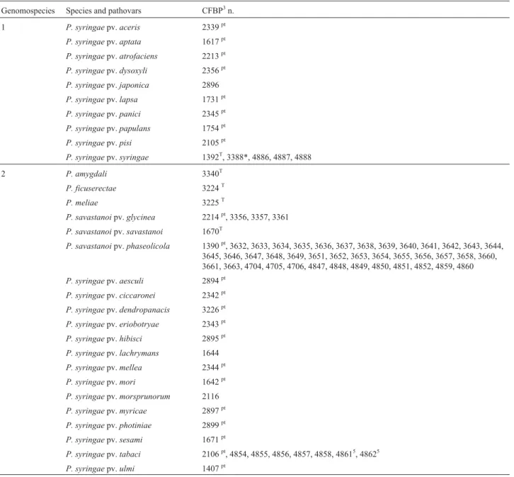
Table 1- Strains ofPseudomonas syringae1group,P. viridiflavaand related speciesP. savastanoi,P. ficuserectae,P. amygdali,P. tremae,P. meliae,P. avellanaeandP. cannabina, according to the genomospecies designation2.
Genomospecies Species and pathovars CFBP3n.
1 P. syringaepv.aceris 2339pt
P. syringaepv.aptata 1617pt
P. syringaepv.atrofaciens 2213pt
P. syringaepv.dysoxyli 2356pt
P. syringaepv.japonica 2896
P. syringaepv.lapsa 1731pt
P. syringaepv.panici 2345pt
P. syringaepv.papulans 1754pt
P. syringaepv.pisi 2105pt
P. syringaepv.syringae 1392T, 3388*, 4886, 4887, 4888
2 P. amygdali 3340T
P. ficuserectae 3224T
P. meliae 3225T
P. savastanoipv.glycinea 2214pt, 3356, 3357, 3361
P. savastanoipv.savastanoi 1670T
P. savastanoipv.phaseolicola 1390pt, 3632, 3633, 3634, 3635, 3636, 3637, 3638, 3639, 3640, 3641, 3642, 3643, 3644,
3645, 3646, 3647, 3648, 3649, 3651, 3652, 3653, 3654, 3655, 3656, 3657, 3658, 3660, 3661, 3663, 4704, 4705, 4706, 4847, 4848, 4849, 4850, 4851, 4852, 4859, 4860
P. syringaepv.aesculi 2894pt
P. syringaepv.ciccaronei 2342pt
P. syringaepv.dendropanacis 3226pt
P. syringaepv.eriobotryae 2343pt
P. syringaepv.hibisci 2895pt P. syringaepv.lachrymans 1644
P. syringaepv.mellea 2344pt
P. syringaepv.mori 1642pt
P. syringaepv.morsprunorum 2116
P. syringaepv.myricae 2897pt
P. syringaepv.photiniae 2899pt
P. syringaepv.sesami 1671pt
P. syringaepv.tabaci 2106pt, 4854, 4855, 4856, 4857, 4858, 48615, 48625
will be designated by the abbreviationP. syr. syringae
in-stead ofP. syringaepv.syringae.
Nutritional characterization
Assimilation of 99 carbon sources (sugars, alcohols, amino acids and organic acids) was performed with the Biotype 100 system (BioMérieux, La Balme-les-Grottes, France). The strips were inoculated with Biotype Medium 1
as recommended by the manufacturer, and the results read visually at two, four and six days after incubation at 28 °C. In addition, 20 conventional biochemical tests were carried out: arginine dihydrolase, oxidase, gelatin, nitrate reduc-tion, levan, fluorescence, hypersensitive reaction (HR) on tobacco, esculin, pectinolysis on calcium pectinate, Tween esterase, DNAse, polypectate hydrolysis at pH 5 and 8.3, and the utilization of sucrose, lactate, L(+)tartrate,
Table 1 (cont.)
Genomospecies Species and pathovars CFBP3n.
3 P. syringaepv.antirrhini 1620pt
P. syringaepv.apii 2103pt
P. syringaepv.berberidis 1727pt
P. syringaepv.delphinii 2215pt
P. syringaepv.lachrymans 2440pt
P. syringaepv.maculicola 1657pt
P. syringaepv.morsprunorum 2351pt
P. syringaepv.passiflorae 2346pt
P. syringaepv.persicae 1573pt P. syringaepv.philadelphii 2898pt
P. syringaepv.primulae 4091
P. syringaepv.ribicola 4068
P. syringaepv.tomato 2212pt
P. syringaepv.viburni 1702pt
4 P. syringaepv.atropurpurea 2340pt
P. syringaepv.coronafaciens 2216pt
P. syringaepv.garçae 1634pt
P. syringaepv.oryzae 3228pt
P. syringaepv.porri4 1908pt
P. syringaepv.striafaciens 1674pt, 1686
P. syringaepv.zizaniae 4117T
5 P. tremae 3229T
6 P. syringaepv.primulae 1660pt
P. syringaepv.ribicola 2348pt
P. viridiflava 2107T
7 P. syringaepv.helianthi 2067pt
P. syringaepv.tagetis 1694pt
8 P. avellanae4 4060T
P. syringaepv.theae 2353pt
9 P. cannabina 2341T
Unknown genomospecies
P. syringaepv.actinidiae 4909pt, 4911, 5095
Unknown genomospecies and pathovar
P. syringae 3650, 3662
1
D(-)tartrate, erythritol, mannitol, and sorbitol in ARJ
me-dium (Gardanet al., 1999).
Bacterial cultures and genomic DNA preparation
Strains were grown at 26-28 °C on King’s medium B for 24 h. From these cultures, cells were washed with sterile distilled water, and a suspension was prepared, which was
adjusted to an O.D.560of 0.2, corresponding to a bacterial
cell suspension at 108cfu mL-1. Aliquots of 500µL in 2 mL
cryotubes were stored at -20 °C. For utilization, after lique-fying the suspension at room temperature, cells were lysed for 10 min in a boiling water bath, and the cryotubes kept on ice before use.
BOX primer and BOX-PCR reaction
The 22-mer BOXA1R oligonucleotide (Bioprobe Systems/Quantum, France) was used to generate
BOX-PCR profiles (Versalovicet al., 1991; Martinet al., 1992).
Amplification reactions were performed in volumes of
25µL, containing 2µM of the single BOX primer, 200µM
each of dATP, dCTP, dGTP and dTTP (Bioprobe Sys-tems/Quantum, France), PCR reaction buffer (10 mM
TrisHCl [pH 9.0], 50 mM KCl, 1.5 mM MgCl2, 0.1%
TritonX100 and 0.2 mg mL-1 bovine serum albumin),
1.5 units ofTaqDNA polymerase (Appligene-Oncor,
Fran-ce) and, as template DNA, 5µL of a bacterial cell
suspen-sion at 108cfu mL-1. Amplification was performed in an MJ
Research, Inc. PTC-100 Thermal Cycler programmed for an initial denaturation step of 7 min at 95 °C, followed by 30 cycles of 1 min at 94 °C, 1 min at 53 °C and 8 min at 65 °C with a final elongation step of 15 min at 65 °C. PCR amplification products were detected by electrophoresis of
12µL aliquots through 1.4% agarose gels in
Tris-borate-EDTA (TBE) buffer (Sambrook and Russell, 2001), which were stained with ethidium bromide (EtBr 1.25 mg/L), vi-sualized under UV light, and printed image through Bio-Print (Vilber Lourmat, France). DNA standards (1-kb DNA ladder Gibco BRL) were included in each electrophoresis gel. All of the amplifications were performed at least twice in separate assays, to ensure the reproducibility of the pat-terns, and only bands common to the replicate amplifica-tions were scored. DNA fingerprints of strains were first compared for similarity by visual inspection of band pat-terns. They were considered identical when all scored bands in each pattern had the same apparent migration dis-tance, even if a slightly different molecular weight was as-signed to the same band over two or three different electrophoreses. Variations in intensity were not taken as differences.
Following the visual inspection, the patterns of all of the isolates were analyzed more rigorously using the Bio-Profil software (Vilber Lourmat, France). Band sizes were assigned by direct comparison to concurrently run DNA standards (1 kb). This information was used to construct a matrix table where each isolate was matched with a
nota-tion +/-, where (+) represents the identical presence and position of a band in the fingerprints to be compared.
Data analysis
Dendrograms were established using TAXONUM, software developed by G. Hunault and L. Gardan (Faculté des Sciences d’Angers, Angers, France). Cluster analysis was carried out using the unweighted pair-group method with averages (UPGMA) with the complement of Jaccard’s similarity coefficient (Sneath and Sokal, 1973). Each frag-ment was considered as a separate marker in pairwise com-parisons.
The BOX fragments as well as the biochemical tests characteristic of each cluster were identified by assessing the amount of information provided by each fragment or character, obtained by calculating the diagnostic ability co-efficient (DAC) (Descamps and Véron, 1981).
Results
Nutritional characteristics
All of the 120 bacterial strains (Table 1) fitted with
the general characteristics ofP. syringae sensu latoandP.
viridiflava.They were obligate aerobes, presenting
oxida-tive metabolism of glucose, and being posioxida-tive for levan
(except P. viridiflavaT, P. syr. ribicolapt and P. syr.
primulaept) and tobacco hypersensitivity and negative for
oxidase and arginine tests, produced fluorescent pigment
on King’s medium B (except forP. syr.actinidiaestrains,
P. sav. glycinea strain 3356, and P. sav. phaseolicola
strains 3653 and 4706).
The numerical analysis of 119 nutritional characteris-tics (data not shown) evidenced two clusters. The first
clus-ter containedP. sav.phaseolicola,P. sav.glycinea,P. syr.
tabaci(two strains),P. syr.moriandP. syr.sesami, all
be-longing to genomospecies 2, and was distinguishable by two substrates only: sorbitol and meso-tartrate. The second
cluster comprised all of the other strains evaluated,i.e.all
nine genomospecies including other strains ofP. syr.tabaci
and other pathovars of genomospecies 2.
Comparing BOX fingerprints of 120Pseudomonas
strains
The amplification of genomic DNA of 88
Pseudomo-nas strains, followed by gel electrophoresis of resulting
PCR products, showed 12 to 22 bands for the whole set of strains, and a total of 133 discrete bands were scored, rang-ing in size from 220 bp to 3.6 kb. From the first list of 89
strains, we were not able to amplifyP. syr. theaeDNA. The
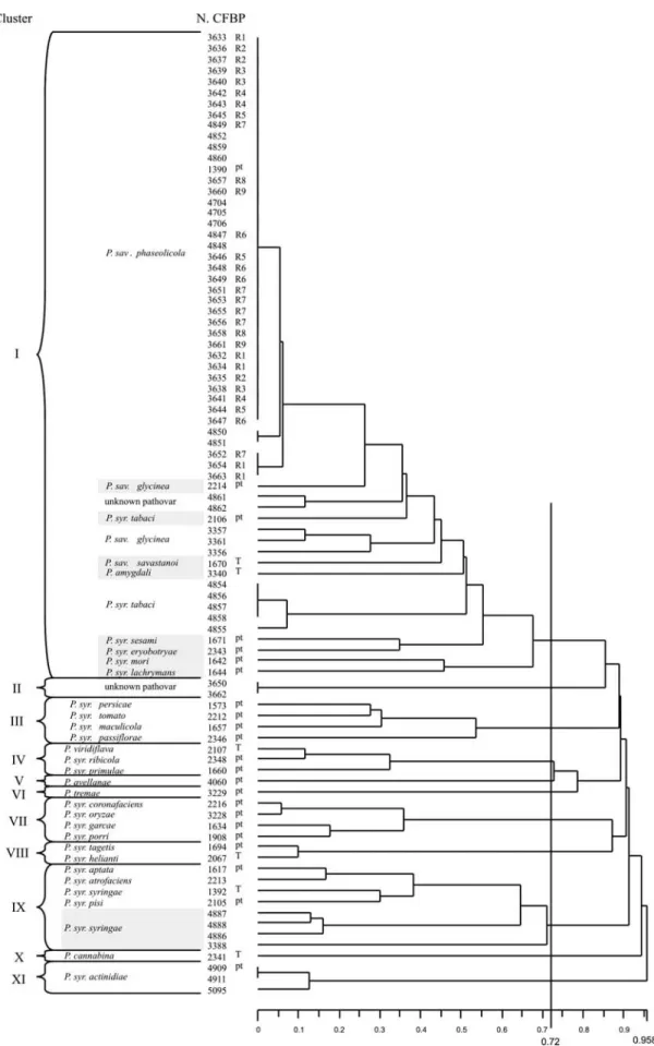
data matrix showing presence or absence of these 133 bands was analyzed by Jaccard coefficient and UPGMA, and a dendrogram displaying the distances between the 88 strains is shown in Figure 1.
At a distance of 0.72, all pathovars belonging to each
Figure 1- Dendrogram obtained by comparison of BOX-PCR fingerprinting patterns from 88 bacterial strains belonging toPseudomonas savastanoi
(1999) were clustered together. Cluster I included all of the strains of genomospecies 2. Into this group the great
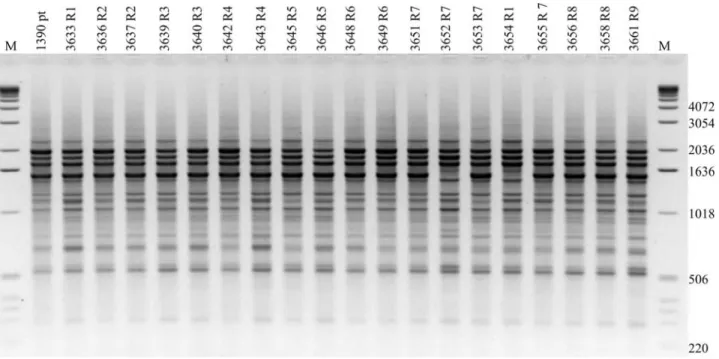
homo-geneity ofP. sav. phaseolicolastrains obtained by
BOX-PCR fingerprinting is illustrated in Figure 2, where only one different band is found and for only two strains. The eight remaining clusters, III, IV, V, VI, VII, VIII, IX and X corresponded to genomospecies 3, 6, 8, 5, 4, 7, 1 and 9,
re-spectively (Gardanet al., 1999). Inside cluster X, three
ad-ditional strains of P. syr. cannabinawere evaluated and
showed the same fingerprint as CFPB 2341 (data not
shown). The twoP. syringaestrains of unknown pathovars
(3650 and 3662) and the three P. syr. actinidiae strains
were grouped in clusters II and XI, respectively.
The second step of analyses originated from the re-sults shown in Figure 2: the homogeneity of a given pathovar. In order to confirm the utility of BOX-PCR to identify the genomospecies, a second analysis was
per-formed. From the total of 41 strains ofP. sav. phaseolicola,
only the type strain was maintained representing the pathovar, based on its fingerprint homogeneity. This analy-sis was performed upon 60 pathovar-type strains and one
more strain ofP. syr. striafaciens. A total of 61 strains were
included in this step of analysis.
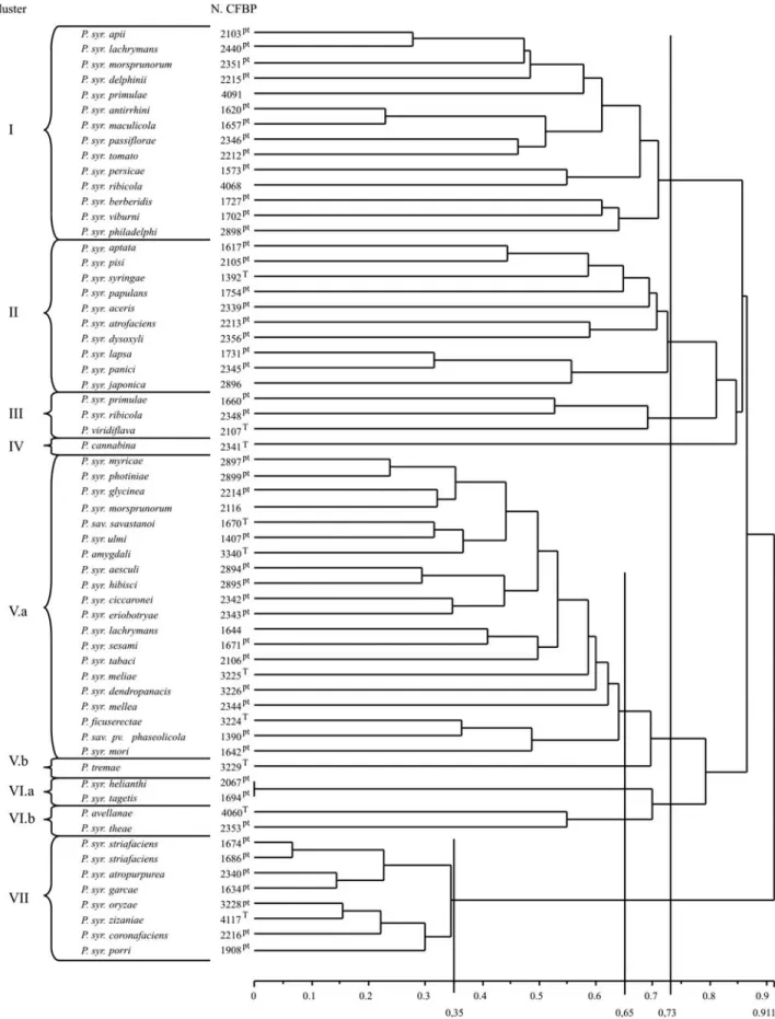
The dendrogram displaying the distance relationships between the strains is shown in Figure 3. At a distance of 0.73 seven clusters were shown (I to VII), where five of them (I, II, III, IV and VII) corresponded strictly to geno-mospecies 3, 1, 6, 9 and 4. The remaining two clusters (V and VI) clustered together at the mentioned distance, the genomospecies 2 and 5 (cluster V) and 7 and 8 (cluster VI). Despite this fact, when analyzing the two groups at a
dis-tance of 0.65 the four genomospecies are separated in tight groups: V.a at 0.65 clustered together all the pathovars of genomospecies 2, V.b clustered all the pathovars of geno-mospecies 5, VI.a clustered all the pathovars of genomos-pecies 7 and VI.b all the pathovars of genomosgenomos-pecies 8. Figure 4 shows this similarity in fingerprints between iso-lates from the same genomospecies, but from different pathovars and the great homogeneity inside genomospecies 4, clustered at a distance of 0.35.
The results of the present study support the observa-tion that BOX-PCR seems to be able to identify bacterial strains at species level.
The two clusters (II and XI, Figure 1) not included
into the first description of the genomospecies (Gardanet
al., 1999), might eventually constitute two new species.
The bands that most discriminated the ten clusters in the first study were selected by DAC analysis (Table 2).
Discussion
The data obtained from the analyses of phenotypic
and genetic diversity of a collection ofP. syringae sensu
lato group,P. viridiflava,and reference bacteria showed
that pathovars belonging to the genomospecies designed
after Gardanet al. (1999) could be separated at species
level by BOX-PCR pattern.
Although bacteria are traditionally identified by phenotypic descriptions, it is not possible to obtain from nutritional studies a battery of discriminating substrates to
the genomospecies as related by Gardanet al.(1999), and
confirmed in this study.
Figure 2- Agarose gel electrophoresis of BOX-PCR fingerprinting patterns from genomic DNA of very homogeneousPseudomonas savastanoipv.
Figure 3- Dendrogram obtained by comparison of BOX-PCR fingerprinting patterns from 61 bacterial type strains belonging toPseudomonas syringae
A multiphasic approach has been proposed as being a reliable method of integrating different types of informa-tion, such as genotypic, phenotypic and phylogenetic data
(Vandammeet al., 1996). Methods of fingerprinting based
on the analysis of the total genome may constitute a valu-able complement (Rademaker and Bruijn, 1997).
Amplification of Box primer by PCR from DNA of
88 strains belonging to 31 pathovars ofP. syringae sensu
latogroup and P. viridiflava, led to the establishment of
patterns that allowed the distinction of 11 BOX clusters, where nine of them cut the nine genomospecies described
inside those pseudomonads (Gardanet al., 1999). When
in-cluding 30 other strains representing the remaining species and pathovars for this first group, the genomospecies dis-crimination was confirmed, despite the necessity of cutting the dendrogram at different but close distances. Geno-mospecies 2 and 5 are very closely related, and the strain
CFBP3229 ofP. syr. tremae,originally included in
geno-mospecies 5, could be clustered with genogeno-mospecies 2. A similar situation was found with genomospecies 7 and 8,
whose strains could be separated at 0.65. In this study,P.
syr. avellanaewas clustered together with P. syr. theae,
both corresponding to genomospecies 8. Using different molecular techniques, other authors found that the strains
described by Gardanet al.(1999) as genomospecies 8 and 3
should be clustered together: Sarkar and Guttman (2004, utilizing multilocus sequencing typing, MLST), Inoue and
Takikawa (2006, comparing thehrpZandhrpAgenes
se-quences). The present analysis, which used BOX-PCR fin-gerprinting, is capable of separating genomospecies 3 and
Figure 4- Agarose gel electrophoresis of BOX-PCR fingerprinting pat-terns from genomic DNA of different pathovars belonging to Pseudomo-nas syringae(pathovarsphotiniae,striafaciens,coronafaciens,garçae,
atropurpurea, primulae, tomato, apii, delphinii and aptata) and P. savastanoipv.phaseolicola, which represent some of the nine known genomic species (GS), assigned after Gardanet al.(1999). Isolates ob-tained from the “Collection Française des Bactéries Phytopathogènes” (CFBP, Angers, France). The molecular size marker is a 1-kb ladder (Gibco BRL Life Technologies Inc.) and the sizes are indicated in base pairs. Shown is a negative image of an ethidium bromide gel.pt: pathotype
strain.
Table 2- Discriminating BOX fragments, permitting distinction of clusters I to XI (Figure 1).
Presence (+) or absence (-) of discriminating fragment
Group
Number
of
strains
BOX-35 BOX-69 BOX-15 BOX-86 BOX-68 BOX-130 BOX-30 BOX-46 BOX-45 BOX-71 BOX-101 BOX-127 BOX-66 Genomospecies
1
I2 37 + + - - - - - - - - - - - 2
II 2 - + - - - + - - - - ?3
III 4 - + - - - + - - - 3
IV 3 - - - + - - - + 6
V 1 - - - + + - - + - - + 8
VI 1 - - - + - - + - - - 5
VII 4 - + - + - - + + - - - + - 4
VIII 2 - - + + - + - + - + + - - 7
IX 5 - - + - + - - - 1
X 1 - - - + - + - 9
XI 3 - - - + - + - - ?4
1
8. It also shows a remarkable homogeneity inside geno-mospecies 4, whose isolates are maintained together until a distance of 0.35.
Long after Louwset al.(1994), claimed BOX
analy-sis could discriminate pathovars ofP. syringae, it now
ap-pears that the authors were dealing with three different
genomospecies: pv.morsprunorum(genomospecies 2), pv.
syringae (genomospecies 1), and pv. tomato
(geno-mospecies 3). According to Louwset al.(1995) BOX-PCR
was able to distinguish strains A and B ofX. campestrispv.
vesicatoria, separated by DNA-DNA hybridization by Stall
et al. (1994), and further named X. axonopodis pv.
vesicatoria(ex A) andX. vesicatoria(ex B) (Vauterinet al.,
1995).
Regarding the two extra groups obtained in this study (clusters II and XI, Figure 1), they are not included in the
nine known genomic species of Gardanet al.(1999), and
might constitute two new species. Cluster II is composed of two bacterial strains isolated from beans (CFBP 3650 and
3662), for which hybridization rate with type strain ofP.
sav. phaseolicola(CFPB 1390pt) was only 50% and 51%,
respectively (data not shown). In addition, they did not pos-sess the phaseolotoxin gene, their esterase isozyme patterns were very distinct and they were aesculine positive
(Mar-ques et al., 2000). If there were no mistakes during the
work, they might constitute another distinct bean pathogen
belonging to the P. syringae group. Cluster XI of the
dendrogram is composed of three strains of P. syr.
actinidiae (CFPB 4909 the pathotype strain, 4911 and
5095). In order to define their inclusion as new genomos-pecies it would be necessary to provide quantitative
DNA-DNA hybridization (Wayneet al., 1987).
Quantitative DNA-DNA hybridization still consti-tutes the reference for the description of a new species, but it is not adapted to a routine base identification of bacteria. It seems very important to propose alternative techniques, which could reproduce identical results, in order to identify bacterial species.
Since our data basis is constituted from BOX-PCR
profiles of 31 or 61 pathovars ofP. syringae-P. viridiflava,
new pathovars will be easily assigned to a known genomic species. Furthermore, considering the hypothesis that it may be possible to verify that discriminating bands de-tected by BOX-PCR technique could amplify specific DNA fragments of each genomic species, a new route
would be offered to help identify theP. syringaegroup of
plant bacteria at species level.
Acknowledgments
We are grateful to Marion Le Saux for helping to up-date information and to Gláucia S.C. Buso for critical read-ing of the manuscript; to J.D. Taylor for kindly providread-ing
the great majority ofP. savastanoipv.phaseolicolastrains;
to Alain Huard and Wesley R. Souza for preparing the fig-ures. This study was supported in part by Région Pays de la
Loire (France). Abi S.A. Marques was supported by Em-brapa (Empresa Brasileira de Pesquisa Agropecuária, Brazil).
References
Catara V, Sutra L, Morineau A, Achouak W, Christen R and Gardan L (2002) Phenotypic and genomic evidence for the revision ofPseudomonas corrugataand proposal of Pseu-domonas mediterraneasp. nov. Int. J Syst Evolution Micro-biol 52:1749-1758.
Cross JE, Kennedy BW, Lambert JW and Cooper RL (1966) Pathogenic races of the bacterial blight pathogen of soy-beans,Pseudomonas glycinea. Plant Dis Rep 50:557-560. Descamps P and Véron M (1981) Une méthode de choix des
caractères d’identification basée sur le théorème de Bayes et la mesure de l’information. Ann Inst Pasteur/Microbiol 132B:157-170.
El Tassa SOM, Moraes MG and Duarte V (1999) Identificação de
Pseudomonas syringaepv.coronafaciensatravés de ERIC-e BOX-PCR. Fito Bras 24:503-508. (Abstract in English). Galal AA, Kehil YEI, el Daoudi YH, Shihata ZA and Ouf MF
(2003) A comparative study on the identification of races and biovars of some Egyptian isolates of Ralstonia solanacearum. Egypt J Phytopathol 31:103-117.
Gardan L, Shafik H, Belouin S, Brosch R, Grimont F and Grimont PAD (1999) DNA relatedness among the pathovars of Pseu-domonas syringaeand description ofPseudomonas tremae
sp. nov. andPseudomonas cannabinasp. nov. (ex. Sutic and Dowson 1959). Int J Syst Bacteriol 49:469-478.
Greco S, Bella P, Tessitori M and Catara V (2004) Indagini sulla disseminazione in vivaio di Xanthomonas hortorum pv.
pelargonii. Colture Protette 33:65-68. (Abstract in English). Inoue Y and Takikawa Y (2006) ThehrpZandhrpAgenes are
variable, and useful for grouping Pseudomonas syringae
bacteria. J Gen Plant Pathol 72:26-33.
Lelliott RA, Billing E and Hayward AC (1966) A determinative scheme for the fluorescent plant pathogenic pseudomonads. J Appl Bacteriol 29:470-489.
Louws FJ, Fulbright DW, Stephens CT and DeBruijn FJ (1994) Specific genomic fingerprints of phytopathogenic
XanthomonasandPseudomonaspathovars and strains gen-erated with repetitive sequences and PCR. Appl Environ Microbiol 60:2286-2295.
Louws FJ, Fulbright DW, Taylor SE and DeBruijn FJ (1995) Dif-ferentiation of genomic structure by rep-PCR fingerprinting to rapidly classifyXanthomonas campestrispv.vesicatoria. Phytopathology 85:528-536.
Louws FJ, Bell J, Medina-Mora CM, Smart CD, Opgenorth D, Ishimaru CA, Hausbeck MK, Bruijn FJ and Fulbright DW (1998) rep-PCR-mediated genomic fingerprinting: a rapid and effective method to identifyClavibacter michiganensis. Phytopathology 88:862-868.
Marques ASA, Corbière R, Gardan L, Tourte C, Manceau C, Tay-lor JD and Samson R (2000) Multiphasic approach for the identification of the different classification levels of Pseudo-monas savastanoi pv. phaseolicola. Eur J Plant Pathol 106:715-734.
in the chromosome ofSteptococcus pneumoniae. Nucleic Acids Res 20:3479-3483.
Onfroy C, Tivoli B, Corbière R and Bouznad Z (1999) Cultural, molecular and pathogenic variability of Mycospharella pinodesandPhoma medicaginisvar.pinodellaisolates from dried pea (Pisum sativum) in France. Plant Pathol 48:218-229.
Palleroni NJ (1984) Genus I. Pseudomonas Migula 1894. In: Krieg NR and Holt JG (eds) Bergey’s Manual of Systematic Bacteriology. v. 1. Williams and Wilkins, Baltimore, pp 141-199.
Rademaker JLW and DeBruijn FJ (1997) Characterization and classification of microbes by rep-PCR genomic fingerprint-ing and computer assisted pattern analysis. In: Gaetano-Anolles G (ed) DNA Markers: Protocols, Applications and Overviews. Willey & sons, New York, pp 151-171. Sambrook J and Russell DW (2001) Molecular Cloning: A
Labo-ratory Manual. 3rd edition. Cold Spring Harbor LaboLabo-ratory Press, New York, 999 pp.
Sarkar SF and Guttman DS (2004) Evolution of the core genome of Pseudomonas syringae, a highly clonal, endemic plant pathogen. Appl Environ Microbiol 70:1999-2012.
Sneath PHA and Sokal RP (1973) Numerical Taxonomy: The Principles and Practice of Numerical Classification. WH Freeman and Company, San Francisco, 573 pp.
Stall RE, Beaulieu C, Egel D, Hodge NC, Leite RP, Minsavage GV, Bouzar H, Jones JB, Alvarez AM and Benedict AA (1994) Two genetically diverse groups of strains are in-cluded in Xanthomonas campestrispv. vesicatoria. Int J Syst Bacteriol 44:47-53.
Tacao M, Alves A, Saavedra MJ and Correia A (2005) BOX-PCR is an adequate tool for typingAeromonasspp. Antoine-van-Leeuwenhoek 88:173-179.
Tamura K, Takikawa S, Tsuyumu S and Goto M (1989) Charac-terization of the toxin produced byPseudomonas syringae
pv.actinidiae, the causal bacterium of kiwifruit canker. Ann Phytopathol Soc Japan 55:512
Taylor JD, Teverson DM, Allen DJ and Pastor-Corrales MA (1996) Identification and origin of races ofPseudomonas syringaepv.phaseolicolafrom Africa and other bean grow-ing areas. Plant Pathol 45:469-478.
Tourte C and Manceau C (1995) A strain of Pseudomonas syringae which does not belong to pathovarphaseolicola
produces phaseolotoxin. Eur J Plant Pathol 101:483-490. Vandamme P, Pot B, Gillis M, de Vos P, Kersters K and Swings J
(1996) Polyphasic taxonomy, a consensus approach to bac-terial systematics. Microbiol Rev 60:407-438.
Vauterin L, Hoste B, Kersters K and Swings J (1995) Reclassifi-cation ofXanthomonas. Int J Syst Bacteriol 45:472-489. Versalovic J, Koeuth T and Lupski JR (1991) Distribution of
re-petitive DNA sequences in eubacteria and application to fin-gerprinting of bacterial genomes. Nucleic Acids Res 19:6823-6831.
Versalovic J, Schneider M, de Bruijn FJ and Lupski JR (1994) Genomic fingerprinting of bacteria using repetitive sequen-ce-based Polymerase Chain Reaction. Methods Mol Cell Biol 5:25-40.
Wayne LG, Brenner DJ, Colwell RR, Grimont PAD, Kandler O, Krichevsky MI, Moore LH, Moore WEC, Murray RGE, Stackebrandt E, Starr MP and Trüpper HG (1987) Interna-tional Committee on Systematic Bacteriology. Report of the ad hoc committee on reconciliation of approaches to bacte-rial systematics. Int J Syst Bacteriol 37:463-464.
Williams JGK, Kubelik AR, Livak KJ, Rafalski JA and Tingey SV (1990) DNA polymorphisms amplified by arbitrary pri-mers are useful as genetic markers. Nucleic Acids Res 18:6531-6535.
Young JM, Saddler GS, Takikawa Y, DeBoer SH, Vauterin L, Gardan L, Gvozdyak RI and Stead DE (1996) Names of plant pathogenic bacteria 1864-1995. Rev Plant Pathol 75:721-762.