ISSN 1806-3713 © 2017 Sociedade Brasileira de Pneumologia e Tisiologia
http://dx.doi.org/10.1590/S1806-37562015000000269
ABSTRACT
We describe the case of a 33-year-old man, a chronic user of powder cocaine, who presented with dyspnea, fever, night sweats, and signiicant weight loss. Chest HRCT revealed centrilobular nodules, giving an initial impression of miliary tuberculosis. Therefore, he was started on an empirical, four-drug antituberculosis treatment regimen. Four weeks later, despite the tuberculosis treatment, he continued to have the same symptoms. We then performed transbronchial lung biopsy. Histopathological analysis of the biopsy sample revealed birefringent foreign body granuloma. A corroborative history of cocaine snorting, the presence of centrilobular nodules, and the foreign body-related histopathological indings led to a diagnosis of pulmonary foreign body granulomatosis. This report underscores the fact that pulmonary foreign body granulomatosis should be included in the differential diagnosis of clinical proiles resembling tuberculosis. Keywords: Lung; Granuloma, foreign-body; Cocaine-related disorders.
Pulmonary foreign body granulomatosis in
a chronic user of powder cocaine
Shruti Khurana1, Ankit Chhoda2, Sandeep Sahay3, Priyanka Pathania4
Correspondence to:
Sandeep Sahay. Houston Methodist Lung Center, Suite 1001, Smith Tower, 6550 Fannin Street, ZIP 77030, Houston, TX, USA. Tel.: 1 713 363-9587. E-mail: [email protected]
Financial support: None. INTRODUCTION
Physicians frequently encounter cocaine abuse in clinical practice. It is the leading cause of illicit drug-related deaths worldwide.(1) Cocaine is abused by multiple methods, the snorting of powder cocaine being the most common. Pulmonary complications, such as alveolitis, barotrauma, talcosis, organizing pneumonia, bullous emphysema,
and pulmonary ibrosis, are frequently reported as a
result of crack cocaine smoking or intravenous cocaine use. (2) However, there has been only one report to date of pulmonary foreign body granulomatosis (PFBG) secondary to cocaine snorting.(3)
CASE REPORT
A 33-year-old Hispanic male of average build was admitted to our facility, complaining of dyspnea, fever, night sweats, and rapid weight loss (18 kg over a
four-month period). A pertinent positive inding on the initial
history taking was his having traveled to a country where tuberculosis is endemic. Physical examination revealed no acute distress and no stigmata of intravenous drug
abuse. He was febrile (≤ 38.5°C), with a blood pressure of
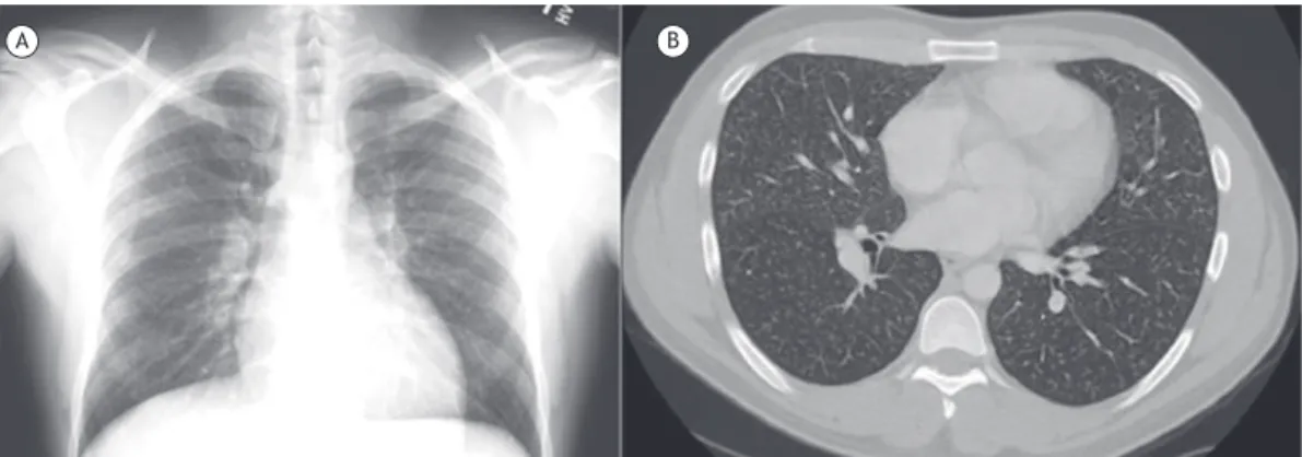
124/72 mmHg, a heart rate of 76 bpm, a respiratory rate of 18 breaths/min, and a constant SpO2 at rest of 98%. The initial total leukocyte count was 8,200 cells/µL, with a predominant neutrophilic reaction. Blood culture was negative for aerobic and anaerobic microorganisms. Urine toxicology and HIV tests were negative. Chest X-ray and HRCT showed micronodules (1-3 mm in size) bilaterally
in centrilobular distribution (Figure 1). An IFN-γ assay
and sputum smear microscopy for acid-fast bacilli were carried out in order to rule out mycobacterial infection. Although the results were negative for tuberculosis, the high clinical suspicion of the disease prompted empirical
initiation of the four-drug antituberculous therapy. Four weeks later, the patient returned to the emergency room with worsening of the shortness of breath. A repeat HRCT revealed similar centrilobular nodules with no radiological improvement (Figure 2). At that time, bronchoscopy with transbronchial lung biopsy was performed for further evaluation of the pulmonary micronodules. Examination
of the BAL luid, with Ziehl-Neelsen staining, revealed
no acid-fast bacilli. Histopathological analysis of the transbronchial lung biopsy specimen showed multiple granulomas with birefringent material in the center
(Figure 3), conirming the diagnosis of PFBG. The patient
strongly denied intravenous drug use. Unfortunately, he experienced a progressive course with a further decline
in his lung function. After progressive ibrosis over the
following one-year period, the patient died of chronic respiratory failure.
DISCUSSION
The lungs are frequently affected by cocaine abuse, regardless of the delivery method. Clinical presentation and
radiological indings are varied and highly nonspeciic.(4) PFBG is a rare condition, and its true incidence is unknown. It is commonly encountered secondary to intravenous injection of pulverized pharmaceutical tablets containing insoluble binders, such as talc, cellulose, starch, and other street adulterants. The tiny particles lodge in the vascular bed and interstitium to cause granulomatous reactions
and ibrosis.(5) Prior to this report, there had been only
one reported case of PFBG in a user of powder cocaine (intranasal route), which was determined to be caused
by cellulose iller.(3) Radiologically, PFBG can present as small diffuse centrilobular nodules, conglomerated masses, diffuse ground-glass opacities, and lower-lobe 1. Lady Hardinge Medical College,
New Delhi, India.
2. Maulana Azad Medical College, Department of Internal Medicine, New Delhi, India.
3. Houston Methodist Lung Center, Houston (TX) USA.
4. Jack C. Montgomery VA Medical Center, Department of Pulmonary Medicine, Muskogee (OK) USA.
Submitted: 1 September 2016. Accepted: 31 October 2016.
Study carried out at the Houston Methodist Lung Center, Houston (TX) USA.
J Bras Pneumol. 2017;43(4):320-321
320
Khurana S, Chhoda A, Sahay S, Pathania P
panlobular emphysema.(6) Because our patient was inhaling cocaine, the appearance of centrilobular nodules on chest CT was consistent with small airway disease, as opposed to miliary tuberculosis, which usually presents as random nodules. It is prudent to consider rare granulomatous conditions such as PFBG in patients with a history of intravenous or inhaled drug abuse. The course of PFBG can be subacute (with fever, weight loss, or hemoptysis) or chronic (with dyspnea and a progressive decline in lung function).(7) Late
complications include pulmonary hypertension, cor pulmonale, panlobular emphysema, and, rarely, res-piratory failure requiring lung transplantation.(8,9) There
is no speciic treatment for PFBG. A few patients have
experienced stabilization of symptoms after cessation of drug use and resolution of acute symptoms with corticosteroid use.(10) Avoidance of exposure continues to be the cornerstone of management.
Figure 3. Photomicrograph of the transbronchial biopsy specimen (lung tissue) showing birefringent foreign body material (left arrow), together with granuloma formation (right arrow) around the foreign body material (H&E; magniication, ×40).
A B
Figure 1. In A, a chest X-ray showing micronodular shadows. In B, a CT scan of the chest, showing centrilobular nodules in both lung ields.
Figure 2. A CT scan of the chest showing the persistence of centrilobular nodules in bilateral lung ields after four weeks of treatment with antituberculosis drugs.
REFERENCES
1. Restrepo CS, Carrillo JA, Martínez S, Ojeda P, Rivera AL, Hatta A. Pulmonary complications from cocaine and cocaine-based substances: imaging manifestations. Radiographics. 2007;27(4):941-56. https://doi.org/10.1148/rg.274065144
2. Tseng W, Sutter ME, Albertson TE. Stimulants and the lung: review of literature. Clin Rev Allergy Immunol. 2014;46(1):82-100. https:// doi.org/10.1007/s12016-013-8376-9
3. Cooper CB, Bai TR, Heyderman E, Corrin B. Cellulose granuloma in the lungs of a cocaine sniffer. Br Med J (Clin Res Ed). 1983;286(6383):2021-2. https://doi.org/10.1136/ bmj.286.6383.2021-a
4. Almeida RR, Zanetti G, Souza AS Jr, Souza LS, Silva JL, Escuissato DL, et. al. Cocaine-induced pulmonary changes: HRCT indings. J Bras Pneumol. 2015;41(4):323-30. https://doi.org/10.1590/S1806-37132015000000025
5. Ellis SJ, Cleverley JR, M̈ller NL. Drug-induced lung disease: high-resolution CT indings. AJR Am J Roentgenol. 2000;175(4):1019-24. https://doi.org/10.2214/ajr.175.4.1751019
6. Marchiori E, Loureņo S, Gasparetto TD, Zanetti G, Mano CM, Nobre LF. Pulmonary talcosis: imaging indings. Lung. 2010;188(2):165-71. https://doi.org/10.1007/s00408-010-9230-y
7. Paŕ JP, Cote G, Fraser RS. Long-term follow-up of drug abusers with intravenous talcosis. Am Rev Respir Dis. 1989;139(1):233-41. https://doi.org/10.1164/ajrccm/139.1.233
8. Shlomi D, Shitrit D, Bendayan D, Sahar G, Shechtman Y, Kramer MR.
Successful lung transplantation for talcosis secondary to intravenous
abuse of oral drug. Int J Chron Obstruct Pulmon Dis. 2008;3(2):327-30.
9. Weinkauf JG, Puttagunta L, Nador R, Jackson K, LaBranche K, Kapasi A, et al. Long-term outcome of lung transplantation in previous intravenous drug users with talc lung granulomatosis. Transplant Proc. 2013;45(6):2375-7. https://doi.org/10.1016/j. transproceed.2012.11.004
10. Smith RH, Graf MS, Silverman JF. Successful management of drug-induced talc granulomatosis with corticosteroids. Chest. 1978;73(4):552-4. https://doi.org/10.1378/chest.73.4.552