Chronic bacterial infection and echocardiographic parameters indicative
of pulmonary hypertension in patients with cystic fibrosis*
Infecção bacteriana crônica e indicadores ecocardiográficos de hipertensão pulmonar em pacientes com fibrose cística*
Paula Maria Eidt Rovedder1, Bruna Ziegler2, Lilian Rech Pasin3, Antônio Fernando Furlan Pinotti4,
Sérgio Saldanha Menna Barreto5, Paulo de Tarso Roth Dalcin6
Abstract
Objectives: To examine the relationship between chronic bacterial infection and pulmonary hypertension, using Doppler echocardiography,
in patients with cystic fibrosis (CF). Methods: A prospective cross-sectional study involving CF patients (≥ 16 years of age) admitted to
a program for adults with the disease. The study included 40 patients with a mean age of 23.7 ± 6.3 years. Patients were submitted to clinical evaluation, Doppler echocardiography, pulmonary function tests, chest X-rays and sputum cultures of Pseudomonas aeruginosa and Burkholderia cepacia. Results: In terms of the following variables, no significant differences were found between P.
aeruginosa-positive patients and P. aeruginosa-negative patients: clinical score (p = 0.472); forced expiratory volume in one second (FEV1; p = 0.693); radiological score (p = 0.760); tricuspid regurgitant jet velocity (TRV, p = 0.330); diameter of the right ventricle (DRV, p = 0.191); and right ventricular/pulmonary artery (RV/PA) systolic acceleration time (SAT, p = 0.330). B. cepacia-positive patients presented significantly lower FEV1 than did B. cepacia-negative patients (p = 0.011). No significant differences were found between B. cepacia-positive patients and B. cepacia-negative patients regarding the following variables: clinical score (p = 0.080); radiological score (p = 0.760); TRV (p = 0.613); DRV (p = 0.429); and RV/PA SAT (p = 0.149). Conclusions: Chronic infection with P. aeruginosa or B. cepacia presented no association with
pulmonary hypertension in adult CF patients. Pulmonary function was worse in B. cepacia-positive patients than in P. aeruginosa-positive patients.
Keywords: Cystic fibrosis; Bacterial infections; Hypertension, pulmonary; Echocardiography, Doppler.
Resumo
Objetivo: Determinar as relações entre infecção bacteriana crônica e hipertensão pulmonar, avaliada por ecocardiografia Doppler, em pacientes com fibrose cística (FC). Métodos: Estudo transversal e prospectivo em pacientes com FC (idade ≥ 16 anos) atendidos por um
programa para adultos com a doença. O estudo incluiu 40 pacientes com média de idade de 23,7 ± 6,3 anos. Os pacientes foram submetidos a avaliação clínica, ecocardiografia Doppler, testes de função pulmonar, exame radiológico do tórax e exames culturais do escarro de Pseudomonas aeruginosa e Burkholderia cepacia. Resultados: Não foram observadas diferenças entre os casos positivos para P. aeruginosa
e os negativos para P. aeruginosa quanto às seguintes variáveis: escore clínico (p = 0,472); volume expiratório forçado no primeiro segundo (VEF1; p = 0,693); escore radiológico (p = 0,760); velocidade de regurgitação tricúspide (VRT, p = 0,330); diâmetro do ventrículo direito (DVD, p = 0,191); e tempo de aceleração sistólica (TAS) do ventrículo direito/artéria pulmonar (VD/AP, p = 0,330). O VEF1 foi significativa-mente menor nos casos positivos para B. cepacia do que nos casos negativos para B. cepacia (p = 0,011). Não foram observadas diferenças entre os casos positivos para B. cepacia e os casos negativos para B. cepacia quanto às seguintes variáveis: escore clínico (p = 0,080); escore radiológico (p = 0,760); VRT (p = 0,613); DVD (p = 0,429); e TAS do VD/AP (p = 0,149). Conclusões: Não foi observada relação entre
infecção crônica por P. aeruginosa e por B. cepacia com hipertensão pulmonar em pacientes adultos com FC. A função pulmonar foi pior nos pacientes positivos para B. cepacia do que nos pacientes positivos para P. aeruginosa.
Descritores: Fibrose cística; Infecções bacterianas; Hipertensão pulmonar; Ecocardiografia Doppler.
* Study carried out in the Department of Pulmonology of the Porto Alegre Hospital de Clínicas, Porto Alegre, Brazil.
1. Assistant Professor in the School of Physical Therapy. Methodist University Center Instituto Porto Alegre – IPA, Porto Alegre Institute – Porto Alegre, Brazil. 2. Physical Therapist. Program for Adults with Cystic Fibrosis of the Porto Alegre Hospital de Clínicas, Porto Alegre, Brazil.
3. Degree in Medicine from the Federal University of Rio Grande do Sul School of Medicine, Porto Alegre, Brazil. 4. Cardiologist in the Department of Cardiology. Porto Alegre Hospital de Clínicas, Porto Alegre, Brazil.
5. Full Professor in the Department of Internal Medicine. Universidade Federal do Rio Grande do Sul – UFRGS, Federal University of Rio Grande do Sul – School of Medicine, Porto Alegre, Brazil.
6. Adjunct Professor in the Department of Internal Medicine. Universidade Federal do Rio Grande do Sul – UFRGS, Federal University of Rio Grande do Sul – School of Medicine, Porto Alegre, Brazil.
Correspondence to: Paula Maria Eidt Rovedder. Rua Domingos Crescêncio, 185/502, Bairro Santana, CEP 90650-090, Porto Alegre, RS, Brasil. Tel 55 51 3210-8241. E-mail: [email protected]
Financial support: This study received financial support from the Fundo de Incentivo à Pesquisa (FIPE, Research Incentive Fund) and the Coordenação de Aperfeiçoamento de Pessoal de Nível Superior (CAPES, Coordination of the Advancement of Higher Education).
tion of CF patients who present advanced pulmonary disease.(17,19,20)
A previous study showed that infection with B. cepacia in CF patients was significantly asso-ciated with PH and increased the mortality rate among such patients.(19)
The objective of this study was to examine the relationship between chronic bacterial infection (with P. aeruginosa or B. cepacia) and echocar-diographic parameters indicative of PH in patients admitted to a program for adults with CF.
Methods
This was a cross-sectional study involving CF patients admitted to the Program for Adults with Cystic Fibrosis of the Hospital de Clinicas de Porto Alegre (HCPA, Porto Alegre Hospital de Clínicas) from September of 2004 to December of 2005.
The project was submitted to and approved by the HCPA Ethics and Research Committee. All participating patients aged 18 or older gave written informed consent, as did the legal guardians of those under 18 years of age.
Study population
We included patients aged 16 or older who had been diagnosed with CF in accordance with the consensus criteria.(4) The inclusion criterion
was having been clinically stable for the preceding 30 days. Clinical stability was defined as presenting no clinical evidence of exacerbations, no modi-fications in the therapeutic regimen and no hospitalizations.
Patients who refused to give written informed consent were excluded, as were those submitted to less than three sputum cultures in specific culture media for P. aeruginosa and B. cepacia during the year prior to their inclusion in the study.
Measurements and instruments
Specific questionnaires were used in order to record clinical variables. The Shwachman-Kulczycki clinical evaluation score was used.(21) In the present
study, the score was determined by the most quali-fied member of the team.
The patients in the study were submitted to at least three sputum cultures in specific culture media for P. aeruginosa and B. cepacia complex. Sputum
Introduction
Cystic fibrosis (CF) is an autosomal recessive genetic disease of progressive evolution, which is more prevalent in Caucasians. The primary defect affects the cystic fibrosis transmembrane conduct-ance regulator protein, resulting in an increase in the viscosity of exocrine gland fluids. These altera-tions cause a decrease in mucociliary clearance in the respiratory epithelium, inflammatory alterations and a predisposition to respiratory infections.(1,2)
Consequently, this disease is associated with recurrent or persistent respiratory infections. The vicious cycle among inflammation, infection and lung injury results in the progressive impairment of pulmonary function and premature death. The pathogens most commonly found in initial evalu-ation of the lung fluid are Staphylococcus aureus and Haemophilus influenzae. With the progres-sion of the disease, Pseudomonas aeruginosa and, in some cases, Burkholderia cepacia start to play a relevant pathogenic role in the disease.(3,4)
The acquisition and the persistence of P. aeruginosa in the lower respiratory tract of CF patients are associated with higher rates of morbidity and mortality.(5,6) The strains initially isolated have
a nonmucoid appearance and are sensitive to multiple antibiotics.(7) These strains of recent
infec-tion can be eradicated with aggressive antibiotic therapy.(8,9) However, over time, P. aeruginosa strains
of the mucoid phenotype, which are associated with a more accelerated decline in lung function and a greater risk of death, emerge.(10-12) Therefore, when
P. aeruginosa is the bacterium initially identified, early and aggressive treatment is recommended in order to eradicate the pathogen and to prevent chronic infection.(13,14)
The progressive damage to the lung parenchyma leads to the destruction of pulmonary vasculature, as well as to hypoxemia and reflex pulmonary vaso-constriction. This process triggers the increase in pulmonary vascular resistance, and consequently, the onset of pulmonary hypertension (PH).(15,16) The
evidence of PH in CF patients is indicative of worse prognosis.(17,18)
evalua-(TRV): Dp = 4v2 where Dp is delta pressure, and v is
velocity. For the purpose of analysis, PH was defined as TRV > 3 m/s. Patients whose echocardiography showed no evidence of tricuspid regurgitation were considered normal. By tracing the anterograde pulmonary flow, we calculated the right ventricular/ pulmonary artery systolic acceleration time (SAT), which represents the interval between the onset of the flow in the pulmonary artery and the peak flow velocity (normal SAT, ≥ 120 m/s). The right ventricular diameter (RVD) was measured at the end of the diastole by tracing M-mode (normal RVD, 0.8-2.6 cm).
The pulmonary function tests were performed using a computerized spirometer (Jaeger-v4.31; Jaeger, Würzburg, Germany). Three maneuvers were performed, and the value for the best of the three was registered. The parameters studied were forced vital capacity (FVC), forced expiratory volume in one second (FEV1) and FEV1/CVF ratio. All param-eters were expressed in percentage of predicted for age, height and gender.(23) Airflows were analyzed
in accordance with the Pulmonary Function Test Guidelines published by the Brazilian Thoracic Society.(24)
Evaluation of peripheral oxygen saturation (SpO2) was performed with the patient at rest, using a model NPB-40 pulse oximeter (Nellcor Puritan Bennett, Pleasanton, CA, USA).
All of the patients were submitted to Doppler echocardiogram, pulmonary function tests and SpO2 test within the same week.
Statistical analysis
Data were entered into a Microsoft Excel 2000 database, after which they were analyzed using the Statistical Package for the Social Sciences program, version 13.0 (SPSS Inc., Chicago, IL, USA). Quantitative data are expressed as mean ± standard deviation or as median and interquartile range. Qualitative data are expressed as number and percentage of all cases.
Quantitative data with normal distribution were analyzed using the t-test for independent samples. Continuous data with non-normal distribution were analyzed using the Mann-Whitney U test. Qualitative data were analyzed using the chi-square test and, when necessary, Yates’ correction or Fisher’s exact test.
cultures were performed in the HCPA Department of Microbiology. Routine sputum culture involved the collection of a sputum sample at every medical visit (in general, every 60 days) or during every hospitali-zation. Sputum culture was performed according to the routine described below. Sputum samples were smeared onto six agar culture media: Brucella agar, azide blood agar, chocolate agar, MacConkey agar, B. cepacia selective agar and P. aeruginosa selec-tive agar. After a 24-h incubation period, plates were checked for bacterial growth. As a rule, a 48-h incubation period is ideal for colony growth in Brucella, azide blood and chocolate agars; a 72-h incubation period is ideal in MacConkey, B. cepacia selective and P. aeruginosa selective agars. Gram-negative strains were submitted to biochemical tests and antibiogram. Nonfermenting strains were submitted to biochemical tests using a semi-auto-mated specific unit (Mini-API system; bioMérieux, Marcy l’Étoile, France). The last resource to rule out B. cepacia and P. aeruginosa was to perform tests using polymerase chain reaction, which were carried out in the molecular biology laboratory. Patients were considered infected with B. cepacia or P. aeruginosa when at least two sputum samples tested positive for one of these bacteria in the preceding 12 months. Patients were considered not infected with B. cepacia and P. aeruginosa when no sputum samples (at least three samples) tested positive for any of these bacteria in the preceding 12 months.
All patients underwent transthoracic two-dimensional M-mode Doppler echocardiography (ATL HDI 5000; Philips Medical Systems, Bothell, WA, USA). The echocardiographic studies were performed, through the standardization of the parasternal, apical and subcostal windows, with the patient at rest, positioned in semi-supine position on the right side of the body, by a single observer who was blinded to patient clinical status. The echocardiographic measurements were performed in accordance with the American Society of Cardiology guidelines.(22) Tricuspid regurgitation
p = 0.008, respectively). No significant differences were found in the remaining variables between the two groups of patients (p > 0.05).
Discussion
This study showed that the chronic infection with P. aeruginosa or B. cepacia complex presented no association with indicators of PH, using Doppler echocardiography, in patients enrolled in a program for adults with CF.
P. aeruginosa is one of the most relevant agents in the chronic infection of adult CF patients. The onset of this infection can occur very early in the life of CF patients, and it is associated with a decline in pulmonary function that is more rapid than that seen in non-infected patients. Studies conducted in the United States have shown that up to 80% of adult CF patients are chronically infected with this bacterium.(25)
Infection with B. cepacia has a great impact on the evolution of the disease. Recent studies have shown that B. cepacia is not a single species but a group of related species known as genomovars. Therefore, this organism should be designated B. cepacia complex. At least nine genomovars of B. cepacia have been identified. Patients infected with this complex can present three different types of clinical evolution: one group of patients (approx-The level of statistical significance was set at
p < 0.05, and all tests were two-tailed.
Results
In the period from September of 2004 to December of 2005, 40 of the 41 patients enrolled in the adult CF program at the HCPA were included in the study. One patient refused to participate in the study. A total of 22 females and 18 males were studied, with a mean age of 23.7 ± 6.3 years (range, 16-47 years).
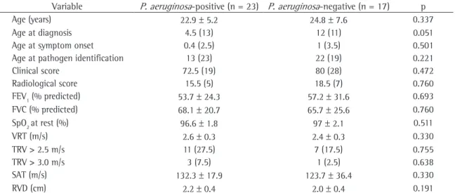
Table 1 summarizes the clinical, pulmonary function, radiographic and echocardiographic characteristics of P. aeruginosa-positive and P. aeruginosa-negative patients. Of the 40 patients, 23 were infected with P. aeruginosa. No statisti-cally significant differences were found between P. positive patients and P. aeruginosa-negative patients regarding the clinical, spirometric, radiographic or echocardiographic variables (p > 0.05).
Table 2 summarizes the clinical, pulmonary function, radiographic and echocardiographic characteristics of B. cepacia-positive patients and B. cepacia-negative patients. Of the 40 patients, 10 were infected with B. cepacia. B. cepacia-positive patients presented significantly lower FEV1 and FVC than did B. cepacia-negative patients (p = 0.011 and
Table 1 - Characteristics of Pseudomonas aeruginosa-positive and P. aeruginosa-negative patients.a
Variable P. aeruginosa-positive (n = 23) P. aeruginosa-negative (n = 17) p
Age (years) 22.9 ± 5.2 24.8 ± 7.6 0.337
Age at diagnosis 4.5 (13) 12 (11) 0.051
Age at symptom onset 0.4 (2.5) 1 (3.5) 0.501
Age at pathogen identification 13 (23) 22 (19) 0.221
Clinical score 72.5 (19) 80 (28) 0.472
Radiological score 15.5 (5) 18.5 (7) 0.760
FEV1 (% predicted) 53.7 ± 24.3 57.2 ± 31.6 0.693
FVC (% predicted) 68.1 ± 20.7 65.7 ± 25.6 0.760
SpO2 at rest (%) 96.6 ± 1.8 97 ± 2.1 0.511
VRT (m/s) 2.6 ± 0.3 2.4 ± 0.3 0.330
TRV > 2.5 m/s 11 (27.5) 7 (17.5) 0.755
TRV > 3.0 m/s 3 (7.5) 1 (2.5) 0.638
SAT (m/s) 132.3 ± 17.9 123.7 ± 36.4 0.330
RVD (cm) 2.2 ± 0.4 2.0 ± 0.4 0.191
FEV1: forced expiratory volume in one second; FVC: forced vital capacity; SpO2: peripheral oxygen saturation, determined using
pulse oximetry; TRV: tricuspid regurgitant jet velocity; SAT: right ventricular/pulmonary artery systolic acceleration time; RVD: and right ventricular diameter. aContinuous variables are expressed as mean ± standard deviation or as median and interquartile range.
urements of B. cepacia-positive patients with CF to those of B. cepacia-negative patients with CF. That study comprised 38 patients with a mean age of 14.6 years and identified 7 patients chroni-cally infected with B. cepacia. B. cepacia-positive patients, in comparison with B. cepacia-negative patients, presented higher systolic pulmonary artery pressure (61.6 ± 17.2 mmHg vs. 37.3 ± 13.9 mmHg), as well as higher right ventricular/pulmonary artery SAT (77 ± 33 m/s vs. 108 ± 25 m/s). In contrast, the present cross-sectional study comprised 40 of the 41 patients enrolled in a program for adults, with a mean age of 23 years. Chronic infection with B. cepacia presented no significant association with echocardiographic measurements.
In our study, the pulmonary disease was signifi-cantly more severe in B. cepacia-positive patients than in B. cepacia-negative patients. Nevertheless, the HP parameters were comparable between these two groups of patients.
Two factors that can affect the clinical charac-teristics of the patients admitted to a program for adults, who are different than those treated by pedi-atrics teams, must be emphasized. The first factor is the survival bias. Patients with more severe forms of the disease, including those chronically infected with P. aeruginosa and B. cepacia, typically die during childhood or adolescence, and the adult survivors imately 20% of the cases) develops the so-called
B. cepacia syndrome, which is characterized by fever, bacteremia, more rapid pulmonary disease progres-sion, necrotic pneumonia and death; a second group of patients presents a more rapid decline in pulmo-nary function but without the dramatic evolution caused by B. cepacia syndrome; and a third group, in which the evolution of CF is not affected by the infection.(25,26)
In our study, 23 patients (57.5%) were infected with P. aeruginosa, and 10 patients (25%) were infected with B. cepacia. In a study comprising 105 CF patients monitored at the University of Campinas, Brazil,(27) the authors reported a
preva-lence of P. aeruginosa-positivity similar to that found in our study (51%). However, the prevalence of B. cepacia-positivity was lower than that found in our study (5.2%). In another study comprising 36 CF patients and carried out in Recife, Brazil,(28)
the prevalence of infection with B. cepacia was 29.2%, which is similar to that found in our study.
Despite the considerable knowledge of the physiopathological role of chronic bacterial infec-tion in the evoluinfec-tion of the pulmonary disease in CF patients, few studies have investigated the relationship between chronic bacterial infection and HP. A case-control study was carried out in order to compare the echocardiographic
meas-Table 2 - Characteristics of Burkholderia cepacia-positive patients and B. cepacia-negative patients.a
Variable B. cepacia-positive (n = 10) B. cepacia-negative (n = 30) p
Age (years) 25.5 ± 5.8 23.2 ± 6.5 0.303
Age at diagnosis 10.5 (25.5) 9 (15) 0.488
Age at symptom onset 2 (3) 0.4 (3.5) 0.521
Age at pathogen identification 28 (17) 9 (19) 0.036
Clinical score 70 (15) 75 (25) 0.080
Radiological score 14 (5) 18 (6) 0.368
FEV1 (% predicted) 37.8 ± 21.1 61.1 ± 26.9 0.011
FVC (% predicted) 51.5 ± 18.3 72.4 ± 21.8 0.008
SpO2 at rest (%) 95.9 ± 2.5 97.1 ± 1.5 0.186
VRT (m/s) 2.6 ± 0.4 2.5 ± 0.3 0.613
TRV > 2.5 m/s 5 (12.5) 13 (32.5) 0.731
TRV > 3.0 m/s 2 (5) 2 (5) 0.256
SAT (m/s) 120.3 ± 18.5 131.8 ± 29.1 0.149
RVD (cm) 2.1 ± 0.4 2.2 ± 0.4 0.429
FEV1: forced expiratory volume in one second; FVC: forced vital capacity; SpO2: peripheral oxygen saturation, determined using
pulse oximetry; TRV: tricuspid regurgitant jet velocity; SAT: right ventricular/pulmonary artery systolic acceleration time; and RVD: right ventricular diameter. aContinuous variables are expressed as mean ± standard deviation or as median and interquartile range.
6. Emerson J, Rosenfeld M, McNamara S, Ramsey B, Gibson RL. Pseudomonas aeruginosa and other predictors of mortality and morbidity in young children with cystic fibrosis. Pediatr Pulmonol. 2002;34(2):91-100.
7. Burns JL, Gibson RL, McNamara S, Yim D, Emerson J, Rosenfeld M, et al. Longitudinal assessment of Pseudomonas aeruginosa in young children with cystic fibrosis. J Infect Dis. 2001;183(3):444-52.
8. Frederiksen B, Koch C, Høiby N. Antibiotic treatment of initial colonization with Pseudomonas aeruginosa postpones chronic infection and prevents deterioration of pulmonary function in cystic fibrosis. Pediatr Pulmonol. 1997;23(5):330-5. 9. Nixon GM, Armstrong DS, Carzino R, Carlin JB, Olinsky
A, Robertson CF, et al. Clinical outcome after early Pseudomonas aeruginosa infection in cystic fibrosis. J Pediatr. 2001;138(5):699-704.
10. Navarro J, Rainisio M, Harms HK, Hodson ME, Koch C, Mastella G, et al. Factors associated with poor pulmonary function: cross-sectional analysis of data from the ERCF. European Epidemiologic Registry of Cystic Fibrosis. Eur Respir J. 2001;18(2):298-305.
11. Pedersen SS, Høiby N, Espersen F, Koch C. Role of alginate in infection with mucoid Pseudomonas aeruginosa in cystic fibrosis. Thorax. 1992;47(1):6-13.
12. Henry RL, Mellis CM, Petrovic L. Mucoid Pseudomonas aeruginosa is a marker of poor survival in cystic fibrosis. Pediatr Pulmonol. 1992;12(3):158-61.
13. Döring G, Conway SP, Heijerman HG, Hodson ME, Høiby N, Smyth A, et al. Antibiotic therapy against Pseudomonas aeruginosa in cystic fibrosis: a European consensus. Eur Respir J. 2000;16(4):749-67.
14. Gibson RL, Burns JL, Ramsey BW. Pathophysiology and management of pulmonary infections in cystic fibrosis. Am J Respir Crit Care Med. 2003;168(8):918-51.
15. MacNee W. Pathophysiology of cor pulmonale in chronic obstructive pulmonary disease. Part two. Am J Respir Crit Care Med. 1994;150(4):1158-68.
16. MacNee W. Pathophysiology of cor pulmonale in chronic obstructive pulmonary disease. Part One. Am J Respir Crit Care Med. 1994;150(3):833-52.
17. Fraser KL, Tullis DE, Sasson Z, Hyland RH, Thornley KS, Hanly PJ. Pulmonary hypertension and cardiac function in adult cystic fibrosis: role of hypoxemia. Chest. 1999;115(5):1321-8.
18. Yankaskas JR, Egan T.M., Mauro M. Major complications. In: Yankaskas JR, Knowles MR, editors. Cystic fibrosis in adults. Philadelphia: Lippincott-Raven; 1999. p. 175-94.
19. Fauroux B, Hart N, Belfar S, Boulé M, Tillous-Borde I, Bonnet D, et al. Burkholderia cepacia is associated with pulmonary hypertension and increased mortality among cystic fibrosis patients. J Clin Microbiol. 2004;42(12):5537-41.
20. Ionescu AA, Ionescu AA, Payne N, Obieta-Fresnedo I, Fraser AG, Shale DJ. Subclinical right ventricular dysfunction in cystic fibrosis. A study using tissue Doppler echocardiography. Am J Respir Crit Care Med. 2001;163(5):1212-8.
21. Shwachman H, Kulczycki LL. Long-term study of one hundred five patients with cystic fibrosis; studies made over a five- to fourteen-year period. AMA J Dis Child. 1958;96(1):6-15. 22. Quiñones MA, Otto CM, Stoddard M, Waggoner A, Zoghbi
WA; Doppler Quantification Task Force of the Nomenclature and Standards Committee of the American Society of Echocardiography. Recommendations for quantification of Doppler echocardiography: a report from the Doppler
present aspects associated with lower severity of the disease. The second factor is the higher proportion of atypical cases, with late diagnosis and less severe pulmonary disease.(29) These factors might have
contributed to the differences between the findings of our study and those of the study conducted by Faroux et al.(19)
The main limiting factors in our study were the small sample and the possibility of a type II error. However, our sample was limited to 41 patients, only 40 of whom were evaluated. Another possible limiting factor was the lack of identification of the B. cepacia complex genomovars. Since some of the genomovars have been associated with worse clin-ical evolution, their identification would have been relevant.
In conclusion, in a group of patients admitted to a program for adults with CF, chronic infection with P. aeruginosa or B. cepacia presented no association with PH, as diagnosed using Doppler echocardi-ography. The pulmonary disease was more severe in B. positive patients than in B. cepacia-negative patients. Further studies involving larger samples are needed in order to determine the role of chronic bacterial infection in the development of HP in CF patients.
Acknowledgments
We would like to thank Vânia Naomi Hirakata and Daniela Benzano for the statistical analysis, as well as all of the staff members of the HCPA Program for Adolescents with Cystic Fibrosis and the HCPA Program for Adults with Cystic Fibrosis for their collaboration.
References
1. McKone EF, Emerson SS, Edwards KL, Aitken ML. Effect of genotype on phenotype and mortality in cystic fibrosis: a retrospective cohort study. Lancet. 2003;361(9370):1671-6. 2. Santos CI, Ribeiro JD, Ribeiro AF, Hessel G. Critical
analysis of scoring systems used in the assessment of Cystic Fibrosis severity: State of the art. J Bras Pneumol. 2005;30(3):286-98.
3. Lyczak JB, Cannon CL, Pier GB. Lung infections associated with cystic fibrosis. Clin Microbiol Rev. 2002;15(2):194-222. 4. Yankaskas JR, Marshall BC, Sufian B, Simon RH, Rodman
D. Cystic fibrosis adult care: consensus conference report. Chest. 2004;125(1 Suppl):S1-S39.
27. Alvarez AE, Ribeiro AF, Hessel G, Bertuzzo CS, Ribeiro JD. Cystic fibrosis at a Brazilian center of excellence: clinical and laboratory characteristics of 104 patients and their association with genotype and disease severity J Pediatr. (Rio J). 2004;80(5):371-379.
28. Magalhães M, Britto MC, Bezerra PG, Veras A. Prevalência de bactérias potencialmente patogênicas em espécimes respiratórios de fibrocísticos do Recife. J Bras Patol Med Lab. 2004;40(4):223-227.
29. Gilljam M, Ellis L, Corey M, Zielenski J, Durie P, Tullis DE. Clinical manifestations of cystic fibrosis among patients with diagnosis in adulthood. Chest. 2004;126(4):1215-24. Quantification Task Force of the Nomenclature and Standards
Committee of the American Society of Echocardiography. J Am Soc Echocardiogr. 2002;15(2):167-84.
23. Miller MR, Hankinson J, Brusasco V, Burgos F, Casaburi R, Coates A, et al. Standardisation of spirometry. Eur Respir J. 2005;26(2):319-38.
24. Sociedade Brasileira de Pneumologia e Tisiologia. Diretrizes para Testes de Função Pulmonar. J Pneumol. 2002;28(Supl 3):S1-S238.
25. Gilligan PH. Microbiology of Cystic Fibrosis Lung Disease. In: Yankaskas JR, Knowles MR, editors. Cystic fibrosis in adults. Philadelphia: Lippincott-Raven; 1999. p. 93-114.