Incidence of viral infection of the respiratory tract in
acute asthma patients treated in the emergency room*
IVETE TEREZINHA MACHADO DA ROCHA1, DIEGO MENEGOTTO2, CRISTINE FELICIATI HOFFMANN2, SERGIO SALDANHA MENNA-BARRETO3, PAULO DE TARSO ROTH DALCIN4, SELIR MARIA STRALIOTTO5,
SUZIE HYONA KANG2, LILIAN RECH PASIN2, JOSIANE FISCHER2, FABIANE NIETO2
Keywords: Emergency Service, Hospital; Vírus diseases/prevention & control; Asthma; Respiratory tract infections; Influenza A virus, human
*Study carried out at the Hospital de Clínicas de Porto Alegre (HCPA, Porto Alegre Hospital de Clínicas) and the Fundação Estadual de Produção e Pesquisa em Saúde/Instituto de Pesquisas Biológicas - Laboratório Central (FEPPS/IPB-LACEN, Health Production and Research State Foundation - Institute of Biological Research - Central Laboratory), Porto Alegre, Rio Grande do Sul, Brazil
1. Graduate student at the Faculdade de Medicina da Universidade Federal do Rio Grande do Sul (UFRGS, University of Rio Grande do Sul School of Medicine), Porto Alegre, Rio Grande do Sul, Brazil.
2. Recipient of a scientific initiation scholarship, Programa Institucional de Bolsas de Iniciação Científica (PIBIC, Institutional Program for Scientific Initiation Scholarships), Universidade Federal do Rio Grande do Sul (UFRGS, University of Rio Grande do Sul), Porto Alegre, Rio Grande do Sul, Brazil
3. Full Professor in the Department of Internal Medicine at the Universidade Federal do Rio Grande do Sul (UFRGS, University of Rio Grande do Sul), Porto Alegre, Rio Grande do Sul, Brazil
4. Adjunct Professor at the Faculdade de Medicina da Universidade Federal do Rio Grande do Sul (UFRGS, University of Rio Grande do Sul School of Medicine), Porto Alegre, Rio Grande do Sul, Brazil
5. Biochemist in the Laboratório de Vírus Respiratório (Respiratory Virus Laboratory) at FEPS/IPB-LACEN
Correspondence to: Ivete Terezinha Machado da Rocha. R. Sg. Manoel Raimundo Soares 753 casa 25, Porto Alegre - RS Brazil. CEP: 91430-380. Phone: 55 51 3338-7382. E-mail: [email protected]
Submitted: 4 February 2005. Accepted, after review: 26 April 2005.
ABSTRACT
INTRODUCTION
A s t h m a e x a c e r b a t i o n s a r e r e l a t e d t o predisposing factors of various natures. Exposure to inhaled allergens, especially mould, pollen and dust mites, has received considerable well-deserved attention as a trigger for extrinsic asthma.(1) Other
significant precipitating factors are: exercise, drug use, air pollution, weather changes and exposure to cold.(1-3) In addition, respiratory tract infections
have also been correlated with asthma attacks.(2)
It has recently been suggested that the significance of viral infections as an inducing factor for acute exacerbations of asthma has been underestimated, primarily due to limitations of the available d i a g n o s t i c m e t h o d o l o g y u s e d t o i d e n t i f y respiratory viruses.(1,4-5)
In children, viral infections increase airway responsiveness and are responsible for 26% to 42% of acute asthma episodes.(6) In adults, the role of
viral infections as a cause for asthma exacerbations has not yet been well defined.(7-8) In three recent
studies, the frequency of identification of the respiratory virus related to asthma attacks ranged from 0% to 44%.(1,9)
Studies using new diagnostic techniques such as polymerase chain reaction have increased the frequency at which viral infections are identified in adults with asthma exacerbations.(10)
The incidence of respiratory viruses as inducing factors of acute asthma in patients treated in emergency rooms has not been studied in our milieu. Consequently, the elucidation of the role of the predisposing factors in severe acute asthma may contribute to optimizing the management of the prevention of this disease.(11-12)
The objective of this study was to evaluate the incidence of respiratory viruses in patients with acute asthma treated in the emergency room of the Hospital de Clínicas de Porto Alegre (Porto Alegre Hospital das Clínicas) in the city of Porto Alegre, Brazil, comparing the characteristics of patients presenting samples that test positive for respiratory viruses with those of patients presenting samples that test negative.
METHODS
This was a study of the incidence of respiratory viruses (respiratory syncytial virus, adenovirus and
influenza, as well as parainfluenza types 1, 2, 3 and 4), with convenience samples, making the diagnosis based on positive results for viral antigens through indirect immunofluorescence assay (IFA) of nasopharyngeal fluid obtained from patients aged 12 and over with severe acute asthma and treated in the emergency room of the Porto Alegre Hospital das Clínicas.
Inclusion criteria were previous medical diagnosis of bronchial asthma; age 12 or above, and being for asthma-related symptoms indicating exacerbation of the disease (dyspnea, wheezing or cough). Exclusion criteria were chronic pulmonary disease, cardiac insufficiency and lack of written informed consent (from the patient or legal guardian).
Sample selection consisted of evaluation of patients who had been consecutively treated during a restricted period of availability for carrying out the IFA for identification of viruses. The research team visited the emergency room in the morning and in the afternoon looking for patients with acute asthma. Since the diagnostic test was not available in the evening, on weekends or on holidays, patients treated during these periods were not considered for inclusion.
identification of inducing factors for the attack, patients were first asked whether they could perceive any inducing event for the worsening of asthma. Subsequently, we tried to determine whether the triggering factor was a respiratory infection (flu, cold or sinusitis), an inhaled allergen (dust, mold, animal fur, pollen or other), exercise or a weather change. Diagnostic evaluation for the identification of respiratory viruses involved the use of IFA of nasopharyngeal fluid, using viral antigens of respiratory syncytial virus, adenovirus and influenza, as well as of parainfluenza types 1, 2, 3 and 4. Nasopharyngeal fluid was collected using a urethral tube attached to a Bioject needle-free CO2-powered injector (Bioject Medical Technologies, Portland, OR, USA). The tube was introduced through the nostril up to the nasopharynx, after which 20 cm of H2O were aspirated and the secretion was obtained using a vacuum aspirator (Fanen). Samples were transported in a Styrofoam container with reusable ice packs (4°C) to the Instituto de Pesquisas Biológicas, Laboratório Central (Central Laboratory of the Institute of Biological Research).
In the laboratory, samples were transferred to a previously identified sterile centrifuge tube containing 2 mL of phosphate buffered saline. Samples were suspended for mucus dilution/cell release and then centrifuged at 1500 rpm at room temperature. Three slides were prepared from each sample under analysis. One was used for the triage of positive cases and one for the determination of the agent if the triage was positive. The third slide was stored in a freezer (-70°C) in case it became necessary to repeat the IFA. Sample material was distributed in seven circles, each approximately 0.05 cm in diameter, on each slide. This material was dried and fixed in acetone at 4°C for 10 minutes. For the staining of slides, 25 mL of specific monoclonal antibodies for each virus (respiratory syncytial virus, adenovirus, influenza A and influenza B, as well as anti-parainfluenza types 1, 2, 3 and 4) were introduced into each circle. We used the Respiratory Panel 1 Viral Screening & Identification Kit (Chemicon International, Temecula, CA, USA). Subsequently, the slides were incubated in a humid chamber at 37°C for 30 minutes, subjected to three 5-minute washes in phosphate buffered saline and dried with cool air. Subsequently, a drop of the specific conjugate was added, followed by incubation in a humid
chamber at 37°C, rinsing and drying. Slides were prepared with buffered glycerol and read under fluorescence microscopy.
The data were entered into a Microsoft Excel, version 2000, database. Subsequently, the Statistical Package for the Social Sciences program, version 12.0 was used to process and analyze the data. Descriptive and comparative analyses between the group of patients whose samples tested positive for respiratory viruses and the group of those whose samples tested negative were carried out. We used Student's t-test for the analysis of continuous variables presenting normal distribution for independent samples. We used the Mann-Whitney U test for the analysis of continuous variables presenting abnormal distribution. We used the chi-square test for the analysis of categorical variables and, when necessary, Yates's correction or Fisher's exact test. The alpha was set at 0.05.
The Porto Alegre Hospital de Clínicas Ethics Committee and Research Committee approved the study. All participating patients or (for patients younger than 18) their legal guardians gave written informed consent.
RESULTS
From March to July 2004, 353 patients diagnosed with acute asthma were treated in the emergency room of the Porto Alegre Hospital de Clínicas. Of those 353 patients, 142 were evaluated for possible inclusion in the study, 52 of which were excluded because they were diagnosed with chronic pulmonary diseases, 11 because they were diagnosed with cardiac insufficiency and 24 because they declined to participate in the study. For the remaining 55 patients, clinical data and nasopharyngeal aspirate samples were collected. The aspirate samples from 6 patients were insufficient for the IFA, and the final sample therefore comprised 49 patients.
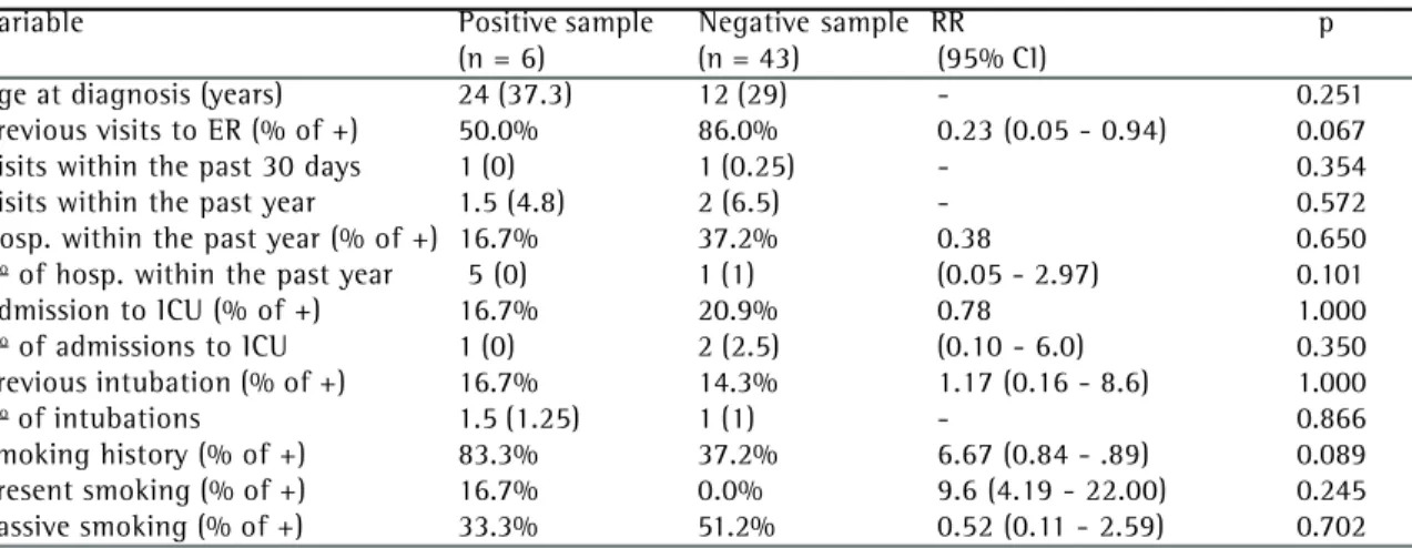
Table 1 shows the general characteristics of the patients in the study. A statistically significant difference was found for age. Patients presenting negative samples were younger (41.7 ± 20.9 years) than those presenting positive samples (61.7 ± 11.5 years) (p = 0.027). No statistically significant difference was found for any of the other variables shown.
Table 2 shows the analysis of data concerning patient history. There was no statistically significant difference (p > 0.05) between the two groups for
the following variables: age at which asthma was diagnosed, previous visits to an emergency room (% of positive responses), number of visits to the emergency room in the past 30 days and in the past year, hospitalizations within the past year (% of positive responses), number of hospitalizations in the past year, admissions to an intensive care unit (% of positive responses), number of admissions to an intensive care unit, previous intubations (% of positive responses), number of previous intubations, history of smoking (% of positive responses), current
Categorical variables presented as %; continuous variables presented as mean ± standard deviation; chi-square test for categorical variables; Student's t-test for independent samples for quantitative variables
TABLE 1
General characteristics of the patients studied
Variable Positive sample Negative sample RR p
(n = 6) (n = 43) (95% CI)
Age at diagnosis (years) 24 (37.3) 12 (29) - 0.251
Previous visits to ER (% of +) 50.0% 86.0% 0.23 (0.05 - 0.94) 0.067
Visits within the past 30 days 1 (0) 1 (0.25) - 0.354
Visits within the past year 1.5 (4.8) 2 (6.5) - 0.572
Hosp. within the past year (% of +) 16.7% 37.2% 0.38 0.650
No of hosp. within the past year 5 (0) 1 (1) (0.05 - 2.97) 0.101
Admission to ICU (% of +) 16.7% 20.9% 0.78 1.000
No of admissions to ICU 1 (0) 2 (2.5) (0.10 - 6.0) 0.350
Previous intubation (% of +) 16.7% 14.3% 1.17 (0.16 - 8.6) 1.000
No of intubations 1.5 (1.25) 1 (1) - 0.866
Smoking history (% of +) 83.3% 37.2% 6.67 (0.84 - .89) 0.089
Present smoking (% of +) 16.7% 0.0% 9.6 (4.19 - 22.00) 0.245
Passive smoking (% of +) 33.3% 51.2% 0.52 (0.11 - 2.59) 0.702
RR: relative risk for positive identification of viruses; 95% CI: 95% confidence interval; ER: emergency room; % of +: % of positive responses; hosp.: hospitalizations; #: number; ICU: intensive care unit; Categorical variables presented as %; continuous variables presented as median (interquartile range) Mann-Whitney U test for quantitative variables; chi-square test for categorical variables
TABLE 2
Data regarding patient history
Variable Total Positive sample Negative sample p
(n = 49) (n = 6) (n = 43)
Gender 0.302
Male 18.4% 4.1% 14.3%
Female 81.6% 8.2% 73.5%
Age (years) 44.1 ± 20.9 61.7 ± 11.5 41.7 ± 20.9 0.027
Race 0.262
Caucasian 61.2% 4.1% 57.1%
Non-Caucasian 38.7% 8.2% 30.6%
Marital status 0.398
Single 58.3% 10.4% 47.9%
Married 35.4% 2.1% 33.3%
Widow(er) 6.3% 0.0% 6.3%
Weight (kg) 61.2 ± 17.6 73.2 ± 9.9 59.4 ± 17.9 0.074
TABLE 3
Use of medications and nebulization by the patients studied
RR: relative risk for positive identification of viruses; 95% CI: 95% confidence interval; Categorical variables presented as % of positive responses; Mann-Whitney U test for quantitative variables; chi-square test for categorical variables
RR: relative risk for positive identification of viruses; 95% CI: 95% confidence interval; % of +: % of positive responses; HR: heart rate; RF: respiratory frequency; SpO2: oxygen saturation measured by digital pulse oximetry
Categorical variables presented as %; continuous variables presented as median (interquartile range) or as mean ± standard deviation Student's t-test for independent samples or Mann-Whitney U test for quantitative variables; Chi-square test for categorical variables
TABLE 4
Data regarding the current attacks experienced by the patients studied
Variable Positive sample Negative sample RR p
(n = 6) (n = 43) (95% CI)
Oral corticosteroids past month 66.7% 53.5% 1.63 (0.33 - 8.08) 0.865
Inhaled corticosteroids 50.0%) 44.2% 1.23 (0.27 - 5.49) 1.000
Long-acting beta-2
adrenergic agonists 33.3% 44.2% 0.67 (0.14 - 3.30) 0.950
Oral xanthine 16.7% 20.9% 0.78 (0.10 - 5.95) 1.000
Short-acting beta-2
adrenergic agonists 50.0% 72.1% 0.44 (0.10 - 1.94) 0.531
Inhaled ipratropium 50.0% 48.8% 1.04 (0.23 - 4.66) 1.000
Use of nebulization 50.0% 79.1% 0.32 (0.08 - 1.40) 0.296
Variable Positive sample Negative sample RR p
(n = 6) (n = 43) (95% CI)
Duration of attack (hours) 42 (43.5) 12 (44.0) 0.145
Identification of triggering factor
Infection (% of +) 66.7% 32.6 3.44 (0.70 - 16.97) 0.241
Allergens (% of +) 0% 23.3% 1.18 (1.03 - 1.35) 0.433
Exercise (% of +) 0% 7.0% 1.15 (1.03 - 1.29) 1.000
Weather changes (% of +) 0% 39.5% 1.23 (1.04 - 1.45) 0.148
Attack/use of systemic
corticosteroids (% of +) 66.7% 44.2% 2.26 (0.46 - 11.22) 0.550
Symptoms
Headache (% of +) 66.7% 69.8% 0.88 (0.18 - 4.31) 1.000
Rhinorrhea (% of +) 50.0% 62.8% 0.63 (0.14 - 2.82) 0.877
Nasal congestion (% of +) 50.0% 60.5% 0.69 (0.16 - 3.08) 0.964
Sore throat (% of +) 50.0% 25.6% 2.4 (0.57 - 10.93) 0.448
Earache (% of +) 16.7% 20.9% 0.78 (0.10 - 5.95) 1.000
Dysphonia (% of +) 50.0% 37.2% 1.58 (0.36 - 7.03) 0.877
Myalgia (% of +) 83.3% 62.8 2.66 (0.34 - 20.94) 0.594
Cough (% of +) 100.0% 88.4% 0.86 (0.77 - 0.97) 0.872
Expectoration (% of +) 100.0% 81.4% 0.53 (0.10 - 2.78) 0.572
Fever at home (% of +) 83.3% 37.2% 6.67 (0.84- 52.89) 0.089
Flu symptoms in the
family (% of +) 50.0% 58.1 0.75 (0.17 - 3.35) 1.000
Signs
Axillary temperature
37.8°C (% of +) 40% 11.9% 3.81 (0.77 - 18.85) 0.316
HR (beats/min) 109.00 ± 8.3 109.5 ± 20.5 0.961
RF (breaths/min) 28.0 ± 3.5 26.2 ± 6.0 0.514
Wheezing (% of +) 100.0 % 93.0% 0.87 (0.78 - 0.97) 1.000
Rales (% of +) 0.0% 14.0% 0.53 (0.10 - 1.49) 0.755
Crackles (% of +) 16.7% 25.6 0.62 (0.08 - 4.77) 1.000
smoking (% of positive responses) and passive smoking (% of positive responses).
Table 3 shows data concerning the use of medications (oral corticosteroids in the past month, inhaled corticosteroids, long-acting inhaled beta-2 adrenergic agonists, oral xanthine, short-acting beta 2-adrenergic agonists and inhaled ipratropium bromide) and the use of nebulization. There was no statistically significant difference (p > 0.05) for these variables between the two groups.
Table 4 shows the analysis of data concerning the present attack. Mean duration of the attack was 42 hours in the group presenting positive samples (interquartile range of 43.5 h) and 12 hours in the group presenting negative samples (interquartile range of 44 h) (p = 0.145). In the group presenting positive samples, 66.7% of the patients had the subjective impression that the asthma attack was triggered by a respiratory infection, whereas only 32.6% of the patients in the group presenting negative samples related the asthma attack to a respiratory infection (p = 0.241). In the group presenting positive samples, no patients related the triggering of the attack to being exposed to inhaled allergens, exercise or weather changes, whereas 23.3% of the patients in the group presenting negative samples related the attack to exposure to inhaled allergens (p = 0.433), 7% to exercise (p = 1.00) and 39.5% to weather changes (p = 0.148). In the group presenting positive samples, 66.7% of the patients were making use of oral corticosteroids at the moment of the attack, whereas, in the group presenting negative samples, 44.2% of the patients were using corticosteroids (p = 0.550). Comparing the two groups, we detected the following incidences of, respectively, symptoms and signs: headache (66.7% and 69.8%; p = 1.000), rhinorrhea (50% and 62.8%; p = 0.877), nasal congestion (50% and 60.5%; p = 0.964), sore throat (50% and
25.6%; p = 0.448), earache (16.7% and 20.9%; p = 1.000), dysphonia (50% and 37.2%; p = 0.877), myalgia (83.3% and 62.8%; p = 0.549), cough (100% and 88.4%; p = 0.872), expectoration (100% and 81.4%; p = 0.572), fever at home (83.3% and 37.2%; p = 0.089), symptoms of flu in the family (50% and 59.1%; p = 1.000) and axillary temperature = 37.8°C (40% and 11.9%; p = 0.316). There was no statistically significant difference between the two groups in terms of heart rate, respiratory frequency, wheezing, rales, crackles or oxygen saturation measured by digital pulse oximetry.
Table 5 shows the outcomes of the asthma attack. Comparing the group of patients presenting positive samples to that of those presenting negative samples, 50% and 76.7%, respectively, were discharged directly from the emergency room (p = 0.321); 50% and 23.3%, respectively, were hospitalized (p = 0.321); and the mean duration of the emergency room stay was 4.67 h and 9.93 h, respectively (p = 0.440).
DISCUSSION
In the present study, the incidence of respiratory viruses in patients with acute asthma treated in the emergency room was 12.2%. Of all clinical characteristics studied, only age was found to correlate with the identification of respiratory viruses, and patients presenting positive samples were, on average, older than those presenting negative samples.
The incidence found in the present study was similar to that found in other studies using the IFA technique as a diagnostic method. In those studies, the frequency of the identification of respiratory viruses ranged from 10% to 21%.(8,13)
Various factors might have contributed to
TABLE 5
Outcomes of the attacks experienced by the patients studied
Outcome Positive sample Negative sample RR p
(n = 6) (n = 43) (95% CI)
Discharge from ER (%) 50.0% 76.7% 0.65 (0.29 - 1.48) 0.321
Hospitalization (%) 50.0% 23.3%
Stay in ER (hours; mean ± SD) 4.67 ± 6.37 9.93 ± 11.34 - 0.440
underestimating the identification of viruses in the present study. Principal among such factors is the very sensitivity of the IFA method. In addition, the duration of the viral profile might have interfered with the identification of viruses since the IFA technique is most efficient when carried within 24 to 48 hours after the onset of symptoms.(14) In our
study, the median duration of symptoms of the asthma attacks was 12 hours, with an interquartile range of 44 hours. Therefore, the frequency found in the identification of the virus during the asthma attack depended directly on the methodology in use. Some techniques, such as viral isolation in cell culture, may increase the viral load and improve the sensitivity of the IFA test, but they are difficult to perform and are not available in our practice. (15-16) Furthermore, respiratory infections caused by
other types of viruses, such as rhinoviruses and coronaviruses, were not considered in this study, and such viruses might have been triggering factors for the asthma attacks.(17-19) The identification of
these viruses requires special techniques such as cell culture and polymerase chain reaction.(4)
Studies using these new diagnostic techniques have shown an increase in the frequency of viral infections in adults with asthma exacerbations. (15-20) Therefore, the results in our study were limited
by the technique used (IFA).
Despite its inherent flaws, the IFA technique it is a rapid and effective method for the detection of viruses. In addition, IFA is affordable and has proven useful for the diagnosis of respiratory infections in clinical practice. Nevertheless, further complementary studies, using polymerase chain reaction, are warranted.
The technical difficulty encountered in this study was the small amount of material collected from the nasopharyngeal aspirate, even after this procedure had been routinely carried out through both nostrils. This was illustrated by the fact that 6 of the 55 patient samples were considered insufficient for viral testing in the study.
It is important to highlight that, in the analysis of data concerning patient histories, the relative risk (RR) for positive identification of viruses related to a history of smoking and current smoking, although less than statistically significant (p = 0.089 and p = 0.245, respectively), was high (RR = 6.67 and RR = 9.6, respectively). This finding may express the deleterious effect of smoking,
which predisposes patients to viral infections and asthma exacerbations.
The data presented in Table 4 concerning the present attack show that, although less than statistically significant (p = 0.089), the frequency of fever at home was higher (83.3%) among patients presenting positive samples than among those presenting negative samples (37.2%). In addition, 40% of the patients presenting positive samples had an axillary temperature equal to or greater than 37.8°C at admission, and this only happened to 11.9% of the patients presenting negative samples (p = 0.316). This finding may, together with the increase in the sample size, confirm the expression of fever as a clinical marker of an infectious process (in this case, of a viral infection). The fact that all patients presented high frequencies of symptoms consistent with viral infection (headache, rhinorrhea, nasal congestion, sore throat, earache, dysphonia and myalgia) also deserves attention. Furthermore, the subjective identification of respiratory infection (cold, flu or sinusitis) as a triggering factor for the attack did not differ between the two groups. Even if the method used to identify viral infection had underestimated the real incidence of respiratory viruses in this sample, the high frequency of these symptoms in the group presenting negative samples very likely provides evidence of the lack of specificity of these clinical parameters as indicators of viral respiratory infection. In contrast, although the difference between the two groups was less than statistically significant, 23.3% of the patients presenting negative samples identified exposure to inhaled allergens as the triggering factor for the attack, whereas none of the patients presenting positive samples associated their asthma attacks to such exposure.
The sample selection technique and the small sample size can be considered methodological limitations. The sample selection technique, known as convenience sampling,(21) made the execution
the fact that the present study was the first step in a broader study that will try to provide a better definition of the incidence of respiratory viruses in acute asthma, as well as to profile the seasonality of acute asthma and to identify potential correlations between identification of viruses and clinical characteristics.
In conclusion, the incidence of respiratory viruses in patients aged 12 and older presenting to the emergency room with acute asthma was 12.2%, which confirmed viral infections as a contributing factor in the triggering of asthma attacks, although it was found to be less frequent than in the pediatric age bracket. The only clinical characteristic correlated with the identification of respiratory viruses was age, and patients presenting samples that tested positive for these viruses were, on average, older than those who presented samples that tested negative.
ACKNOWLEDGMENTS
We would like to thank Volf Aguiar for the statistical analysis, the technical team of the Laboratório de Vírus Respiratório (Respiratory Virus Laboratory) of the Fundação Estadual de Produção e Pesquisa em Saúde (Health Production and Research State Foundation), at the Biological Research Institute Central Laboratory, for their contribution in performing the viral identification tests, and to the medical, administrative and nursing staff of the Porto Alegre Hospital de Clínicas emergency room for their collaboration in the study.
REFERENCES
1. Busse WW. Respiratory infections: their role in airway responsiveness and the pathogenesis of asthma. J Allergy Clin Immunol.1990; 85(4):671-83. Review. 2. Nicholson KG, Kent J, Ireland DC. Respiratory viruses
and exacerbations of asthma in adults. BMJ. 1993; 307(6910):982-6.
3. Cerwenka A, Carter LL, Reome JB, Swain SL, Dutton RW. In vivo persistence of CD8 polarized T cell subsets producing type 1 or type 2 cytokines. J Immunol. 1998;161(1):97-105.
4. Johnston SL, Pattemore PK, Sanderson G, Smith S, Lampe F, Josephs L, et al. Community study of role of viral infections in exacerbations of asthma in 9-11 year old children. BMJ. 1995;310(6989):1225-9.
5. Pattemore PK, Johnston SL, Bardin PG. Viruses as precipitants of asthma symptoms. I. Epidemiology. Clin Exp Allergy. 1992;22(3):325-36. Review.
6. Noah TL, Henderson FW, Wortman IA, Devlin RB, Handy J, Koren HS, Becker S. Nasal cytokine production in viral acute upper respiratory infection of childhood. J Infect Dis. 1995;171(3):584-92.
7. Jennings LC, Barns G, Dawson HP. The association of viruses with acute asthma. N Z Med J. 1987;100(829):488-90.
8. Hudgel D W, Langston L Jr, Selner JC, McIntosh K. Viral and bacterial infections in adults with chronic asthma. Am Rev Respir Dis.1979;120(2):393-7.
9. Sokhandan M, McFadden ER Jr, Huang YT, Mazanec MB. The contribution of respiratory viruses to severe exacerbations of asthma in adults. Chest. 1995; 107(6):1570-4.
10. Mertsola J, Ziegler T, Ruuskanen O, Vanto T, Koivikko A, Halonen P. Recurrent wheezy bronchitis and viral respiratory infections. Arch Dis Child. 1991;66(1):124-9. 11 . Openshaw PJ, Lemanske RF. Respiratory viruses and asthma: can the effects be prevented? Eur Respir J Suppl. 1998;27:35S-39S. Review.
1 2 . Tuffaha A, Gern JE, Lemanske RF Jr. The role of respiratory viruses in acute and chronic asthma. Clin Chest Med. 2000;21(2):289-300. Review.
13. Huhti E, Mokka T, Nikoskelainen J, Halonen P. Association of viral and mycoplasma infections with exacerbations of asthma. Ann Allergy. 1974:33(3):145-9.
14. Madeley CR, Peiris JS. Methods in virus diagnosis: immunofluorescence revisited. J Clin Virol. 2002; 25(2):121-34. Review.
1 5 . Atmar RL, Guy E, Guntupalli KK, Zimmerman JL, Bandi VD, Baxter BD, Greenberg SB. Respiratory tract viral infections in inner-a-city asthmatic adults. Arch Intern Med. 1998;158(22):2453-9.
1 6 . Simpson JL, Moric I, Wark PA, Johnston SL, Gibson PG. Use of induced sputum for the diagnosis of influenza and infections in asthma: a comparison of diagnostic techniques. J Clin Virol. 2003;26(3):339-46. 1 7 . Papadopoulos NG, Johnston SL . Viruses and asthma
exacerbations. Thorax. 1998;53(11):913-4.
1 8 . Osur SL. Viral respiratory infections in association with asthma and sinusitis: a review. Ann Allergy Asthma Immunol. 2002;89(6):553-60. Review.
1 9 . Corne JM, Marshall C, Smith S, Schreiber J, Sanderson G, Holgate ST, Johnston SL. Frequency, severity, and duration of rhinovirus infections in asthmatic and non-asthmatic individuals: a longitudinal cohort study. Lancet. 2002;359(9309):831-4.
2 0 . Green RM, Custovic A, Sanderson G, Hunter J, Johnston SL, Woodcock A.. Synergism between allergens and viruses and risk of hospital admission with asthma: case-control study. BMJ. 2002;324(7340):763. 21. Hulley SB, Newman TB, Cummings SR. Escolhendo os