O
RIGINALA
RTICLE|
A
RTIGOO
RIGINALAuthors
Denise Maria do Nascimento Costa 1,2 Lucila Maria Valente 1
Pedro Alves da Cruz Gouveia 1
Filipe Wanick Sarinho 1
Gisele Vajgel Fernandes 1 Maria Alina Gomes de Mattos Cavalcante 1 Camila Barbosa Lyra de Oliveira 1,2
Carolina de Andrade Jordão de Vasconcelos 2
Emanuel Sávio Cavalcante Sarinho 2
1 Universidade Federal de Pernambuco.
2 Instituto de Medicina Integral Prof. Fernando Figueira.
Submitted on: 10/14/2016. Approved on: 1/11/2017.
Correspondence to:
Denise Costa. Hospital das Clínicas. Av. Professor Moraes Rêgo, nº 1235, Cidade Universitária, Recife, PE, Brazil.
CEP: 50670-901
E-mail: [email protected]
Comparative analysis of primary and secondary
glomerulopathies in the northeast of Brazil: data from the
Pernambuco Registry of Glomerulopathies - REPEG
Análise comparativa das glomerulopatias primárias e secundárias
no nordeste do Brasil: dados do Registro Pernambucano de
Glomerulopatias - REPEG
Introdução: No Brasil, glomerulopatias
são a terceira causa de doença renal crô-nica terminal, responsáveis por 11% dos pacientes em diálise. Entretanto, estudos sobre a prevalência desta patologia no nordeste do Brasil são escassos. Objetivo:
O objetivo foi descrever os achados das biópsias e analisar comparativamente a apresentação clínico laboratorial en-tre as glomerulopatias primárias (GP) e as glomerulopatias secundárias (GS).
Métodos: Estudo retrospectivo, realizado em dois hospitais públicos de ensino do estado de Pernambuco, nordeste do Bra-sil. Resultados: Foram avaliadas 1.151 biópsias, de 1998 a 2016. A amostra foi composta por 670 biópsias de rins nati-vos, após exclusão de patologias extra glomerulares e materiais inadequados. GP foram mais frequentes do que GS (58% × 42%). Dentre as GP, houve predomínio de glomeruloesclerose segmentar e focal (GESF). Glomerulonefrite membranopro-liferativa e glomerulopatia colapsante fo-ram responsáveis por 9% e 3% das GP, respectivamente. Das GS, as etiologias principais foram nefrite lúpica (67%) e infecciosas (10%). Sexo feminino, hema-túria e nível elevado de creatinina esti-veram relacionadas a uma maior chance de GS na análise multivariada. Síndrome nefrótica foi mais comum dentre as GP, já anormalidades urinárias e síndrome nefrítica prevaleceram nos pacientes com GS. Conclusões: Este é o primeiro registro de glomerulopatias do nordeste do Brasil. Demonstrou-se também uma análise com-parativa das principais alterações clínico laboratoriais das GP e GS, com classifi-cações atualizadas das doenças glomeru-lares.
RESUMO
Palavras-chave: epidemiologia;
glomeru-lonefrite; glomérulos renais; patologia.
Introduction: In Brazil, glomerulopathies are the third leading cause of chronic renal disease, accounting for 11% of di-alysis patients. Studies on the prevalence of this disease in Northeastern Brazil are scarce. Objective: The aim was to describe the findings of biopsies and to conduct a comparative analysis on the clinical laboratory presentation of pri-mary glomerulopathies (PG) and
second-ary glomerulopathies (SG). Methods:
This was a retrospective study conduct-ed at two public teaching hospitals in the state of Pernambuco, Northeastern Brazil. Results: A total of 1151 biopsies performed between 1998 and 2016 were analyzed. The sample consisted of 670 bi-opsies of native kidneys, after excluding extra glomerular diseases and unsuitable material. PG were more frequent than SG
(58% vs. 42%). There was a prevalence
among PG of focal segmental glomerulo-sclerosis (43%). Membranoproliferative glomerulonephritis and collapsing glo-merulopathy, accounted for 9% and 3% of the PG, respectively. For SG, the main etiologies were lupus nephritis (67%) and infections (10%). Female sex, hematuria and an elevated level of creatinine were related to a greater chance of SG, at mul-tivariate analysis. An increase of protein-uria reduced this chance. Nephrotic syn-drome was more common among the PG, while urinary abnormalities and nephritic syndrome prevailed in patients with SG.
Conclusion: This is the first registry of glomerulopathies in Northeastern Brazil. It also presents a comparative analysis of the main clinical laboratory abnormalities of PG and SG, and includes the current classifications of glomerular diseases.
ABSTRACT
Keywords: epidemiology; glomerulone-phritis; kidney glomerulus; pathology.
INTRODUCTION
Glomerulopathies are renal diseases with different histopathological subtypes. In addition to being crucial for diagnosis, microscopic evaluation can also offer prognostic data and serve as a guide for treatment.1
Once records have been collected and analyzed, biopsies may provide epidemiological information such as etiology, prevalence and incidence, clinical manifestations and other relevant data regarding renal pathologies.2 However, glomerulopathies are
uncommon diseases and are often asymptomatic, accidentally discovered through routine tests. Thus, in general, records of these disorders are scarce.3
In Brazil, glomerulopathies are a major cause of end-stage renal disease, accounting for 11% of dialysis patients. According to the 2014 Dialysis Census by the Brazilian Society of Nephrology, chronic glomerulonephritis are the third main cause of chronic kidney disease in patients on dialysis, after hypertension and diabetes mellitus.4 However, this
diagnosis is often presumed, since it is based on clinical and laboratory presentation without performing a renal biopsy, especially when patients present with end-stage renal disease at their first consultation.
The most representative data on glomerulopathies in Brazil are the Paulista Registry of Glomerulopathies, and the biopsy registry at the Kidney and Hypertension Hospital of São Paulo.2,5 The first study evaluated patients
in the state of São Paulo, and the second covered biopsies from all over Brazil, which had been sent to a single center in Southeast Brazil for analysis. There are also local studies that have evaluated specific populations, either by city or by age group, with fewer biopsies.6-8 Due to the wide
ethnic and socioeconomic diversity throughout Brazil, it is of considerable interest to gain knowledge regarding the regional peculiarities of glomerular diseases.
Pernambuco, located in Northeastern Brazil, is the seventh most populous state in the country, with more than 9 million people. The incidence of poverty is 52% and the mean household monthly income is U$ 210,00.9
While there is a noticeable distinction between the socio-economic reality of this state and those of European and Asian countries, where most of the largest registries of glomerulopathies are located,3,10-13 it is however
somewhat closer to other countries in Latin America.14,15
This is the first study on glomerulopathies conducted in Pernambuco, in Northeastern Brazil. The aim of this retrospective study was to describe the main pathological clinical findings at the time of biopsy and compare this data to other available date in the literature. A comparative analysis was also conducted
between primary glomerulopathies (PG) and secondary glomerulopathies (SG), regarding the epidemiological characteristics and clinical presentation.
METHODS
Data on patients monitored in referral glomerulopathy outpatient clinics at two public teaching hospitals, from February 1998 to January 2016, were evaluated and assembled, and hence, the Pernambuco Registry of Glomerulopathies (REPEG) was initiated. The two centers, Hospital das Clínicas da Universidade Federal de Pernambuco (HC-UFPE) and Instituto de Medicina Integral Professor Fernando Figueira (IMIP), are located in Recife, the capital of Pernambuco.
The following data were obtained: name of patient, age, sex, clinical and laboratory presentation, indication for renal biopsy, and histopathological and etiological diagnosis. The registry included results from light microscopy (LM) and immunofluorescence (IF), with or without electron microscopy (EM), compatible with glomerulopathies. Renal biopsies were evaluated by local nephropathologists, as well as from the southeast of Brazil and from North Carolina (USA). Biopsies of transplanted kidneys and rebiopsies were not included in the study.
Indications for renal biopsies were categorized into five clinical syndromes: urinary abnormalities (UA); nephrotic syndrome (NS); nephritic syndrome (NepS); acute kidney injury (AKI) and chronic kidney disease (CKD). UA were defined as hematuria and/or non-nephrotic proteinuria, with no other signs or symptoms of kidney disease.
Hematuria was established by the presence of five or more red blood cells per field in urinalysis and non-nephrotic proteinuria when proteinuria < 3.5 g/ day. NS was defined by proteinuria > 3.5 g/day. NepS encompassed rapidly progressive glomerulonephritis, and was defined by hematuria, hypertension and increased creatinine. AKI was defined as a rapid deterioration of renal function, with changes that did not fit with a definition of NepS. CKD was established by the persistent reduction over more than three months of the glomerular filtration rate < 60 ml/min/m2, with no other
changes compatible with the previous definitions.
The subcategorization of histopathologic findings was established for PG as follows: (a) focal segmental glomerulosclerosis (FSGS); (b) membranous nephropa-thy (MN); (c) minimal change disease (MCD); (d) IgA nephropathy (IgAN); (e) membranoproliferative glomer-ulonephritis (MPGN); (f) non-IgA mesangial glomeru-lonephritis (MesGN); (g) collapsing glomeruglomeru-lonephritis (CG); (h) others, including IgM nephropathy (predomi-nance of IgM deposits ≥ 2+ in more than 50% of the mesangial region of non-sclerotic glomeruli), C1q ne-phropathy (glomerular deposition of C1q ≥ 2+ in mesan-gial region, with corresponding electron dense deposit in EM and no clinical, laboratory or pathology findings of systemic lupus erythematosus) and advanced chronic glomerulonephritis. MPGN was defined when there was no identified secondary etiology, with the possibility of being immune-complex-mediated or complement-medi-ated (C3 ≥ 2+ when compared to the other components of the IF) or with IF negative.
SG were subcategorized as follows: (a) lupus nephritis (LN); (b) related to paraproteinemias (associated with amyloidosis and multiple myeloma); (c) associated to infectious diseases (post-streptococcal glomerulonephritis and other bacterial, viral and parasitic causes); (d) related to metabolic disorders (diabetic nephropathy and secondary to storage diseases); (e) hereditary disorders (Alport’s disease, thin basement membrane disease or other hereditary diseases); (f) vasculitis (microscopic polyangiitis and granulomatosis with polyangiitis, or kidney isolated vasculitis); (g) cryoglobulinemias (h) others (including MPGN with identified underlying etiology); (i) unclassified cases (no definite diagnosis even with renal biopsy).
As in previous studies, patients aged 19 years or under were considered as children. Those aged 20 to 39 years were classified as young adults, those between 40 and 59 years as adults and 60 years and over as older people.5
STATISTICALANALYSIS
Data were stored in a Microsoft Excel® database,
which was exported to SPSS® 18, in which the
analysis was performed. An evaluation of the influence of personal and clinical factors in classifying the etiology was conducted using the Chi-square test for independence, when the variable was qualitative. The Kolmogorov-Smirnov test was used to assess the normality of the quantitative variables, through which the non-normality was verified. The Mann-Whitney test was used to compare the distribution of quantitative variables between the primary and secondary etiology groups. A p-value less than 0.05
(by two-tailed testing) was considered to indicate statistical significance.
Factors included in the multivariate analysis were those that presented p-value less than 0.20 in the univariate analysis. The Poisson regression model with robust variance was applied to assess the risk of secondary etiological classification. A p-value less than 0.05 was considered for factors to remain in the model. The confidence intervals for the ratio of prevalence were also calculated and the Wald test was used to compare the risks for secondary classification of the etiology between the levels of the evaluated factors.
RESULTS
GLOBALFINDINGS
The REPEG contains data on 1918 patients monitored at glomerular disease outpatient clinics in the state of Pernambuco, over the last 18 years. Of the 1151 kidney biopsies, 481 were inadequate for analysis, since there was no LM and/or IF or they only contained renal medullary tissue, and were consequently discarded from the study. Of the 670 biopsies evaluated, seven with extra-glomerular diseases were excluded so that only glomerular diseases were evaluated. EM had been performed in 7% of all cases.
As demonstrated in Table 1, there was a predominance of renal biopsies in young adult (50%) and female (59%) patients. The main indication for performing the procedure was NS, in over 50% of the samples analyzed, without renal failure (68%). Around 37% of patients had waited more than six months in order to perform a renal biopsy.
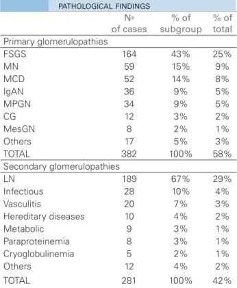
The frequency of different glomerular pathological findings is demonstrated in Table 2. Primary etiologies were more frequent (58%) with the predominance of FSGS (43%). Other causes of PG were MN (15%) and MCD (14%), followed by IgAN (9%) and MPGN (9%). LN represented 67% of secondary glomerular diseases with a predominance of class IV (29%), followed by class IV + V (22%) and V (21%).
Nº of cases Percentage %
Age
0-19 years 112 18%
20-39 years 312 50%
40-59 years 161 25%
> 60 years 45 7%
Unknown 33
Sex
Male 269 41%
Female 394 59%
Clinical syndrome (reason for renal biopsy)
UA 124 19%
NS 407 63%
NepS 83 13%
AKI 11 2%
CKD 18 3%
Unknown 20
Time between symptoms and biopsy
< 6 months 357 63%
6-12 months 78 14%
> 12 months 135 23%
Unknown 93
Proteinuria
< 1 g/day 40 7%
1-3.5g/day 182 30%
> 3.5g/day 375 63%
Unknown 66
Creatinine
≤ 1.5g/dl 415 68%
> 1.5g/dl 194 32%
Unknown 54
Hypertension
Yes 294 51%
No 280 49%
Unknown 89
TABLE 1 DISTRIBUTION OF CASES ACCORDING TO AGE,
SEX, INITIAL LABORATORY FINDINGS, CLINICAL SYNDROMES, TIME BEFORE PERFORMING RENAL BIOPSY AND HYPERTENSION
UA: urinary abnormalities; NS: nephrotic syndrome; NepS: Nephritic syndrome; AKI: acute kidney injury; CKD: chronic kidney disease.
Nº
of cases
% of subgroup
% of total
Primary glomerulopathies
FSGS 164 43% 25%
MN 59 15% 9%
MCD 52 14% 8%
IgAN 36 9% 5%
MPGN 34 9% 5%
CG 12 3% 2%
MesGN 8 2% 1%
Others 17 5% 3%
TOTAL 382 100% 58%
Secondary glomerulopathies
LN 189 67% 29%
Infectious 28 10% 4%
Vasculitis 20 7% 3%
Hereditary diseases 10 4% 2%
Metabolic 9 3% 1%
Paraproteinemia 8 3% 1%
Cryoglobulinemia 5 2% 1%
Others 12 4% 2%
TOTAL 281 100% 42%
FSGS: focal segmental glomerulosclerosis; MN: membranous nephropathy; MCD: minimal change disease; IgAN: IgA nephropathy; MPGN: membrane proliferative glomerulonephritis; CG: collapsing glomerulopathy; MesGN: non-IgA mesangial glomerulonephritis; LN: lupus nephritis.
TABLE 2 FREQUENCY OF DIFFERENT GLOMERULAR
PATHOLOGICAL FINDINGS
Vasculitis accounted for 7% of the secondary etiologies (15 patients with isolated renal vasculitis and 5 patients with ANCA-related vasculitis). Other secondary causes (4%) identified in this registry were: anti-glomerular basement membrane disease (2), cancer (1), methimazole-induced vasculitis (1), sickle cell anemia (1) and Crohn’s disease (1).
MPGN accounted for 6% of all biopsies. Of these, 81% were immune-complex-mediated, 12% were complement-mediated and 7% presented negative IF. Of the 35 patients with immune-complex-mediated MPGN, the etiologies of ten were identified: cryoglobulinemia (2), HIV (1), chronic lymphocytic leukemia (1) and
hepatosplenic schistosomiasis (6). CG represented 2% of all biopsies, wich two etiologies were identified (parvovirus and anabolic) and 12 were idiopathic.
The three main indications for renal biopsy were evaluated according to the histopathological findings (Figure 1). Among the patients biopsied for NS, the main histopathological findings were FSGS (33%), followed by LN (19%). For patients who were only evaluated if they presented with initial symptoms of NepS, the main diagnosis was LN (44%), followed by vasculitis (19%) and IgAN (14%). When the initial presentation of renal disease was UA, there was a predominance of LN (46%), followed by FSGS (14%) and IgAN (11%).
COMPARATIVE ANALYSIS OF PRIMARY AND SECONDARY GLOMERULOPATHIES
Table 3 presents evaluations of the epidemiological and clinical laboratory profiles of patients, according to the etiology of the disease. In the univariate analysis, there is a difference between the primary and secondary groups regarding sex, hypertension, duration of symptoms, hematuria, proteinuria and serum creatinine.
Factor evaluated Etiology p-value Primary Secondary
Age ± SD (years) * 35.0 ± 15.3 34.0 ± 14.1 0.709 2
Sex, N (%)
Male 198 (74%) 71 (26%)
< 0.001 1
Female 184 (47%) 210 (53%)
Hypertension, N (%)
Yes 167 (57%) 127 (43%)
0.273 1
No 172 (61%) 108 (39%)
Time with symptoms, N (%)
< 6 months 198 (55%) 159 (45%)
0.023 1
6 to 12 months 56 (73%) 21 (27%)
> 12 months 83 (61%) 53 (39%)
Hematuria, N (%)
Yes 190 (51%) 182 (49%)
< 0.001 1
No 144 (69%) 64 (31%)
Proteinuria (Q1-Q3), (g/24h) #
5.9 (3.5 - 9.0)
3.4
(2.0 - 6.0) < 0.001 2
Albumina (Q1-Q3), (g/dl) #
2.0 (1.5 - 3.0)
2.8
(2.1 - 3.4) < 0.001 2
Creatinina (Q1-Q3), (mg/dl) #
1.0 (0.7 - 1.6)
1.2
(2.0 - 6.0) 0.005
2
¹ p-value of the Chi-square test; 2 p-value of the Mann-Whitney test.
*mean ± standard variation; # median (Q1-Q3).
TABLE 3 DISTRIBUTION OF ETIOLOGIES ACCORDING
TO THE FACTORS OF PERSONAL, CLINICAL AND LABORATORY PROFILES OF THE EVALUATED PATIENTS
Factor
evaluated OR 95% CI p-value *
Sex
Male 1.000 -
-Female 1.722 1.353 - 2.191 < 0.001
Hematuria
Yes 1.337 1.070 - 1.670 0.011
No 1.000 -
-Proteinuria 0.944 0.916 - 0.972 < 0.001
Creatinine 1.079 1.009 - 1.153 0.027
OR: odds ratio; * p-value of the Wald Chi-square test.
TABLE 4 POISSON MODEL FOR THE SECONDARY
ETIOLOGY Figure 1. Clinical/pathological correlations observed in main primary
and secondary glomerular diseases. NS: nephrotic syndrome; NepS: nephritic syndrome; UA: urinary abnormalities; FSGS: focal segmental glomerulosclerosis; LN: lupic nephritis; MN: membranous nephropathy; MCD: minimal change disease; MPGN: membranoproliferative glomerulonephritis; Vasc: vasculitis; IgAN: IgA nephropathy; Infect: infectious; Hered: hereditaries.
secondary etiology with an OR = 1.7 (95% CI = 1.35 to 2.19), as well as the presence of hematuria OR = 1.3 (95% CI = 1.07 to 1.67). An increase in each 1g/day of proteinuria was related to a reduced chance of 6% of
being classified as SG (p < 0.001). An increase in creatinine by 1g/dl, increased the risk of a secondary classification of the etiology of glomerulopathy by around 8% (p = 0.027). Analyzing the etiology according to age groups after applying Pearson’s test, younger patients presented a significantly higher risk for PG (p = 0.001).
Comparing the reasons for renal biopsy (Figure 2), there was a predominance of NS for most patients. However, this presentation was more common in the PG group when compared to the SG (79% vs. 41%, p < 0.001). There was a predominant manifestation of UA and NepS in patients with SG (p < 0.001). Within this series, AKI and CKD were not common indications for renal biopsy, and for both presentations there was a predominance of SG.
Figure 2. Frequency of different forms of biopsy-proven primary and secondary glomerulopathies, according to the clinical syndromes.
DISCUSSION
The REPEG is the first registry to have involved the biopsies of patients with glomerular diseases from two referral centers in the state of Pernambuco. Similar to other studies, the registry demonstrates a predominance of primary glomerular diseases, with a prevalence ranging from 54% to 69%.2,11 However, our prevalence of SG
The higher prevalence of SG relative to other studies may have resulted from the diversity of classifications for these glomerulopathies, for example, the inclusion of hereditary changes and diabetes mellitus as secondary causes in this study. Mesquita et al.17 used definitions for
SG similar to this study, and encountered a prevalence of 57%, only considering renal biopsies with glomerular diseases. However, regional differences may also have influenced these results. Among the SG, LN represented 67% of biopsies, similar to several other samples.1,2,11,18
Amongst the PG, there was a predominance of FSGS, followed by MN, MCD, IgAN and MPGN. Several studies of biopsy registries, especially in Latin America, have demonstrated a predominance of FSGS, ranging from 25% to 35%.2,5,7,15,19 A high prevalence such as found
in REPEG (43%), although unusual, was also observed in Mexico, and represented 47% of the biopsies.14CG in this study accounted for 2% of all biopsies, while in other samples ranged from 0.3% to 1.8%.20,21
The most common indication for biopsy was NS, of which the main findings were FSGS and LN. Some studies presented a prevalence of FSGS and MCD among the causes of NS.5 Rivera et al.12
also encountered this prevalence in children under 15 years, although in adults, in accordance with Gesualdo et al.,10 MN was more prevalent.
In the present study, the prevalence of LN among NS was higher when compared to other studies, for which there was a prevalence ranging from 5 to 10% of NS.5,12,13 The Japanese registry demonstrates that
among the cases of NS there was a predominance of PG (particularly MCD, when IgAN was excluded), followed by diabetic nephropathy (9%).13 The discordant results
from these epidemiological studies may be a consequence of individual indications of renal biopsies by the services.
Among the biopsies performed for NepS and UA there was a predominance of LN (44% and 46%, respectively), which is unlike results encountered by other authors,5,10 who have reported IgAN as the
main glomerulopathy related to both presentations. In study by Rivera et al.,12 there was a predominance of
IgAN among the causes of UA in all age groups. In the Italian registry, there was predominance of IgAN in PG with NepS and UA, and immune-complex-mediated diseases among SG with the same presentations.10
In the REPEG, IgAN was the third leading cause of NepS and UA. A lower proportion of IgAN in Pernambuco may have arisen because routine renal biopsies were not performed in cases of isolated hematuria, that is with no systemic manifestations of disease and with no renal failure or proteinuria > 500 mg/day.
A comparative analysis of PG and SG, with regard to patients’ main clinical laboratory findings encountered a similar mean age among the groups. In the subanalysis by age, in accordance with a Brazilian study by Polito et al.,5 PG prevailed in all age groups with a significant
difference for patients aged 40 years and under.
The multivariate analysis of the present study investigated the association of clinical laboratory factors with primary or secondary etiologies and encountered a predominance of males among the PG and female among the SG. The predominance of women with SG is due to the high prevalence of immune-mediated glomerulonephritis, including LN, a pathology predominantly encountered in women. Polito et al.5 and
Gesualdo et al.10 obtained similar results, also due to the
high prevalence of LN among SG.
On the other hand, Ferraz et al.7 and Kutlugun
et al.22 found no gender differences among the etiologies.
However, in the latter study there was a high prevalence of AA amyloidosis (43%) as a secondary cause, which may have been responsible for this difference in the results. The REPEG revealed higher proteinuria in patients with PG and an inversely proportional relationship between the increase in proteinuria and the chance of a obtaining a diagnosis of SG.
An earlier study encountered no difference in the mean proteinuria between the etiologies.22 The
worsening of renal function and the presence of hematuria were also related to secondary etiology in the final model of this study. In fact, Kutlugun et al.22
demonstrated that renal dysfunction (creatinine > 1.5 mg/dl) was significantly more related to SG.
One advantage of this study is the comparative analysis of the clinical and laboratory manifestations of the primary and secondary etiologies of glomerulopathy. It also has the distinction of assess MPGN according to its new classification.23 This current categorization, based
on the findings of IF, allows a greater understanding of the pathophysiology of the disease, which thus facilitates the search for the etiologic mechanisms involved. Furthermore, it is the first registry to evaluate CG as a distinct entity from FSGS.
As in the Barisoni et al.,24 this would be a more
appropriate nomenclature, since CG presents more podocyte proliferation than depletion. However, although advantageous, this characteristic prevents any comparison of our data with other glomerulopathies registries that have not yet incorporated these classifications.
state of Pernambuco demonstrated that assessing race based solely on skin color can be extremely imprecise in our population.25 Some of the disadvantages of this
study are the fact that is a retrospective analysis and there was no EM in some biopsies.
CONCLUSION
This is the first registry of glomerulopathies in Northeastern Brazil. As well as providing important information on these conditions according to regional differences, it has also provided a comparative analysis of the major clinical laboratory changes of the affected patients. Since establishing the REPEG, the data gathered herein are extremely relevant for obtaining a greater understanding of these diseases within our environment, thus helping to provide better assistance to patients and also the ability to serve as a database for future studies.
ACKNOWLEDGMENTS
The authors would like to express their thanks to the renal pathologists: Marcello F. Franco; Luiz Antonio Moura; Luiz Antonio da Fonte; J. Charles Jennette; Suzana Moraes de Oliveira Melo; Roberto Neiva.
REFERENCES
1. Naumovic R, Pavlovic S, Stojkovic D, Basta-Jovanovic G, Ne-sic V. Renal biopsy registry from a single centre in Serbia: 20 years of experience. Nephrol Dial Transplant 2009;24:877-85. DOI: http://dx.doi.org/10.1093/ndt/gfn564
2. Malafronte P, Mastroianni-Kirsztajn G, Betônico GN, Romão JE Jr, Alves MA, Carvalho MF, et al. Paulista Registry of glo-merulonephritis: 5-year data report. Nephrol Dial Transplant 2006;21:3098-105. DOI: http://dx.doi.org/10.1093/ndt/gfl237 3. Rychlík I, Jancová E, Tesar V, Kolsky A, Lácha J, Stejskal J, et al.
The Czech registry of renal biopsies. Occurrence of renal diseases in the years 1994-2000. Nephrol Dial Transplant 2004;19:3040-9. DOI: http://dx.doi.org/10.1093/ndt/gfh521
4. Sociedade Brasileira de Nefrologia. Censo de diálise. 2014. [ci-ted 2016 Jan 6] Available from: http://www.censo-sbn.org.br/ censosAnteriores
5. Polito MG, de Moura LA, Kirsztajn GM. An overview on frequen-cy of renal biopsy diagnosis in Brazil: clinical and pathological pat-terns based on 9.617 native kidney biopsies. Nephrol Dial Trans-plant 2010;25:490-6. DOI: http://dx.doi.org/10.1093/ndt/gfp355 6. Crensiglova C, Rehme BB, Kinasz LR, Chula DC,
Nasci-mento MM, Soares MF. Frequency and clinical histological analysis of glomerular diseases in a tertiary hospital in sou-thern Brazil. J Bras Nefrol 2016;38:42-8. DOI: http://dx.doi. org/10.5935/0101-2800.20160007
7. Ferraz FH, Martins CG, Cavalcanti JC, Oliveira FL, Quirino RM, Chicon R, et al. Profile of glomerular diseases in a public hospital of Federal District, Brazil. J Bras Nefrol 2010;32:249-56. 8. Oliveira LB, Cobo Ede C, Machado JR, Custódio FB, da
Sil-va MV, de Oliveira FA, et al. Clinical and epidemiological prevalence of glomerulopathies elderly in the city of Ubera-ba - MG. J Bras Nefrol 2015;37:166-70. DOI: http://dx.doi. org/10.5935/0101-2800.20150027
9. Instituto Brasileiro de Geografia e Estatística. Estados [cited 2016 Jan 6]. Available from: http://www.ibge.gov.br/estadosat/ perfil.php?sigla=pe
10. Gesualdo L, Di Palma AM, Morrone LF, Strippoli GF, Sche-na FP; Italian Immunopathology Group, Italian Society of Nephrology. The Italian experience of the national registry of renal biopsies. Kidney Int 2004;66:890-4. PMID: 15327376 DOI: http://dx.doi.org/10.1111/j.1523-1755.2004.00831.x 11. Li LS, Liu ZH. Epidemiologic data of renal diseases from a
single unit in China: analysis based on 13.519 renal biopsies. Kidney Int 2004;66:920-3. PMID: 15327382
12. Rivera F, López-Gómez JM, Pérez-García R; Spanish Registry of Glomerulonephritis. Clinicopathologic correlations of renal pa-thology in Spain. Kidney Int 2004;66:898-904. PMID: 15327378 DOI: http://dx.doi.org/10.1111/j.1523-1755.2004.00833.x 13. Sugiyama H, Yokoyama H, Sato H, Saito T, Kohda Y, Nishi
S, et al.; Committee for Standardization of Renal Pathological Diagnosis; Committee for Kidney Disease Registry; Japanese Society of Nephrology. Japan Renal Biopsy Registry and Ja-pan Kidney Disease Registry: Committee Report for 2009 and 2010. Clin Exp Nephrol 2013;17:155-73. DOI: http://dx.doi. org/10.1007/s10157-012-0746-8
14. Chávez Valencia V, Orizaga de La Cruz C, Becerra Fuentes JG, Fuentes Ramírez F, Parra Michel R, Aragaki Y, et al. Epide-miology of glomerular disease in adults: a database review. Gac Med Mex 2014;150:403-8. PMID: 25275842
15. Mazzuchi N, Acosta N, Caorsi H, Schwedt E, Di Martino LA, Mautone M, et al.; Programa de Prevención y Tratamiento de las Glomerulopatías. Frequency of diagnosis and clinic presentation of glomerulopathies in Uruguay. Nefrologia 2005;25:113-20. 16. Zaza G, Bernich P, Lupo A; ‘Triveneto’ Register of Renal
Biop-sies (TVRRB). Incidence of primary glomerulonephritis in a large North-Eastern Italian area: a 13-year renal biopsy study. Nephrol Dial Transplant 2013;28:367-72. DOI: http://dx.doi. org/10.1093/ndt/gfs437
17. Mesquita M, Fosso C, Bakoto Sol E, Libertalis M, Corazza F, Vanden Houte K, et al. Renal biopsy findings in Belgium: a retrospective single center analysis. Acta Clin Belg 2011;66:104-9. PMID: 21630606 18. Swaminathan S, Leung N, Lager DJ, Melton LJ 3rd, Bergstralh
EJ, Rohlinger A, et al. Changing incidence of glomerular disea-se in Olmsted County, Minnesota: a 30-year renal biopsy stu-dy. Clin J Am Soc Nephrol 2006;1:483-7. DOI: http://dx.doi. org/10.2215/CJN.00710805
19. Arias LF, Henao J, Giraldo RD, Carvajal N, Rodelo J, Arbeláez M. Glomerular diseases in a Hispanic population: review of a re-gional renal biopsy database. Sao Paulo Med J 2009;127:140-4. DOI: http://dx.doi.org/10.1590/S1516-31802009000300006 20. Grcevska L, Polenakovik M. Collapsing glomerulopathy:
clini-cal characteristics and follow-up. Am J Kidney Dis 1999;33:652-7. DOI: http://dx.doi.org/10.1016/S0272-6386(99)70215-5 21. Ferreira AC, Carvalho D, Carvalho F, Galvão MJ, Nolasco F.
Collapsing glomerulopathy in Portugal: a review of the his-tological and clinical findings in HIV and non-HIV patients. Nephrol Dial Transplant 2011;26:2209-15. DOI: http://dx.doi. org/10.1093/ndt/gfq686
22. Kutlugun AA, Tokgoz B, Sipahioglu MH, Oymak O, Utas C. Comparison of the clinical and laboratory presentations of primary and secondary glomerular diseases. Ren Fail 2011;33:781-4. DOI: http://dx.doi.org/10.3109/088602 2X.2011.600495
23. Sethi S, Fervenza FC. Membranoproliferative glomerulonephri-tis: pathogenetic heterogeneity and proposal for a new classi-fication. Semin Nephrol 2011;31:341-8. DOI: http://dx.doi. org/10.1016/j.semnephrol.2011.06.005
24. Barisoni L, Schnaper HW, Kopp JB. A proposed taxonomy for the podocytopathies: a reassessment of the primary nephrotic diseases. Clin J Am Soc Nephrol 2007;2:529-42. DOI: http:// dx.doi.org/10.2215/CJN.04121206