. . . .
. . . .
CLINICAL RESEARCH
Rivaroxaban and dabigatran in patients undergoing
catheter ablation of atrial fibrillation
Rui Provideˆncia
1,2*
, Eloi Marijon
3, Jean-Paul Albenque
1, Ste´phane Combes
1,
Nicolas Combes
1, Franc¸ois Jourda
1, Hassiba Hireche
1, Joa˜o Morais
4, and Serge Boveda
11De´partement de Rythmologie, Clinique Pasteur, 45 avenue de Lombez, BP 27617, 31076 Toulouse Cedex 3, France;2Faculty of Medicine, University of Coimbra, 3004-504 Coimbra,
Portugal;3Paris Cardiovascular Research Center, 75015 Paris, France; and4Servic¸o de Cardiologia, Hospital de Santo Andre´, Centro Hospitalar Leiria-Pombal, 2410-197 Leiria, Portugal
Received 26 November 2013; accepted after revision 6 January 2014
Aims The recent availability of the novel oral anticoagulants (NOACs) may have led to a change in the anticoagulation regimens
of patients referred to catheter ablation of atrial fibrillation (AF). Preliminary data exist concerning dabigatran, but infor-mation regarding the safety and efficacy of rivaroxaban in this setting is currently scarce.
Methods and results
Of the 556 consecutive eligible patients (age 61.0+9.6; 74.6% men; 61.2% paroxysmal AF) undergoing AF catheter
ab-lation in our centre (October 2012 to September 2013) and enroled in a systematic standardized 30-day follow-up period: 192 patients were under vitamin K antagonists (VKAs), 188 under rivaroxaban, and 176 under dabigatran. Peri-procedural mortality and significant systemic or pulmonary thromboembolism (efficacy outcome), as well as bleed-ing events (safety outcome) durbleed-ing the 30 days followbleed-ing the ablation were evaluated accordbleed-ing to anticoagulation
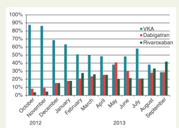
regimen. During a 12-month time interval, the use of the NOACs in this population rose from,10 to 70%. Overall,
the rate of events was low with no significant differences regarding: thrombo-embolic events in 1.3% (VKA 2.1%;
rivar-oxaban 1.1%; dabigatran 0.6%;P¼0.410); major bleeding in 2.3% (VKA 4.2%; rivaroxaban 1.6%; dabigatran 1.1%;
P¼0.112), and minor bleeding 1.4% (VKA 2.1%; rivaroxaban 1.6%; dabigatran 0.6%;P¼0.464). No fatal events were
observed.
Conclusion The use of the NOAC in patients undergoing catheter ablation of AF has rapidly evolved (seven-fold) over 1 year. These
preliminary data suggest that rivaroxaban and dabigatran in the setting of catheter ablation of AF are efficient and safe, compared with the traditional VKA.
-Keywords Atrial fibrillation † Rivaroxaban † Dabigatran † Vitamin K antagonists † Fluindione † Stroke † Cryoablation †
Thromboembolism † Bleeding † Arrhythmia
Introduction
Atrial fibrillation (AF) is the most frequent sustained arrhythmia in
clin-ical practice.1Catheter ablation of AF has been established as the most
effective therapy for the treatment of symptoms in these patients.2
However, this procedure is associated with a significant thrombo-embolic risk during and shortly after the procedure, requiring an
effective anticoagulation.3 Vitamin K antagonists (VKA) have been
traditionally used to prevent procedure-related thromboembolism.4
Recently, noval oral anticoagulants (NOACs) offering important advantages beyond their easiness of administration, like less interac-tions and no need of laboratory monitoring, have become available and appear as an attractive alternative in this setting. The impact of
the wide availability of these NOACs in preventive anticoagulant treatment of patients that are currently being referred to catheter ab-lation of AF is currently unknown.
Dabigatran (a direct thrombin inhibitor) has displayed reassuring safety and efficacy data, suggesting that it might be used as an
alterna-tive to VKA.5,6However, data are almost absent concerning
rivarox-aban, another NOAC with a different mechanism of action (a factor Xa inhibitor) which is being increasingly used worldwide.
Aim
We aimed to: (i) observe the change in the pattern of anticoagulant prescription in patients referred for catheter ablation of AF in our
*Corresponding author. Tel:+33 5 62 21 16 45; Fax:+33 5 62 21 16 41, Email: [email protected]
Published on behalf of the European Society of Cardiology. All rights reserved.&The Author 2014. For permissions please email: [email protected]. Europace
doi:10.1093/europace/euu007
by guest on February 19, 2014
http://europace.oxfordjournals.org/
centre since the introduction of the NOAC; (ii) assess the efficacy and safety of dabigatran and rivaroxaban in patients referred for cath-eter ablation of AF compared with VKA.
Methods
Prospective, non-randomized, single-centre, observational study evalu-ating the efficacy and safety of different anticoagulation regimens in the setting of catheter ablation of AF.
Study sample
All consecutive patients undergoing catheter ablation of AF from October 2012 to September 2013 in Clinique Pasteur, Toulouse, France, were assessed for possible inclusion in this investigation. All patients with paroxysmal and non-paroxysmal forms, undergoing first or redo procedures and planned for treatment with radiofrequency or cryoballoon ablation were considered eligible.
Participants needed to be on effective oral anticoagulation (VKA, dabi-gatran, or rivaroxaban) for at least 30 days before the procedure to be considered for inclusion. Patients treated only with antiplatelet agents or subcutaneous heparin before the procedure were excluded from the analysis. On the other hand, if patients had been treated with one of the NOACs and due to intolerance to the drug needed to be changed to a different oral anticoagulant (namely if the changing was already planned before the procedure), they were excluded from the analysis.
Patients treated with VKA before the procedure that were changed to dabigatran and rivaroxaban on the day of the procedure, and those treated with VKA, dabigatran, and rivaroxaban before the procedure, and remaining on the same drug after the procedure were considered eligible for analysis.
Information on baseline CHADS2, CHA2DS2-VASc, HAS-BLED, and
other clinical data was retrieved. Vascular disease was defined as having at least one of the following: myocardial infarction, peripheral artery disease, or complex aortic plaque.
Computed tomography scan (64-slice Siemens&dual source CT scan) was performed in 24 h prior to the procedure for assessing pulmonary vein and left atrial anatomy and to exclude the presence of thrombi in the left atrial appendage. Transthoracic echocardiography was also per-formed in all the patients for assessing left atrial dimensions, left ventricle ejection fraction, and the presence of valvular heart disease.
All patients provided an informed consent prior to the procedure. The study complied with the Declaration of Helsinki and the research proto-col was approved by the local ethics committee.
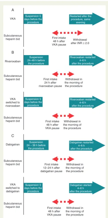
Peri-procedural anticoagulation
According to the type of peri-procedural anticoagulation, the study sam-ple was divided into three groups (Figure 1): Group A—treated with VKA before and after the procedure; Group B—rivaroxaban or VKA before and rivaroxaban started on the day of the procedure; Group C—dabigatran or VKA before and dabigatran started on the day of the procedure.
Vitamin K antagonists were stopped 5 days before the procedure in all the patients and subcutaneous heparin (either calcium heparin 10 000 IU twice a day (bid) or enoxaparin 1 mg/kg bid, or 0.5 mg/kg bid if chronic kidney disease was present) was started 48 h after.
Dabigatran was interrupted 24–36 h before the procedure. The inter-ruption in patients treated with rivaroxaban occurred 24–48 h before the
What’s new?
† We demonstrate that the use of the novel oral anticoagulants
in patients undergoing catheter ablation of atrial fibrillation has rapidly evolved (seven-fold) over 1 year.
† We provide the largest data to date concerning the safety and
efficacy of rivaroxaban in the setting of catheter ablation of atrial fibrillation.
† The incidence of bleeding and thrombo-embolic events was
low with all treatment regimens (rivaroxaban, dabigatran, and vitamin K antagonists), with no significant differences being found.
Suspension 5 days before the
procedure
Suspension 5 days before the
procedure
Suspension 5 days before the
procedure Suspension 24–48 h before
the procedure
Suspension 24 – 36 h before
the procedure
Restarted after the procedure, same
evening
Rivaroxaban restarted 4–6 h after the procedure
Rivaroxaban restarted 4–6 h after the procedure
Dabigatran restarted 4–6 h after the procedure
Dabigatran started 4–6 h after the procedure
First intake 48 h after VKA pause
Withdrawal after INR ≥ 2.0
First intake 24 h after rivaroxaban pause
First intake 12–24 h after dabigatran pause
Withdrawal in the morning of the procedure
First intake 48 h after VKA pause
First intake 48 h after VKA pause
Withdrawal in the morning of the procedure
Withdrawal in the morning of the procedure
Withdrawal in the morning of the procedure VKA Subcutaneous heparin bid Subcutaneous heparin bid Subcutaneous heparin bid Subcutaneous heparin bid Subcutaneous heparin bid Rivaroxaban VKA switched to rivaroxaban VKA switched to dabigatran Dabigatran A B C
Figure 1 Flowchart illustrating the different treatment regimens and timing of drug interruption and restart, as well as bridging heparin therapy.
by guest on February 19, 2014
http://europace.oxfordjournals.org/
ablation. In some patients, an earlier interruption was possible due to the presence of comorbidities (like chronic kidney disease) or physician prefer-ence due to other reasons. Subcutaneous heparin was started 24 h after the interruption of rivaroxaban. In patients treated with dabigatran, subcutane-ous heparin was started 12 h after suspension of the drug except for patients with estimated glomerular filtration rate below 45 mL/min, where subcuta-neous heparin was started only 24 h after dabigatran interruption.
Vitamin K antagonists were restarted in the evening of the procedure. Dabigatran and rivaroxaban were started 4–6 h after the procedure. No protamine was administered at the time of sheath removal. After the pro-cedure, subcutaneous heparin was continued only in patients treated with VKA and until international normalized ratio (INR) values were over 2.0.
Catheter ablation
All patients received general anaesthesia.
Venous punctures were performed: three for radiofrequency proce-dures (a 8Fr sheath and a 8Fr and 7Fr introducer) and two for cryoballoon ablation (a steerable 12Fr sheath (Flexcathw
, Medtronic&, and a 7Fr intro-ducer) and the multipolar irrigated radiofrequency catheter nMARQTM,
Biosense Webster&,) (8.5Fr sheath). By using a transfemoral venous ap-proach, a quadripolar catheter was placed in the coronary sinus. A single transseptal puncture was performed under fluoroscopic guidance (with use of transoesophageal echocardiography only if no success was obtained with fluoroscopy). Upon completion of the transseptal puncture, patients received intravenous heparin to maintain an activated clotting time of.300 s.
Patients underwent standard ablation procedures as directed by the operator. These procedures included pulmonary vein isolation with or without additional substrate modification. Ablation catheters used included cryothermic balloon catheter (Arctic Frontw, Arctic Front Advancew
, Medtronic, Inc.) and open-irrigated catheters: Celsius Ther-mocoolw, EZsteer Thermocoolw, SmartTouchw, nMARQw(Biosense Webster), Tacticathw
(St Jude Medical).
Thrombo-embolic and bleeding events
The following safety and efficacy endpoints were assessed, using the cri-teria proposed bySorgente et al.:7
(1) All-cause peri-procedural death.
(2) Thromboembolism—a composite of stroke, transient ischaemic attack (TIA), systemic or pulmonary embolism. A stroke was defined as a sudden focal neurological deficit of presumed cerebrovascular aeti-ology lasting for.24 h, not due to another identifiable cause and con-firmed by computed tomography or magnetic resonance imaging of the brain. If symptoms were short lasting (,24 h) and no evidence of ne-crosis was found on brain imaging, the event was considered to be a TIA. A systemic embolic event was defined as an abrupt vascular insuf-ficiency associated with clinical or radiological evidence of arterial oc-clusion in the absence of another likely mechanism (e.g. atherosclerosis, instrumentation, or trauma). A pulmonary embolism was diagnosed when dyspnoea or other suggestive clinical presentation was accom-panied by a radiological confirmation of a new pulmonary perfusion of intra-luminal defect, according to the requisites of the latest pulmon-ary embolism European Society of Cardiology guidelines.8
(3) Major bleeding—comprising cardiac tamponade, bleeding necessitat-ing intervention (either thrombin injection or surgery) or transfusion, massive haemoptysis, haemothorax, retroperitoneal bleeding, or any other life-threatening bleed leading to prolongation of hospitalization. (4) Minor bleeding—defined as puncture site bleeding, thigh ecchymosis or haematoma, pericardial effusion with no haemodynamic com-promise, minor gastrointestinal bleeding,epistaxis, or any bleeding treated conservatively with no need for transfusion, surgery, or pro-longed hospitalization.
We highlight that these criteria for defining major and minor bleeding are strongly based on theInternational Society on Thrombosis and Haemostasis,9 but adapted to the catheter ablation of AF setting.
Other complications were also reported when the investigators judged that these might possibly be associated with peri-procedural anticoagulation.
Follow-up was obtained for the first 30 days after the procedure for all the patients and was either obtained after an observation of each patient at our centre or by a report from the patient’s assistant cardiologist con-cerning that time period. Patients and their assistant cardiologists were instructed to report to the study centre immediately if any of these events occurred.
Statistical analysis
Comparisons were performed between the different peri-procedural antic-oagulation regimens.x2was used for the comparison of nominal variables.
One-way analysis of variance (ANOVA), or its non-parametric equivalent, Kruskal–Wallis when appropriate, was used for comparison of continuous variables; the Levene’s test was used to check the homogeneity of the vari-ance. Results withP,0.05 were regarded as significant.
When overall significant differences were found in the overall assess-ment of the three treatassess-ment groups, direct comparisons among the differ-ent groups were performed.Post hoctesting of ANOVA was performed by using the least significant difference test. When the Kruskal–Wallis test was used,post hocwas performed using the Mann–Whitney test. When com-paring two categorical variables inpost hoc, thex2test was used, unless the
observed value in any of the 2×2 contingency table cells was,5. In this case, the Fisher’s exact test was used. The Bonferroni correction was used for estimating the significantPvalue for multiple comparisons. PASW Sta-tistics (SPSS Inc.) version 18.0 was used for descriptive and inferential stat-istical analysis.
Results
Study sample and anticoagulation
Out of a total of 580 AF ablation procedures performed during the inclusion period, the following 24 were excluded from analysis: 6 due to lack of oral anticoagulation in the previous month (2 patients treated with aspirin, 2 with enoxaparin, and 2 with no thrombopro-phylaxis); two patients were changed from the NOAC (one rivarox-aban and one dabigatran) into warfarin at the time of ablation due to previously known drug intolerance; 14 patients with intolerance to dabigatran were changed to rivaroxaban and 2 patients with rivarox-aban intolerance changed the treatment to dabigatran.
Of the remaining 556 patients included in the analysis, 192 were included in Group A (treatment with VKA), 188 in Group B (treat-ment with rivaroxaban), and 176 in Group C (treat(treat-ment with dabiga-tran). Vitamin K antagonists were used before the procedure in 86 of the patients (45.7%) treated with rivaroxaban and 68 (38.6%) of
those treated with dabigatran (Figure2).
Fluindione was the most frequently used VKA (86% in Group A; 40% in Group B; 32% in Group C). Only a minority of patients were treated with other VKA. Warfarin was used in 20 patients in Group A (10.4%), 8 patients in Group B (4.3%), and 10 patients in Group C (5.7%). Acenocumarol was used in seven patients in Group A (3.6%), two patients in Group B (1.1%), and two patients in Group C (1.1%).
Rivaroxaban 15 mg id was used in three patients before and in seven patients after the procedure. The 20 mg id dosage was used
by guest on February 19, 2014
http://europace.oxfordjournals.org/
in all of the remaining patients. Dabigatran 110 mg bid was used in 22 patients before and 17 patients after the procedure. The 150 mg bid dosage was used in all of the remaining patients.
In a small percentage of patients, the NOACs were suspended
earlier (4 – 5 days before the procedure) than described inFigure1
(12 patients treated with dabigatran and 17 patients treated with
rivaroxaban). This occurred due to physician’s preference in the first months of utilization, in procedures that were anticipated to have higher complexity, and in patients with compromised renal func-tion. Despite the small number of patients in this situation, a similar efficacy and safety profile was found in this subset.
Some differences were found at baseline among the three different
treatment groups (Table1). Patients treated with VKA and
rivaroxa-ban only differed in terms of age (a 3-year difference, with VKA patients being older). Patients treated with rivaroxaban were similar at baseline when compared with those treated with dabiga-tran, except for a lower bleeding risk (HAS-BLED) in the dabigatran group. Differences at baseline were more pronounced in the VKA vs. dabigatran comparison: higher prevalence of persistent AF, older patients, higher body mass index, higher thrombo-embolic and bleeding risk, more hypertensive patients, and lower baseline haemoglobin in the VKA group.
No differences were found concerning the use of antiplatelet
agents before and after the procedure (Table 2). All patients
treated with VKA before the ablation performed bridging therapy with subcutaneous heparin (mainly calcium heparin). However, in
patients suspending rivaroxaban (n¼27) and dabigatran (n¼46)
24 h or less before the procedure, no subcutaneous heparin bridging was performed. The differences found in the concomitant use of sub-cutaneous heparin in the different groups are explained by the differ-ent half-life of the three agdiffer-ents and consequdiffer-ently the timing of interruption before the procedure.
100% 90% 80% 70% 60% 50% 40% 30% 20% 10% 0%
October
NovemberDecemberJan uar
y
Febr uar
y
March Apr il
May June July August
September
VKA Dabigatran Rivaroxaban
2012 2013
Figure 2 Evolution of the type of anticoagulants used at the arrival of our centre from October 2012 to September 2013 in patients admitted for AF ablation. VKA, vitamin K antagonists.
. . . .
Table 1 Baseline overall sample and characterization of each treatment group
Overall (n5556)
VKA (n5192)
Rivaroxaban (n5188)
Dabigatran (n5176)
Overall P
Subgroup comparisons
Paroxysmal AF 61.2% (340) 52.6% (101) 63.3% (119) 68.2% (120) 0.007 VKA vs. dabigatran Age 61.0+9.5 62.9+8.3 60.1+9.9 59.8+9.8 0.002 VKA vs. dabigatran, VKA vs.
rivaroxaban Female gender 25.4% (141) 26.0% (50) 26.1% (49) 23.9% (42) 0.859
Body mass index 27.6+4.4 28.3+4.6 27.3+4.2 27.2+4.5 0.031 VKA vs. dabigatran CHADS2 0.8+0.9 0.9+1.0 0.8+1.0 0.5+0.8 0.001 VKA vs. dabigatran
CHA2DS2-VASc 1.5+1.3 1.8+1.4 1.5+1.3 1.2+1.2 0.001 VKA vs. dabigatran
HAS-BLED 1.0+0.9 1.1+0.9 1.0+0.9 0.7+0.8 0.001 VKA vs. dabigatran, rivaroxaban vs. dabigatran Hypertension 39.6% (220) 49.0% (94) 39.4% (74) 30.1% (53) 0.001 VKA vs. dabigatran Diabetes mellitus 8.8% (49) 10.9% (21) 9.0% (17) 6.3% (11) 0.282
Previous stroke of TIA 9.4% (52) 10.4% (20) 11.2% (21) 6.3% (11) 0.225 Glomerular filtration rate
(Cockroft– Gault)
74.3+23.7 73.6+27.3 74.1+20.9 75.3+22.3 0.783
Baseline C-reactive protein (mg/L)
3.7+5.2 3.6+4.7 4.0+5.8 3.4+5.1 0.637
Baseline haemoglobin (g/dL) 14.7+1.3 14.6+1.3 14.7+1.2 15.0+1.2 0.029 VKA vs. dabigatran Indexed left atrial volume
(mL/m2)
45.7+16.5 47.6+17.1 45.3+18.1 44.1+13.9 0.172
Left ventricular ejection fraction (%)
62.4+8.7 61.6+10.2 61.9+8.3 63.9+7.0 0.125
VKA, vitamin K antagonists; AF, atrial fibrillation; TIA, transient ischaemic attack. Reference values for haemoglobin: 13.5 – 17.5 g/dL; C-reactive protein:,3.00 mg/L.
The observed differences were significant for the following subgroup comparisons: VKA vs. dabigatran; VKA vs. rivaroxaban; Rivaroxaban vs. dabigatran.
by guest on February 19, 2014
http://europace.oxfordjournals.org/
The length of the procedure was longer in the VKA group than in the NOACs. However, the time difference (when comparing proce-dures with dabigatran or rivaroxaban vs. VKA) was more pro-nounced in the dabigatran group (20 min) than in patients treated
with rivaroxaban (≏10 min).
Safety and efficacy of the different drug
regimens
A low overall rate of thrombo-embolic events and complications was
observed (Table3). No patients died during the follow-up period. No
significant differences in the rate of events were found among the three treatment groups.
An overall low incidence (0.9%) of stroke was reported. Four strokes were observed in the VKA group (2.1%) and another one was observed in a patient treated with rivaroxaban (0.5%) (see
Supplementary material online, Table S4). These either left no
sequels or led to minor disability at the end of the 30-day follow-up period. However, puncture complications filling major bleeding cri-teria were observed in all the groups, ranging from 1 to 3% of patients. A 1% incidence of pericardial effusion justifying pericardiocentesis or hospitalization/changes in therapy was observed in patients treated VKA and in one patient (0.5%) treated with rivaroxaban. A low inci-dence (1.4%) of minor bleeding (haematoma) was observed.
One patient treated with dabigatran developed an atrio-oesophageal fistula and one patient treated with VKA presented oesophageal ulcer-ation after cryoballoon ablulcer-ation (see Supplementary material online,
Table S4).
The use of cryoballoon ablation was not associated with a
signifi-cant increase in minor bleeding (2.4 vs. 1.0%;P¼0.243) or major
bleeding-related puncture complications (1.8 vs. 1.8%;P¼1.0), in
spite of the larger diameter sheaths required for this technique. Grouping dabigatran and rivaroxaban in one variable, a similar incidence of thrombo-embolic events (0.8% NOAC vs. 2.1% VKA;
P¼0.205) and minor bleeding (1.1% NOAC vs. 2.1% VKA;
P¼0.354) were observed when compared with VKA. Despite the
presence of a higher incidence of major bleeding events in the VKA group on univariate analysis (4.2% VKA vs. 1.4% NOAC;
P¼0.038), after adjusting for all baseline differences, VKA were
not independent predictors of major bleeding on multivariate analysis.
Discussion
We present reassuring preliminary data in support of the efficacy and safety of rivaroxaban in the setting of catheter ablation of AF. Further-more, the low complication rate from patients previously treated with VKA that were changed to the NOAC at the time of catheter ablation of AF seems to provide support to this practice.
Despite the slight differences in some baseline (mainly between the VKA and dabigatran group, impairing possible comparisons between these two agents, which was also not the aim of this work) and intra-procedural aspects, already highlighted in the previ-ous section, our data seem to suggest an overall low event rate with all treatment regimens. Furthermore, similar baseline characteristics allow a legitimate comparison of rivaroxaban with both VKA and dabigatran.
As shown in these data, patients are being referred more frequent-ly to catheter ablation centres under treatment with the NOAC.
. . . .
Table 2 Procedural and medication aspects in the overall sample and each treatment group
Overall (n5556)
VKA (n5192)
Rivaroxaban (n5188)
Dabigatran (n5176)
Overall P
Subgroup comparisons
Antiplatelet agent before the procedure
0.190
Aspirin 7.0% (39) 9.4% (18) 6.9% (13) 4.5% (8) Clopidogrel 0.9% (5) 0.5% (1) 1.6% (3) 0.6% (1) Aspirin+clopidogrel 0.4% (2) 1.0% (2) 0% 0% Antiplatelet agent after the
procedure
0.197
Aspirin 3.8% (21) 5.7% (11) 3.7% (7) 1.7% (3) Aspirin+clopidogrel 0.2% (1) 0.5% (1) 0% 0% Subcutaneous heparin
bridging
0.001 VKA vs. rivaroxaban, VKA vs. dabigatran, rivaroxaban vs. dabigatran
LMWH 15.1% (84) 6.3% (12) 27.7% (52) 11.4% (20) Calcium heparin 71.8% (399) 93.8% (180) 58.0% (109) 62.5% (110)
Cryoablation 29.5% (164) 33.3% (64) 31.9% (60) 22.7% (40) 0.056 Redo procedure 22.8% (127) 25.0% (48) 24.5% (46) 18.8% (33) 0.292
Procedure duration 134.8+47.3 145.3+54.5 132.4+39.1 125.4+44.3 0.001 VKA vs. rivaroxaban, VKA vs. dabigatran
Fluoroscopy duration 24.9+9.7 26.9+10.4 23.8+9.2 23.8+9.0 0.004 VKA vs. rivaroxaban, VKA vs. dabigatran
VKA, vitamin K antagonists; LMWH, low-molecular-weight heparin.
The observed differences were significant for the following subgroup comparisons: VKA vs. rivaroxaban; VKA vs. dabigatran; Rivaroxaban vs. dabigatran.
by guest on February 19, 2014
http://europace.oxfordjournals.org/
However, if a solid body of evidence has been gathered for dabigatran supporting its use as an alternative to VKA (almost 2000 patients in a
recently published meta-analysis5), data had been almost absent for
rivaroxaban until now. To the best of our knowledge, this is the largest series of patients managed with rivaroxaban in the setting of catheter ablation of AF.
Eiteland colleagues have provided data on 16 patients who under-went AF ablation and were treated with rivaroxaban 20 mg id. However, two of these patients had been treated with subcutaneous low-molecular-weight heparin and one patient had no
anticoagula-tion prior to the procedure.10
There is also information from a subanalysis of the ‘Rivaroxaban Once Daily Oral Direct Factor Xa Inhibition Compared with Vitamin K Antagonism for Prevention of Stroke and Embolism Trial in Atrial Fibrillation’ (ROCKET-AF) trial, assessing the out-comes after cardioversion and AF ablation. The incidence of stroke or systemic embolism after electrical cardioversion, pharma-cological cardioversion, and catheter ablation of AF was 0.93%. However, no specific information exists concerning the 79 patients who underwent 85 AF ablation procedures in the trial, peri-procedural drug regimen, and the event rate in each treatment
arm.11
Further data will certainly be provided by the still ongoing ‘A Study
Exploring Two Treatment Strategies in Patients With Atrial Fibrillation Who Undergo Catheter Ablation Therapy’ (VENTURE-AF trial), comparing rivaroxaban and warfarin, which estimates an enrolment of 200
patients.12
Our data show that some thrombo-embolic events occurred in
patients with low risk as illustrated by the CHADS2and CHA2DS2
-VASc scores (see Supplementary material online,Table S4), which
highlights the low acuity of these scores for predicting events in this setting and also suggests that these complications may be more strongly related to the procedure than to the overall baseline thrombo-embolic risk of each patient. Also, it is legitimate to think that the mechanism of thrombus formation after an ablation
procedure may be different to that happening naturally in AF and be more related to the extension of left atrial endothelial lesion/ denuded left atrial wall. Therefore, it is of utmost importance to assure therapeutic anticoagulation levels right after the procedure.
The overall rate of complications in this sample was low and similar
to what has been described in a series of.93 000 procedures from
2000 to 2010, that reported an intra-hospital rate of stroke or TIA of 1.02%, pericardial complications in 1.53%, and vascular complications
in 1.52%.13
Previously published data suggest that prolonged interruption of dabigatran in this setting may lead to an increase in thrombo-embolic
complications.14In our sample, the patient who presented a stroke
on treatment with rivaroxaban had suspended the drug 4 days before the procedure and despite being on subcutaneous calcium heparin, we wonder if the same principle may also apply to this drug. However, the low rate of events and the low number of patients with a long interruption of rivaroxaban in our sample do not allow the making of such inference.
The overall incidence of atrio-oesophageal fistula after catheter
ablation of AF is thought to be rare.15 Despite the presence of
reports of oesophagitis as a possible complication of therapy with dabigatran (out of the context of catheter ablation), due to the
pres-ence of tartrate as an excipient,16no strong conclusions can be drawn
towards a causal effect in the patient presented in our sample, the first reported case of a patient developing an atrio-oesophageal fistula while on treatment with this drug.
In this paper, we assessed the role of the NOACs when used before and after the procedure. However, since 38 – 45% of patients in the dabigatran and the rivaroxaban groups, respectively, were treated with VKA before the procedure and changed to the NOACs at the time of the procedure, more robust data are provided regarding the post-procedural period. Furthermore, despite the scarcity of data regarding the initiation of a direct thrombin inhibitor or a Factor Xa inhibitor after ablation, this has been proposed in the 2012 Heart Rhythm Society/European Heart Rhythm Association/
. . . .
Table 3 Events observed during the 30-day follow-up period
Overall (n5556) VKA (n5192) Rivaroxaban (n5188) Dabigatran (n5176) P
All-cause mortality 0% 0% 0% 0% N.A.
Thromboembolism 1.3% (7) 2.1% (4) 1.1% (2) 0.6% (1) 0.410 Stroke 0.9% (5) 2.1% (4) 0.5% (1) 0%
TIA 0.4% (2) 0% 0.5% (1) 0.6%(1)
Pulmonary embolism 0% 0% 0% 0%
Systemic embolism 0% 0% 0% 0%0
Major bleeding 2.3% (13) 4.2% (8) 1.6% (3) 1.1% (2) 0.112 Puncture complications 1.8% (10) 3.1% (6) 1.1% (2) 1.1% (2)
Pericardial effusion 0.5% (3) 1.0% (2) 0.5% (1) 0%
Minor bleeding 1.4% (8) 2.1% (4) 1.6% (3) 0.6% (1) 0.464 Haematoma 1.4% (8) 2.1% (4) 1.6% (3) 0.6% (1)
Other complications 0.4% (2) 0.5% (1) 0% 0.6% (1) 0.398 Atrio-oesophageal fistula 0.2% (1) 0% 0% 0.6% (1)
Oesophageal ulcer 0.2% (1) 0.5% (1) 0% 0%
VKA, vitamin K antagonist; TIA, transient ischaemic attack; N.A., not applicable.
by guest on February 19, 2014
http://europace.oxfordjournals.org/
European Cardiac Arrhythmia Society Expert Consensus Statement
on catheter and surgical ablation of AF.3
In our study, we have interrupted the NOACs 24–48 h before the procedure. However, there seems to be a growing trend for shorter interruption times. In some recent investigations, it has been
inter-rupted either the night before the procedure,17or even not interrupted
at all.18
Limitations
Although we report the fist large prospective evaluation of rivarox-aban in the setting of catheter AF ablation, we agree that this investi-gation has several limitations which need to be discussed. First, our observational and non-randomized single-centre data result from a small sample of patients, but despite this provide the larger body of evidence available until now.
Secondly, subcutaneous calcium heparin and fluindione were used in a relevant number of patients. These drugs are not common-ly used in other countries and no direct comparative studies exist in this context with the enoxaparin and warfarin, the drugs most widely used and with larger evidence. However, the European Society of
Cardiology 2012 update of the AF guidelines19 and the recent
Venice Consensus on AF ablation20refer to VKA as the
recom-mended treatment, not defining if warfarin or other drugs are used. Also, fluindione has been widely used in France for decades
(it constitutes 80% of VKA prescription21) and no data exist that
suggest a higher incidence of events while on this drug. Also, unlike warfarin, fluindione is not a racemic mixture and its longer
half-life is thought to help stabilize INR levels.22 For all these
reasons, this point should not be considered as a true limitation but rather a particularity.
Thirdly, unlike other centres where ablation is performed under uninterrupted VKA anticoagulation due to a possible reduction in
peri-procedural complications,23 our centre has been performing
these procedures using subcutaneous heparin bridging for over 10 years. However, in the aforementioned Consensus, there is no sug-gestion or recommendation for choosing uninterrupted VKA over
the VKA interruption.3,20Furthermore, our practice reflects a local
preference, among most French centres, of interrupted VKA, which is also preferred by our anaesthetists. A possible criticism is that we did not compare rivaroxaban with VKA at their best. However, patients treated with the NOAC also suspended treat-ment at the time of the procedure.
Fourthly, we have used heparin bridging after the interruption of the NOAC. Most available investigations with dabigatran did not use heparin bridging. Therefore, it is not yet known which should be the preferred option, as besides the lack of evidence or compara-tive trials, no recommendations exist regarding this point.
Fifthly, the baseline differences found in some of the three treat-ment groups may impair subgroup comparisons as previously men-tioned. However, if we look carefully to Supplementary material
online,Table S4, we can notice that not all seven patients sustaining
thrombo-embolic events had a high CHADS2score: four patients
had a CHADS score of 0, two patients a CHADS score of 1, and one patient a score of 3. Similarly, regarding the bleeding complica-tions: for example, in the three observed pericardial effusions, two had a HAS-BLED score of 0 and one had a score of 2.
Lastly, the overall low number of events did not allow assessing the association of the different anticoagulation regimens with the out-comes on multivariate analysis.
Conclusion
Since the introduction of the NOAC, patients are being referred for catheter ablation of AF under treatment with these agents with in-creasing frequency: from 10 to 70% over a 1-year period.
Our preliminary data suggest that NOAC in the setting of catheter ablation of AF are as efficient and safe, compared with the traditional VKA management.
However, despite being reassuring, these preliminary data need to be replicated in future randomized trials.
Supplementary material
Supplementary material is available atEuropaceonline.
Conflict of interest:R.P. was a co-investigator of the ENGAGE-AF
TIMI 48 and ATLAS ACS 2-TIMI 51 trials and is speaker and consult-ant for Boehringer Ingelheim; S.C. is investigator and J.-P.A., N.C., and S.B. are co-investigators of the XANTUS trial. J.M. was investigator of the ATLAS ACS 2-TIMI 5 trial and is speaker/consultant for Bayer Healthcare, Boheringher Ingelheim, and Daiichi Sankyo.
References
1. Wilke T, Groth A, Mueller S, Pfannkuche M, Verheyen F, Linder Ret al.Incidence and prevalence of atrial fibrillation: an analysis based on 8.3 million patients.Europace 2013;15:486 – 93.
2. Crawford T, Oral H. Current status and outcomes of catheter ablation for atrial fibrillation.Heart Rhythm2009;6(12 Suppl):S12 – 7.
3. Calkins H, Kuck KH, Cappato R, Brugada J, Camm AJ, Chen SAet al.2012 HRS/ EHRA/ECAS expert consensus statement on catheter and surgical ablation of atrial fibrillation: recommendations for patient selection, procedural techniques, patient management and follow-up, definitions, endpoints, and research trial design.Europace2012;14:528 – 606.
4. Hussein AA, Martin DO, Saliba W, Patel D, Karim S, Batal Oet al.Radiofrequency ablation of atrial fibrillation under therapeutic international normalized ratio: a safe and efficacious periprocedural anticoagulation strategy.Heart Rhythm2009;6: 1425 – 9.
5. Provideˆncia R, Albenque JP, Combes S, Bouzeman A, Casteigt B, Combes Net al. Safety and efficacy of dabigatran versus warfarin in patients undergoing catheter ablation of atrial fibrillation: a systematic review and meta-analysis.Heart2014; 100:324 – 35.
6. Hohnloser SH, Camm AJ. Safety and efficacy of dabigatran etexilate during catheter ablation of atrial fibrillation: a meta-analysis of the literature.Europace2013;15: 1407 – 11.
7. Sorgente A, Chierchia GB, de Asmundis C, Sarkozy A, Capulzini L, Brugada P. Com-plications of atrial fibrillation ablation: when prevention is better than cure.Europace 2011;13:1526 – 32.
8. Torbicki A, Perrier A, Konstantinides S, Agnelli G, Galie` N, Pruszczyk Pet al. Guide-lines on the diagnosis and management of acute pulmonary embolism: the Task Force for the Diagnosis and Management of Acute Pulmonary Embolism of the Euro-pean Society of Cardiology (ESC).Eur Heart J2008;29:2276 – 315.
9. Schulman S, Kearon C; Subcommittee on Control of Anticoagulation of the Scien-tific and Standardization Committee of the International Society on Thrombosis and Haemostasis. Definition of major bleeding in clinical investigations of antihemo-static medicinal products in non-surgical patients.J Thromb Haemost2005;3:692 – 4. 10. Eitel C, Koch J, Sommer P, John S, Kircher S, Bollmann Aet al.Novel oral anticoagu-lants in a real-world cohort of patients undergoing catheter ablation of atrial fibrilla-tion.Europace2013;15:1587 – 93.
11. Piccini JP, Stevens SR, Lokhnygina Y, Patel MR, Halperin JL, Singer DEet al.Outcomes after cardioversion and atrial fibrillation ablation in patients treated with rivaroxaban and warfarin in the ROCKET AF trial.J Am Coll Cardiol2013;61:1998 – 2006.
by guest on February 19, 2014
http://europace.oxfordjournals.org/
12. A study exploring two treatment strategies in patients with atrial fibrillation who undergo catheter ablation therapy (VENTURE-AF). http://clinicaltrials.gov/show/ NCT01729871.
13. Deshmukh A, Patel NJ, Pant S, Shah N, Chothani A, Mehta Ket al.Inhospital compli-cations associated with catheter ablation of atrial fibrillation in the United States between 2000 – 2010: analysis of 93,801 procedures.Circulation2013;128:2104 – 12. 14. Bin Abdulhak AA, Khan AR, Tleyjeh IM, Spertus JA, Sanders SU, Steigerwalt KEet al. Safety and efficacy of interrupted dabigatran for peri-procedural anticoagulation in catheter ablation of atrial fibrillation: a systematic review and meta-analysis.Europace 2013;15:1412 – 20.
15. Ghia KK, Chugh A, Good E, Pelosi F, Jongnarangsin K, Bogun Fet al.A nationwide survey on the prevalence of atrio-esophageal fistula after left atrial radiofrequency catheter ablation.J Interv Card Electrophysiol2009;24:33 – 6.
16. Ootani A, Hayashi Y, Miyagi Y. Dabigatran-induced esophagitis.Clin Gastroenterol Hepatol2013. pii: S1542-3565(13)01307-4. (EPUB ahead of print).
17. Kim JS, She F, Jongnarangsin K, Chugh A, Latchamsetty R, Ghanbari Het al.Dabigatran vs warfarin for radiofrequency catheter ablation of atrial fibrillation.Heart Rhythm 2013;10:483 – 9.
18. Maddox W, Kay GN, Yamada T, Osorio J, Doppalapudi H, Plumb VJet al.Dabigatran versus warfarin therapy for uninterrupted oral anticoagulation during atrial fibrilla-tion ablafibrilla-tion.J Cardiovasc Electrophysiol2013;24:861 – 5.
19. Camm AJ, Lip GY, De Caterina R, Savelieva I, Atar D, Hohnloser SHet al.2012 focused update of the ESC Guidelines for the management of atrial fibrillation: an update of the 2010 ESC Guidelines for the management of atrial fibrillation. Devel-oped with the special contribution of the European Heart Rhythm Association. Euro-pace2012;14:1385 – 413.
20. Raviele A, Natale A, Calkins H, Camm JA, Cappato R, Ann Chen Set al.Venice Chart International Consensus Document on atrial fibrillation ablation: 2011 update. J Cardiovasc Electrophysiol2012;23:890 – 923.
21. Ansell J, Hollowell J, Pengo V, Martinez-Brotons F, Caro J, Drouet L. Descriptive analysis of the process and quality of oral anticoagulation management in real-life practice in patients with chronic non-valvular atrial fibrillation: the International Study of Anticoagulation Management (ISAM).J Thromb Thrombolysis2007;23: 83 – 91.
22. Fihn SD, Gadisseur AA, Pasterkamp E, van der Meer FJ, Breukink-Engbers WG, Geven-Boere LMet al.Comparison of control and stability of oral anticoagulant therapy using acenocoumarol versus phenprocoumon.Thromb Haemost2003;90: 260 – 6.
23. Santangeli P, Di Biase L, Horton R, Burkhardt JD, Sanchez J, Al-Ahmad Aet al. Ablation of atrial fibrillation under therapeutic warfarin reduces periprocedural complications: evidence from a meta-analysis.Circ Arrhythm Electrophysiol2012;5: 302 – 11.
by guest on February 19, 2014
http://europace.oxfordjournals.org/