Original article
Cross-resistance to fluconazole induced by exposure to the
agricultural azole tetraconazole: an environmental resistance
school?
Marcos Fabio Gadelha Rocha,1,2L. P. Alencar,2M. A. N. Paiva,2Luciana Magalh~aes Melo,2 Silviane Praciano Bandeira,1Y. B. Ponte,2Jamille Alencar Sales,2G. M. M. Guedes,1 D. S. C. M. Castelo-Branco,1T. J .P. G. Bandeira,3R. A. Cordeiro,1W. A. Pereira-Neto,1
G. S. Brandine,1Jose Luciano Bezerra Moreira, 1Jose Julio Costa Sidrim 1and Raimunda S^amia Nogueira Brilhante1
1Department of Pathology and Legal Medicine, Postgraduate Program in Medical Microbiology, Specialized Medical Mycology Center, Federal University of
Ceara, Fortaleza, Cear a, Brazil,2School of Veterinary Medicine, Postgraduate Program in Veterinary Sciences, State University of Ceara, Fortaleza, Cear a, Brazil and3School of Medicine, Christus College
–Unichristus, Fortaleza, Ceara, Brazil
Summary
This study aimed to investigate the influence of tetraconazole and malathion, both used in agricultural activities, on resistance to fluconazole, itraconazole and voriconazole inCandida parapsilosisATCC 22019. The susceptibility to tetraconazole, malathion, fluconazole, itraconazole and voriconazole, through broth microdilution. Then, 12 independent replicates, were separated and exposed to four treatment groups, each one containing three replicates: G1: tetraconazole; G2: malathion; G3: fluconazole (positive control); G4: negative control. Replicates from G1, G2 and G3, were exposed to weekly increasing concentrations of tetraconazole, malathion and fluconazole, respectively, ranging from MIC/2 to 329 MIC, throughout 7 weeks.The exposure to tetraconazole, but not malathion, decreased susceptibility to clinical azoles, especially fluconazole. The tetraconazole-induced fluconazole resistance is par-tially mediated by the increased activity of ATP-dependent efflux pumps, considering the increase in antifungal susceptibility after the addition of the efflux pump inhibi-tor, promethazine, and the increase in rhodamine 6G efflux and CDR gene expres-sion in the G1 replicates. Moreover, MDR expression was only detected in G1 and G3 replicates, suggesting that MDR pumps are also involved in tetraconazole-induced fluconazole resistance. It is noteworthy that tetraconazole and fluconazole-treated replicates behaved similarly, therefore, resistance to azoles of clinical use may be a consequence of using azoles in farming activities.
Key words: Candidasp., azole resistance, environmental resistance, tetraconazole, efflux pumps,CDRgenes.
Introduction
The overuse and misuse of antibacterial drugs are most likely responsible for the emergence of antimicro-bial resistance in bacteria, especially in areas where resistant species are commonly isolated, such as hospi-tal waste,1untreated urban wastewater2and livestock production and aquaculture effluent.3,4 Similarly, the detection of antifungal resistance is often associated with patients receiving prolonged antifungal
Correspondence:R. S. N. Brilhante, Department of Pathology and Legal Medicine, Postgraduate Program in Medical Microbiology, Specialized Medical Mycology Center, Federal University of Ceara, Fortaleza, Ceara, Brazil.
Tel.: 55 85 3366-8319. Fax: 55 85 3366-8303. E-mail: brilhante@ufc.br
Submitted for publication10 September 2015 Revised20 November 2015
Accepted for publication9 December 2015
treatment. However, antifungal resistance also affects microbial populations from aquatic and terrestrial environments, commonly involving hosts with no previous history of antifungal exposure.4–7
Although fluconazole resistance is more frequently reported, resistance to other azoles, such as ketocona-zole, itraconazole and voriconaketocona-zole, also occurs.4–7
This phenomenon is phenotypically stable, and in human strains, is the result of the action of several mechanisms, alone or combined.8–10 The increased
expression of transmembrane transporters of the ATP-binding cassette family (ABC) is described as the main mechanism for azole resistance in Candida spp. ABC efflux pumps generally have low substrate specificity, and produce energy through ATP hydrolysis to remove a wide variety of hydrophobic compounds from the cell, including azoles.11–13
In addition to the ABC pumps, the increased expres-sion of the multidrug resistant (MDR) gene of the major facilitator superfamily, which encodes proton-depen-dent efflux pumps, is associated with specific fluconazole resistance.14This gene is controlled by the transcription factor Mrr1p (multidrug resistant regulator), and over-expressed in resistant clinical strains.11,14–16
In addition, resistance may also be mediated by the increased expression of ERG11 gene, which leads to higher concentrations of the target enzyme Erg11p,17– 19 hence, requiring greater amount of drug to inhibit
fungal growth.11,20Moreover, azole resistance may be mediated by mutations inERG11gene, decreasing the affinity between azoles and the enzyme Erg11p.17,18,20
Finally, azole resistance may also be associated with decreased activity of the enzyme D5,6-desaturase (Erg3p), encoded by ERG3 gene, leading to the decreased production of 14a -metilergosta-8,24(28)-dien-3b,6a-diol, a toxic metabolite, hence promoting yeast survivability.11,21
The reports of azole resistance among Candida
strains from animal and environmental sources with little or no contact with human hosts and no previous history of intentional antifungal exposure4,5,7,22,23 drew our attention, especially because this resistance occurs at higher rates, when compared to human strains. Based on the idea that resistance is established through the evolution of microorganisms in response to environmental stresses,24 it was hypothesised that these strains have developed azole resistance in response to the direct or indirect exposure to drugs used in agriculture. Thus, the antifungal susceptibility of one azole-susceptible standard Candida strain was assessed, after continuous exposure to compounds used in agriculture (one azole derivative and one
organophosphate). Hence, the aim of this study was to investigate the contribution of tetraconazole and malathion for the in vitro development of azole resis-tance, using C. parapsilosis ATCC 22019 as a model strain.
Materials and methods
Tested strain
The strain C. parapsilosis ATCC 22019, which is sus-ceptible to azoles of clinical use, was grown on potato dextrose agar for 48 h, from one single colony. The susceptibility of this strain to the antifungal drugs and the tested compounds was determined,25 prior to
beginning the experiments.
Antifungal susceptibility testing
The minimum inhibitory concentrations (MIC) of the antifungal drugs (fluconazole, itraconazole and voriconazole) and the tested agricultural compounds (tetraconazole and malathion) against one isolated col-ony of C. parapsilosis ATCC 22019, after growth on potato dextrose agar, were determined according to the document M27-A3, as recommended by the Clini-cal and Laboratory Standards Institute – CLSI.25 The
susceptibility was investigated through the broth microdilution assay, in 96-well microplates, with a final volume of 200lL of RPMI buffered at pH 7 with 0.165 mol l 1MOPS. The tested concentration ranges
were 0.125–512lg ml 1 for fluconazole (Zoltec, Pfi-zer, Brazil), 0.125–2048lg ml 1 for tetraconazole (Domark 100 EC; Isagro SpA, Segrate, Milano, Italy) and malathion (Malathion 500 EC; De Sangosse Agro-chemical, Curitiba, Parana, Brazil) and 0.015–16 for itraconazole (Janssen Pharmaceutica, Beerse, Antwerp, Belgium) and voriconazole (Pfizer, New York, New York, USA). The plates were read after 24 and 48 h of incubation at 35°C, but the MIC values considered were those obtained after 48 h of growth. MIC for azoles and malathion were the lowest drug concentra-tion capable of inhibiting 50% of growth, when com-pared to the drug-free growth control. C. kruseiATCC 6258 and C. parapsilosis ATCC 22019 grown on potato dextrose agar were included as controls.
Induction of resistance
malathion, fluconazole, itraconazole and voriconazole, respectively, against the strain C. parapsilosis ATCC 22019. It is important to highlight that the obtained clinical antifungal MICs were within the expected range for this control strain.25 Afterwards, this strain was subcultured into 12 tubes containing YEPD broth (0.5% yeast extract, 1% peptone and 2% dextrose). These tubes were, then, subjected to four different treatments, each one in three independent replicates: (i) tetraconazole (agricultural azole); (ii) malathion (agricultural organophosphate), (iii) fluconazole (posi-tive control) and (iv) drug-free YEPD broth (nega(posi-tive control). The initial concentration to which the repli-cates of each treatment group were exposed was equivalent to half the MIC value of each treatment compound, i.e. 4, 4 and 1lg ml 1 for tetraconazole, malathion and fluconazole respectively. The three replicates of each treatment group were exposed to the same drug concentration for 1 week, during which these replicates were subcultured to YEPD broth con-taining the drugs, at a 48-h interval. At the end of each week, fluconazole susceptibility of all three inde-pendent replicates of each treatment group was assessed. Then, the concentration of each treatment drug was doubled and a new cycle of three subcul-tures was started. These procedures were performed until fluconazole MICs stabilised, which occurred when the independent replicates were exposed to drug con-centrations 32 times higher than the initial MIC value of each tested drug. Additionally, at the end of the induction of resistance procedures, three independent replicates of each treatment group were stored at 4°C in 10% glycerol for 10 months. After this period, the independent replicates were subcultured in drug-free YEPD broth, during 2 weeks, and the MICs for the antifungal drugs (fluconazole, itraconazole and voriconazole) and the agricultural drugs (tetraconazole and malathion) were reassessed. It is important to highlight that for the weekly evaluation of fluconazole susceptibility, the three replicates from the negative control treatment and C. krusei ATCC 6258 and C.
parapsilosis ATCC 22019 grown on potato dextrose agar were included as controls. Moreover, all three independent replicates of each treatment group were tested in duplicate throughout this research.
Investigation of resistance mechanisms
Antifungal susceptibility with and without efflux pump inhibitor
Fluconazole, itraconazole and voriconazole MICs, with and without an efflux pump inhibitor, against the
three independent replicates of the groups exposed to tetraconazole and fluconazole, and the drug-free group (negative control) were determined according to the document M27-A3,25 after finishing the protocol of inducing resistance, in order to verify the contribution of these pumps to the resistant phenotype. For such, MIC values were determined in the presence of a subinhibitory concentration (i.e. 32lg ml 1) of promethazine, a CDR1p and CDR2p inhibitor.26 The obtained antifungal MICs before and after the addition of promethazine were observed. Malathion-treated group was not included in this analysis because this compound was not able to induce fluconazole resistance.
Efflux of rhodamine 6G
Since rhodamine 6G (R6G) acts as a substrate for CDR pumps,21,26this fluorescent substance is used as a tool for the evaluation of their efflux activity. Here, the test was performed according to the protocol described by Ivnitski-Steeleet al.[26] and Vale-Silvaet al.[21] with modifications. Briefly, each of the three independent replicates of the groups exposed to tetraconazole and fluconazole, and the drug-free group (negative control) were grown in triplicate (nine replicates for each treat-ment group), overnight in 5 ml of YEPD broth at 37°C for 24 h. Afterwards, the replicates were trans-ferred to 5 ml of fresh YEPD broth at 37°C, under constant agitation, allowing growth until reaching a concentration 29 107 cells ml 1. Cells were
cen-trifuged at 4500gfor 5 min, the supernatant was
dis-carded, and the pellet was washed twice in 2 ml of PBS (pH 7.0). Subsequently, cells were deprived of energy by incubation under constant agitation in 2 ml of PBS at 37°C, for 1 h. Then, R6G was added to obtain a final concentration of 15lmol l 1 and the suspension was incubated at 37°C, for 1 h, in the dark, under constant agitation, in order to allow the influx of R6G into the yeast cells.
After incubation, the cells were washed twice in 2 ml of PBS, at 4°C, and suspended in 300lL of PBS. Then, 12.5lL of the suspension were added to 32.5ll of PBS in a 96-well plate (Hard-Shellâ Low-Profile Thin-Wall) and left to settle for 5 min. Subsequently, glucose was added to reach a final con-centration of 1% and the relative fluorescence units (RFU) were recorded at 1 min intervals, during 60 min at 37°C.27 The fluorescent signal was mea-sured with a CFX96 TouchTM
absence of glucose. Negative controls were prepared with PBS instead of glucose. Replicates of the malathion-treated group were not included in the R6G assay because this compound did not induce flucona-zole resistance.
Ergosterol content
The amount of cellular sterols within the treated yeast cells was quantified, in order to detect phenotypical alterations in ergosterol biosynthesis that could lead to azole resistance. For such, the extraction of total ster-ols was carried out, as described by Cordeiro et al.
[28] The three independent replicates of each treat-ment group were grown overnight in YEPD broth at 37°C and centrifuged at 9660 g for 3 min. Pellets
were washed in PBS and the turbidity was adjusted to 0.5 on McFarland scale (i.e. approximately 1.0– 5.09 106 cells ml 1). Then, 1 ml of this suspension was centrifuged at 9,6609 g for 3 min and the pel-let was resuspended in 0.5 ml of KOH alcoholic solu-tion (0.7 mol l 1KOH in 60% ethanol) and incubated for 1 h at 95°C, in a water bath. After cooling, 0.6 ml of n-hexane was added to the tubes, which were vigorously vortexed for 5 min. Then, the tubes were centrifuged at 13,4169 g for 1 min and the
top organic layer was transferred to a new tube and mixed with 1 ml of n-hexane. The absorbance of this solution was read at 295 nm, in a spectrophotometer. The optical density data were compared to those obtained in a standard curve with ergosterol (Sigma-Aldrich, St. Louis, Missouri, USA). Readings were per-formed in duplicate for each of the three independent replicates of the treatment groups. The data obtained from replicates exposed to tetraconazole and flucona-zole were compared to the negative control. Malathion-treated replicates were not included in this analysis.
CDR, MDR and ERG11 gene expression
The expression of CDR andMDR (efflux pump genes) andERG11 (gene that encodes 14-alpha-demethylase, the target enzyme for azoles) was also assessed in trea-ted groups, since the overexpression of these genes is the most common mechanism of azole resistance. For such, total RNA was extracted, in triplicate, from each of the three independent replicates of the groups exposed to tetraconazole and fluconazole and the drug-free group (negative control), using the RNeasy mini kit (Qiagen, GmbH, Hilden, Germany) according to the manufacturer’s instructions. Briefly, the inde-pendent replicates were grown in YEPD broth at 37°C, for 24 h, until reaching the mid-log growth
phase. Then, 1 ml of each independent replicate, con-taining approximately 59107 cells was centrifuged at 4500 g and the pellet was lysed with RLT buffer
and 0.45–0.55 mm glass beads, using Precellys 24 disrupter (Bertin Technologies, Montigny le Breton-neux, Yvelines, France). The lysate was diluted 1 : 1 with 70% ethanol and transferred to a spin column. Genomic DNA was degraded using RNase-free DNase for 15 min at room temperature. After three washes, the RNA was eluted in RNase-free water and the con-centration was measured using Qubit fluorometer and RNA Assay Kit (Invitrogen, Paisley, UK). Reverse tran-scription was performed with 1lg of RNA combined with 1lL of Improm II (Promega, Madison, Wiscon-sin, USA), 0.5 mmol l 1 of each dNTP, 40 U of
RNaseOUT, 0.5lg of random primers and RNase-free water to a final volume of 20lL. Reverse transcrip-tion was performed at 42°C, for 60 min, followed by 70°C, for 15 min. The first strand cDNA products were stored at –80°C for later use as templates for quantitative real-time PCR (qPCR). Negative controls and RT blanks were prepared under the same condi-tions, but without inclusion of RNA or reverse tran-scriptase respectively.
Expression levels ofCDR,MDR andERG11 and ref-erence endogenousACT1and18Sgenes were assessed through qPCR. The reaction mix contained 0.5lL of cDNA, 1.5lL of primer (2 lmol l 1) and 7.5lL of 29 FastStart Universal SYBR Green Master (Roche,
Indianapolis, IN, USA), in a final volume of 15lL per reaction. The reactions were carried out in a CFX96 TouchTM
Real-Time PCR Detection System (Bio Rad) with the following amplification conditions: enzyme activation at 95°C for 5 min, followed by 40 cycles, each consisting of denaturation at 95°C for 30 sec, annealing at a specific temperature for each primer pair for 30 sec (Table S1) and extension at 72°C for 30 s.29 Fluorescence data were acquired at the end of each extension step. After the final extension, the specificity was ascertained by melting of the ampli-cons, detecting the fluorescence obtained at the annealing temperature of each primer, at an increase of 0.5°C every 5 s, until the temperature reached 95°C. To determine the linearity and reaction effi-ciency for each primer, the standard curve was plotted using serial dilutions of cDNA with qPCR conditions identical to those established by the experimental pro-tocol. Negative control and RT blank were used. The relative quantification of the gene expression was per-formed using the 2 DDCq method.30 Target gene expression was normalised against ACT1 and 18S
levels obtained for the target genes from the replicates of the treated groups (tetraconazole or fluconazole) were statistically higher than those obtained from the negative group. Threshold and Cq (threshold cycle) values were automatically determined by the BioRad CFX manager 3.0 software (BioRad), using default parameters. Replicates of the malathion-treated group were not included in this analysis.
Statistical analysis
The correlation between the exposure to each treat-ment group and fluconazole MICs was compared by using Pearson’s correlation coefficient (r2). The efflux pump activity of different groups was compared through the analysis of R6G efflux with and without glucose, using the Kruskal–Wallis test with Dunn’s multiple comparison test forpost hoccomparisons. The gene expression levels were compared using one-way analysis of variance (ANOVA), with Bonferroni’spost hoc
test for comparisons. In all tests,P <0.05 was consid-ered statistically significant.
Results
Induction of resistance
The three independent replicates of C. parapsilosis
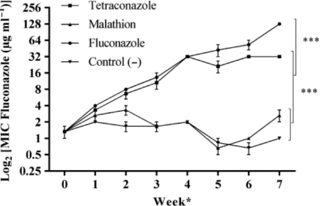
ATCC 22019 of each treatment group, exposed to increasing concentrations of tetraconazole, malathion and fluconazole (positive control) evolved to present higher tolerance to the compound they were exposed. At the end of the experiment, after storage in 10% glycerol, at 4°C, for 10 months, the replicates were grown in drug-free YEPD broth, for 2 weeks, and reas-sessed for their susceptibility to the compounds to which they were previously exposed. All tested com-pounds (tetraconazole, malathion and fluconazole) pre-sented increased MICs against the set of replicates exposed to the respective compound. Tetraconazole MICs increased from 4 to 8 times, malathion MICs increased from 128 to 512 times, and fluconazole MICs increased from 64 to 128.
In the tetraconazole-treated group, a positive corre-lation (r2= 0.97) between the exposure concentration of tetraconazole and fluconazole MICs was observed for all the three independent replicates. Resistance to this clinical azole was rapidly induced by exposure to increasing concentrations of the agricultural drug. In fact, fluconazole resistance started after exposure to tetraconazole concentrations of 29MIC (Fig. 1). Sim-ilar results were observed for the replicates that were
exposed to fluconazole (positive control). On the other hand, susceptibility to fluconazole did not change in the three replicates exposed to malathion or the nega-tive control replicates. Therefore, the malathion-trea-ted group was not included in the subsequent assays, since exposure to this compound did not interfere with fluconazole MICs.
Antifungal susceptibility testing with and without efflux pump inhibitor
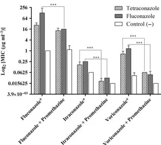
After the induction of resistance, the independent replicates of the groups exposed to tetraconazole and fluconazole, and the drug-free group (negative control) were evaluated for their susceptibility to antifungal agents used in medical practice. In addition to flucona-zole resistance, both itraconaflucona-zole and voriconaflucona-zole pre-sented increased MIC values against the three replicates exposed to tetraconazole, when compared to the negative control, and similar results were obtained for the positive control drug (fluconazole) (Fig. 2 and Table S2).
The antifungal susceptibility testing showed that efflux pump inhibition in the three tetraconazole repli-cates promoted 3-dilution and 4-dilution decrease (P <0.001), in MIC for itraconazole and voriconazole respectively (Fig. 2). However, even thoughCDRefflux
Figure 1 Dynamics of fluconazole susceptibility of independent replicates ofC.parapsilosisATCC 22019 exposed to increasing concentrations of tetraconazole (n=3), malathion (n=3) and fluconazole (n=3). Data are expressed as geometric mean
pump inhibition with promethazine caused a 1-dilu-tion decrease in fluconazole MICs against the three tetraconazole-treated replicates, the resulting MIC values were not statistically different from the flucona-zole MICs obtained without promethazine. This finding demonstrates that other mechanisms contribute to flu-conazole resistance in the three tetraflu-conazole-treated independent replicates. Similar results were also observed for the three fluconazole-treated replicates (positive control), against which the three tested azole drugs (fluconazole, itraconazole and voriconazole) also presented decreased (P <0.001) MIC values when promethazine was added (Fig. 2).
Efflux of rhodamine 6G
The three tetraconazole-independent replicates pre-sented increased efflux of R6G, when glucose was added to give the energetic support for the activity of
CDR efflux pumps. The efflux of R6G in these replicates was similar to that observed in the flucona-zole-treated replicates (positive control), demonstrating
the increased activity of the energy-dependent efflux pumps (Fig. 3).
Ergosterol content
The ergosterol concentrations (meanSE) extracted from the three independent replicates of the groups exposed to tetraconazole and fluconazole, and the drug-free group (negative control) were 4.130.67, 3.830.35 and 3.580.46lmol l 1 respectively. Thus, previous exposure to agricultural or clinical azole was not able to interfere with the ergosterol concentration of yeast cells.
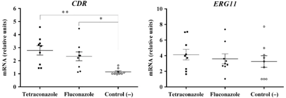
CDR,MDRandERG11gene expression
In the three tetraconazole-treated replicates, the expression of the CDR gene was increased, while the expression ofERG11 gene was not altered, when com-pared to the negative control. These results were simi-lar to those found for the three fluconazole-treated
Figure 2 Antifungal susceptibility of the three independent repli-cates of tetraconazole-treated and fluconazole-treated groups and negative control group, with and without efflux pump inhibitor, after inducing antifungal resistance. Data are expressed as geo-metric meanSD of each treatment group. Unexposed group: Control ( ). Data are compared between replicates of the same treatment group, with and without the addition of promethazine. *Indicates a significant increase (P<0.05) in MIC values of flu-conazole, itraconazole and voriflu-conazole, after exposure of repli-cates to increasing concentrations of tetraconazole (n=3) and fluconazole (n=3) compared to the unexposed group (n=3). ***P<0.001. In theY-axis, antifungal concentration ranges from 0.015lg ml 1[log2(0.015)
= 6] to 256lg ml 1 [log2(256)=8].
replicates (Fig. 4). TheMDR1 gene reached detectable levels only in the replicates exposed to tetraconazole and fluconazole, but not in those submitted to the neg-ative control. Thus, MDR1 expression level was not calculated. It is important to highlight that neither pri-mer-dimers nor unspecific products were detected in qPCR amplifications.
Discussion
Candida parapsilosis ATCC 22019 was chosen due to its phenotypical stability, as it is a quality control strain for antifungal susceptibility testing, according to the recommendations of CLSI, which were followed during the present research.25 In addition, this
partic-ular strain was originally recovered from a human case of sprue and the species C. parapsilosis is com-monly isolated from the microbiota of humans and other animals and from terrestrial and aquatic envi-ronmental sources, often presenting reduced azole sus-ceptibility.4,5,7,31 These characteristics supported the choice of this strain as a model for inducing antifungal resistance through the chronic exposure to compounds used in agricultural practices.
The effect of thein vitroexposure of an azole-suscep-tibleCandida strain to the azole tetraconazole and the organophosphate malathion, both widely used in farming activities, and its consequence on the suscepti-bility of microorganisms to azoles of clinical use were evaluated. The obtained results indicate that exposure ofC. parapsilosis to tetraconazole decreases susceptibil-ity to fluconazole, itraconazole and voriconazole, simi-lar to what was seen in the positive control group exposed to fluconazole. However, exposure to malathion did not change the susceptibility to the tested antifungal drugs; hence, the three
malathion-treated independent replicates were not included in the analyses to investigate the resistance mechanisms.
The stress caused by tetraconazole exposure, as demonstrated in this study, most likely promotes cellu-lar mechanisms to escape from the effects of this agri-cultural antifungal. This allows the development of important characteristics that also promote fungal pro-tection against antifungal agents with similar mecha-nisms of action. The induction of azole resistance in
Candida species has previously been observed due to
in vitro18,32–34 and in vivo35 continuous exposure to
azoles of medical use. In some researches, the MIC val-ues of several azole drugs were simultaneously increased and not only the MIC of the drug used in the induction assay.33,34 Similarly, in this study, the
MIC values of fluconazole, itraconazole and voricona-zole were significantly increased, after the continuous
in vitro exposure of three C. parapsilosis ATCC 22019 independent replicates to increasing concentrations of tetraconazole. These results are equivalent to those obtained for the positive control drug (fluconazole), demonstrating that exposure to clinical azole,18,32–35
as well as to agricultural drugs, may be responsible for the development of cross-resistance to several azoles in strains ofCandidaspp. and other fungal species.36–38
There are several reports of resistance in fungal spe-cies, which involves common mechanisms, regardless of the triggering factor, i.e. azoles used in human and animal medical practice, or in agricultural practice. These indications associated with the recovery of resis-tant fungal strains from the microbiota of animals without prior history of antifungal treatment4,5,7 rein-force the assumption of the existence of a selective pressure for the development of antifungal-resistant strains within the rural and/or wild environment. This environmental adaptation is crucial for the
development of commensal and pathogenic character-istics of Candida spp.39 In this sense, Hube [39] pro-posed the existence of ‘commensal virulence schools’ in which the microorganism develops certain charac-teristics to adapt or successfully infect the host. Simi-larly, the stress caused by the exposure to azoles in agriculture36 (e.g. tetraconazole) could be an ‘environ-mental resistance school’, promoting the development of important features in microorganisms to enhance their survivability in the environment. Finally, the development of these features seems to induce cross-resistance to medical azoles.
The knowledge on azole resistance in C. parapsilosis
is still incipient. Even though this phenomenon proba-bly results from a combination of classical mechanisms studied in otherCandidaspecies,14,35,40,41the high MIC values of azoles againstC. parapsilosismay also be asso-ciated with other molecular mechanisms that still need to be elucidated.40 However, it has been show that in fluconazole-resistantC. parapsilosis,CDRoverexpression is more common than the overexpression of MDR1, and both are more common than overexpression of
ERG11.14,35,40,41 Thus, in this study, we primarily decided to investigate the drug-induced efflux-mediated mechanisms of resistance inC.parapsilosis. In this con-text, it was observed that tetraconazole exposure increased the activity of efflux pumps inC.parapsilosis, suggesting that this induced resistance is mainly asso-ciated with increased drug efflux through ATP-depen-dent pumps. These results are similar to those obtained for the fluconazole-treated (positive control) replicates in this study, and in others.18,32–34,40,41One of the
evi-dences for the involvement of ATP-dependent pumps is the reduction of the antifungal MICs, especially those of itraconazole and voriconazole, against the azole-resistant tetraconazole-treated replicates, when these efflux pumps are inhibited by promethazine.42 Other evidence for the involvement of these pumps in the development of antifungal resistance after exposure to tetraconazole is the increase in R6G efflux, which pre-sented clonal difference between the independent repli-cates, based on the observed efflux outliers and the relative stability in efflux activity among the replicates from the negative control group. These findings demonstrated that the CDR efflux pumps, among other ATP-dependent pumps, are primarily involved in the development of azole resistance. Moreover, this obser-vation was confirmed by the increased CDR gene expression in tetraconazole-treated replicates, similar to what was seen for the fluconazole-treated replicates.
The inhibition of the CDR efflux pumps reduced the azole MICs against the tetraconazole and
fluconazole-treated replicates, but these MICs were still higher than those obtained against the negative control group. Moreover, CDR efflux pump inhibition did not cause significant reduction in fluconazole MICs against tetraconazole-treated replicates, which suggests the involvement of MDR proton pumps in this azole-induced resistance.14,43 Additionally, the detection of
MDR1 transcripts only in tetraconazole and flucona-zole-treated replicates, but not in negative control, strengthens the assumption that these proton pumps are also involved in the development of tetraconazole-induced fluconazole resistance,43 similar to what has been described for clinical strains.14,35,40 The mainte-nance of the amount of ergosterol extracted from the tetraconazole and fluconazole-treated C. parapsilosis
ATCC 22019 replicates, when compared to the negative control, and the undifferentiated ERG11
expression between treated replicates and the negative control, showed that, in this study, increased ergosterol biosynthesis was not involved with the development of resistance.43 However, mutation in genes involved in ergosterol biosynthesis cannot be ruled out, especially, considering the possibility that more than one resistance mechanism may be acting at the same time. Even though we did not perform a sequence analysis of the genes involved in the biosyn-thesis of ergosterol, it is known that the single-nucleo-tide polymorphism (SNP) Y132F in ERG11 nucleotide sequence is the most common alteration in strains of fluconazole-resistantC. parapsilosis.14,40
The investigation of the possible resistance mecha-nisms demonstrated that the tetraconazole-treated replicates presented similar results to those treated with fluconazole. Thus, we have demonstrated that resistance to medical azoles may be a consequence of the exposure to agricultural azoles. In C. parapsilosis
ATCC 22019, for instance, this phenomenon seems to be mainly associated with the increased activity of
CDR efflux pumps, as a response to the continuous exposure to a drug-rich environment. These adaptive changes decrease azole susceptibility through unspeci-fic pathways and seem to be phenotypically stable, after the storage of the strains at 4°C for 10 months in glycerol. Thus, we believe that the misuse or pro-longed use of azoles in humans8,9,16 and also in the
environment may be the cause of resistance to these drugs in strains from diverse origins. The selection of
addition, considering that these agricultural drugs have long residual effect,44 their direct action on the yeast microbiota of humans and animals that feed on products with drug residues may also be speculated. Either or both mechanisms may be involved in the recovery of azole resistantCandidastrains from several animal species, such as prawns, porcupine, raptors, rheas and tortoises, as reported in previous works of our group.4,5,7,45,46
The occurrence of azole resistance in strains from veterinary and environmental sources suggests that these strains can acquire such features in the environ-ment,36,38,47,48 as a response to stress.24 Thus, in allusion to what was described by Hube [39] who defended the existence of ‘commensal virulence schools’, the presence of agricultural azoles in the environment would function as an ‘environmental resistance school’, and, in this case, tetraconazole would act as an intensive school, since it acts on envi-ronmental microbial communities for periods longer than 90 days.44 Thus, these data bring perspectives for further studies on the impact of drugs and other chemical compounds used in agriculture on microbial populations and on the induction of azole resistance in the environment.
Acknowledgments
This work was supported by grants from the National Council for Scientific and Technological Development (CNPq; Brazil; 307606/2013-9; 443167/2014-1; PNPD-UFC/Microbiologia Medica/CAPES).
References
1 Varela AR, Ferro G, Vredenburg Jet al.Vancomycin resistant ente-rococci: from the hospital effluent to the urban wastewater treat-ment plant.Sci Total Environ2013;450–451: 155–61.
2 Birosova L, Mackul’ak T, Bod ık Iet al.Pilot study of seasonal occur-rence and distribution of antibiotics and drug resistant bacteria in wastewater treatment plants in Slovakia.Sci Total Environ2014; 490: 440–4.
3 Reboucßas RH, de Sousa OV, Lima ASet al.Antimicrobial resistance profile ofVibriospecies isolated from marine shrimp farming envi-ronments (Litopenaeus vannamei) at Ceara, Brazil.Environ Res2011; 111: 21–24.
4 Brilhante RSN, Paiva MAN, Sampaio CMSet al.Yeasts from Macro-brachium amazonicum: a focus on antifungal susceptibility and viru-lence factors ofCandidaspp.FEMS Microbiol Ecol2011;76: 268–77.
5 Castelo-Branco DSCM, Brilhante RSN, Paiva MANet al. Azole-resistantCandida albicansfrom a wild Brazilian porcupine (Coendou prehensilis): a sign of an environmental imbalance?Med Mycol2013; 51: 555–60.
6 Cordeiro RA, Teixeira CEC, Brilhante RSNet al.Minimum inhibitory concentrations of amphotericin B, azoles and caspofungin against Candidaspecies are reduced by farnesol.Med Mycol2013;51: 53–9.
7 Brilhante RSN, Alencar LP, Cordeiro RAet al.Detection of Candida species resistant to azoles in the microbiota of rheas (Rhea ameri-cana): possible implications for human and animal health.J Med Microbiol2013;62: 889–95.
8 White TC, Holleman S, Dy Fet al.Resistance mechanisms in clinical isolates of Candida albicans.Antimicrob Agents Chemother2002;46: 1704–13.
9 Lopez-Ribot JL, McAtee RK, Lee LNet al.Distinct patterns of gene expression associated with development of fluconazole resistance in serial candida albicans isolates from human immunodeficiency virus-infected patients with oropharyngeal candidiasis.Antimicrob Agents Chemother1998;42: 2932–7.
10 Cannon RD, Lamping E, Holmes ARet al.Efflux-mediated antifungal drug resistance.Clin Microbiol Rev2009;22: 291–321.
11 Akins RA. An update on antifungal targets and mechanisms of resis-tance in Candida albicans.Med Mycol2005;43: 285–318.
12 Tsao S, Rahkhoodaee F, Raymond M. Relative contributions of the Candida albicansABC transporters Cdr1p and Cdr2p to clinical azole resistance.Antimicrob Agents Chemother2009;53: 1344–52.
13 Coste AT, Turner V, Ischer Fet al.A mutation in Tac1p, a transcrip-tion factor regulating CDR1 and CDR2, is coupled with loss of heterozygosity at chromosome 5 to mediate antifungal resistance in Candida albicans.Genetics2006;172: 2139–56.
14 Grossman NT, Pham CD, Cleveland AAet al.Molecular mechanisms of fluconazole resistance in Candida parapsilosis Isolates from a U.S. Surveillance System.Antimicrob Agents Chemother2015;59: 1030–7.
15 Lohberger A, Coste A, Sanglard D. Distinct roles ofCandida albicans drug resistance transcription factorsTAC1,MRR1, andUPC2in vir-ulence.Eukaryot Cell2014;13: 127–42.
16 Morschh€auser J, Barker KS, Liu TTet al.The transcription factor Mrr1p controls expression of theMDR1efflux pump and mediates multidrug resistance inCandida albicans.PLoS Pathog2007;3: 1603–16.
17 Xie JL, Polvi EJ, Shekhar-Guturja Tet al.Elucidating drug resistance in human fungal pathogens.Future Microbiol2014;9: 523–42.
18 Marr KA, Lyons CN, Ha Ket al.Inducible azole resistance associated with a heterogeneous phenotype in Candida albicans.Antimicrob Agents Chemother2001;45: 52–9.
19 Flowers SA, Barker KS, Berkow ELet al.Gain-of-function mutations in UPC2 are a frequent cause of ERG11 upregulation in azole-resis-tant clinical isolates of Candida albicans.Eukaryot Cell2012;11: 1289–99.
20 Xiang M-J, Liu J-Y, Ni P-Het al. Erg11mutations associated with azole resistance in clinical isolates ofCandida albicans.FEMS Yeast Res2013;13: 386–93.
21 Vale-Silva LA, Coste AT, Ischer Fet al.Azole resistance by loss of function of the sterol∆5,6- desaturase gene (ERG3) in Candida albi-cans does not necessarily decrease virulence.Antimicrob Agents Che-mother2012;56: 1960–8.
22 Jin Y, Lin D. Fungal urinary tract infections in the dog and cat: a retrospective study (2001-2004).J Am Anim Hosp Assoc2005;41: 373–81.
23 Nawange SR, Singh K, Naidu Jet al.Naturally acquired systemic dual infection caused byCandida famata(Debaryomyces hansenii) and Candida catenulatain albino rats bred for sale in the market at Jabal-pur (Madhya Pradesh), India.Mycoses2010;53: 173–5.
24 Cannon RD, Lamping E, Holmes ARet al. Candida albicansdrug resis-tance - Another way to cope with stress.Microbiology2007;153: 3211–7.
25 CLSI.Reference Method for Broth Dilution Antifungal Susceptibility Testing of Yeasts. Approved Standard M27-A3, 3rd edn. Wayne, PA, 2008. 26 Ivnitski-Steele I, Holmes AR, Lamping Eet al.Identification of Nile red as a fluorescent substrate of the Candida albicans ATP-binding cassette transporters Cdr1p and Cdr2p and the major facilitator superfamily transporter Mdr1p.Anal Biochem2009;394: 87–91.
28 Cordeiro RA, Marques FJF, Cordeiro RAet al.Synthesis and antifun-gal activityin vitroof isoniazid derivatives againstHistoplasma capsu-latumvar.capsulatum.Antimicrob Agents Chemother2014;58: 2504–
11.
29 Pulcrano G, Panellis D, De Domenico Get al.Ambroxol influences voriconazole resistance ofCandida parapsilosisbiofilm.FEMS Yeast Res2012;12: 430–8.
30 Livak KJ, Schmittgen TD. Analysis of relative gene expression data using real-time quantitative PCR and the 2(-Delta Delta C(T)) method.Methods2001;25: 402–8.
31 Brilhante RSN, Paiva MAN, Sampaio CMSet al.Surveillance of azole resistance among Candida spp. as a strategy for the indirect monitor-ing of freshwater environments.Water Air Soil Pollut2015;226: 52. 32 Silva CR, Andrade Neto JB, Sidrim JJCet al.Synergistic effects of
amiodarone and fluconazole on Candida tropicalis resistant to flu-conazole.Antimicrob Agents Chemother2013;57: 1691–700.
33 Barchiesi F, Calabrese D, Falconi Let al.Experimental induction of fluconazole resistance inCandida tropicalisATCC 750.Antimicrob Agents Chemother2000;44: 1578–84.
34 Pinto e Silva AT, Costa-De-Oliveira S, Silva-Dias Aet al.Dynamics of in vitro acquisition of resistance by Candida parapsilosis to different azoles.FEMS Yeast Res2009;9: 626–33.
35 Zhang L, Xiao M, Watts MRet al.Development of fluconazole resis-tance in a series ofCandida parapsilosisisolates from a persistent can-didemia patient with prolonged antifungal therapy.BMC Infect Dis 2015;15: 340.
36 Muller F-MC, Staudigel A, Salvenmoser S€ et al.Cross-resistance to medical and agricultural azole drugs in yeasts from the oropharynx of human immunodeficiency virus patients and from environmental Bavarian vine grapes.Antimicrob Agents Chemother2007;51: 3014–6.
37 Verweij PE, Snelders E, Kema GHet al.Azole resistance inAspergillus fumigatus: a side-effect of environmental fungicide use?Lancet Infect Dis2009;9: 789–95.
38 Faria-Ramos I, Tavares PR, Farinha Set al.Environmental azole fungicide, prochloraz, can induce cross-resistance to medical triazoles inCandida glabrata.FEMS Yeast Res2014;14: 1119–23.
39 Hube B. Fungal adaptation to the host environment.Curr Opin Microbiol2009;12: 347–9.
40 Berkow EL, Manigaba K, Parker JEet al.Multidrug transporters and alterations in sterol biosynthesis contribute to azole antifungal resis-tance in Candida parapsilosis.Antimicrob Agents Chemother2015; 59: 5942–50.
41 Souza ACR, Fuchs BB, Pinhati HMSet al.Candida parapsilosis resis-tance to fluconazole: molecular mechanisms andin vivoimpact in
infectedGalleria mellonellalarvae.Antimicrob Agents Chemother2015; 59: 6581–7.
42 Kolaczkowski M, Kolaczkowska A, Motohashi Net al.New high-throughput screening assay to reveal similarities and differences in inhibitory sensitivities of multidrug ATP-binding cassette transporters.Antimicrob Agents Chemother2009;53: 1516–27.
43 Silva AP, Miranda IM, Guida Aet al.Transcriptional profiling of azole-resistant Candida parapsilosis strains.Antimicrob Agents Che-mother2011;55: 3546–56.
44 Zhang W, Xu J, Dong Fet al.Effect of tetraconazole application on the soil microbial community.Environ Sci Pollut Res2014;21: 8323–32.
45 Brilhante RSN, Castelo Branco DSCM, Duarte GPSet al.Yeast micro-biota of raptors: a possible tool for environmental monitoring. Envi-ron Microbiol Rep2012;4: 189–93.
46 Brilhante RSN, de Arag~ao Rodrigues PH, de Alencar LPet al. Evi-dence of fluconazole-resistant Candida species in tortoises and sea turtles.Mycopathologia2015;180: 421–6.
47 Chowdhary A, Kathuria S, Xu Jet al.Emergence of azole-resistant Aspergillus fumigatusstrains due to agricultural azole use creates an increasing threat to human health.PLoS Pathog2013;9: 3–7.
48 Faria-Ramos I, Farinha S, Neves-Maia Jet al.Development of cross-resistance byAspergillus fumigatusto clinical azoles following expo-sure to prochloraz, an agricultural azole.BMC Microbiol2014;14: 155.
Supporting information
Additional supporting information may be found in the online version of this article.
Table S1. Primers used for gene expression analysis
of Candida parapsilosis ATCC 22019 subjected to resis-tance induction assay through qPCR.
Table S2. Antifungal susceptibility of Candida