Printed in Brazil - ©2003 Sociedade Brasileira de Química 0103 - 5053 $6.00+0.00
A
r
ti
c
le
# On leave of absence for a sabbatical year from the Universidade
Federal de Minas Gerais. * e-mail: [email protected]
Synthesis and Characterization of [(CO)
4FeL-L] and [(CO)
4FeL-LFe(CO)
4] wherein L-L
is N(CH
2CH
2)
3N and also P(NMeNMe)
3P in the Latter Complex. Single Crystal X-ray
Molecular Structure of [Fe(CO)
4DABCO]
Robson M. Matos#,a,b and John G. Verkade *,a
a
Chemistry Department, Iowa State University, Gilman Hall, Ames, IA, 50011, USA
b
Departamento de Química, ICEx, Universidade Federal de Minas Gerais, Av. Antônio Carlos, 6627, Pampulha, 31270-901 Belo Horizonte - MG, Brazil
Descrevemos aqui a síntese e caracterização de novos compostos mono- e dinucleares derivados de pentacarbonilferro. Eles foram obtidos através da irradiação UV de [Fe(CO)5] com o ligante apropriado em THF e foram caracterizados por espectroscopia de RMN de 1H, 13C, 13C{1H}, 31P e 31P{1H}, por especroscopia na região do IV e análise elementar. A estrutura molecular do
[Fe(CO)4(DABCO)] determinada por um estudo de difração de raio-X é descrita. O espectro de
RMN de 13C{1H} dos complexos revelam fluxionalidade, provavelmente via pseudorotação de
Berry.
Herein we report the syntheses and characterization of the novel title mono- and dinuclear compounds from pentacarbonyliron. They were obtained in a stepwise manner by UV irradiation of [Fe(CO)5] with the appropriate ligand in THF and were characterized by 1H, 13C, 13C{1H} 31P and 31P{1H} NMR spectroscopy, by IR spectroscopy and by elemental analysis. The single crystal
X-ray molecular structure of [Fe(CO)4(DABCO)] is described. The
13C{1H} NMR spectra of each of
the title complexes reveal their fluxional behavior, which presumably occurs via Berry pseudorotation.
Keywords: DABCO, tetracarbonyliron, NMR spectroscopy
Introduction
With the exception of nitrosyl complexes, mononuclear iron tetracarbonyl complexes bearing nitrogen ligands are relatively rare1 and most of the reported examples involve aromatic nitrogen ligands such as pyridine,2-5 picoline,2 pyrazine,3 quinoline2 and lutidine.2 Several aliphatic trialkylaminetetracarbonyl iron complexes have been reported in which the ligand is NMe3,6,7 NEt
3, 6n-NPr
3, 6n
-NBu3,6 N(PhCH 2)2Me,
6 hexamethylenetetraamine6 or
quinuclidine.6 The instability of aminetetracarbonyl iron complexes has been attributed to the tendency of nitrogen bases to facilitate disproportionation of iron pentacarbonyl.4,8-11 In general, amine tetracarbonyliron complexes must be prepared via indirect routes, as through the use of [Fe2(CO)9],12 the reaction of Fe(CO)
5 with amine
oxide7 and the reaction of HFe(CO) 4
- with NH
2OSO3 to yield H3NFe(CO)4.13
Herein we report the preparation of [Fe(CO)4(DABCO)] [DABCO = N(CH2CH2)3N] (1), and {[Fe(CO)4]2(DABCO)} (2) directly from [Fe(CO)5] and [Fe(CO)4(DABCO)], respectively, under UV photolysis conditions. We also report the preparation of {[Fe(CO)4]2[P(NMeNMe)3P]} (3).
Experimental Section
General procedures
spectra were recorded on a Bruker DRX400 spectrometer operating at the indicated frequency and the chemical shifts are quoted in ppm using TMS as an internal standard or H3PO4 as an external reference as appropriate. Infrared spectra were recorded on a Bio-Rad FTS-7 FTIR spectrometer and the frequencies are given without correction. Elemental analyses were carried out at Iowa State University. The molecular weight was measured by Schwarzkopf Laboratories using the “osmometric” technique.Melting points (uncorrected) were determined on a Thomas-Hoover capillary melting point apparatus. UV irradiation was carried out using an Ace photochemical reactor containing a Hanovia 7825-34 mercury vapor lamp. The crystal and molecular structure of 1 was carried out by Dr. Ilia Guzeiat the University of Wisconsin.
Synthesis of [Fe(CO)4(DABCO)] (1)
Three different ratios of ligand to carbonyl(1:1; 2:1 and 1:2) were employed. The last ratio gave the best yield. Thus
a Schlenk flask was charged with DABCO (2.00g, 17.9
mmol), THF (40 mL) and [Fe(CO)5] (7.00 g, 35.7 mmol). The mixture was irradiated for 4 h and then the excess [Fe(CO)5] and the solvent were evaporated under reduced
pressure to dryness to yield a red solid that was purified by column chromatography on Florisil and eluted with n -hexane. Evaporation of all volatile materials under reduced pressure gave red crystals of the title compound (4.20 g; 84%). mp 98-100 oC, dec., (hexane). 1H NMR data (400.13 MHz, CD3CN 25 oC): δ 2.75 (virt. t,9.2 Hz separation, 6H), 3.04 (virt t, 9.2 Hz separation, 6H). 13C{1H} NMR (100.62 MHz, CD3CN, 25 oC): δ 48.2 (s, CA), 59.8 (s, CB), 217.8 (s,
CO). 13C NMR data (100.62 MHz; CD
3CN, 25
oC): δ 48.2 (tt, 1J(CH) 140.9 Hz, 2J(CH) 5.5 Hz, CA), 59.8 (tt, 1J(CH) 141.8 Hz, 2J(CH) 5.5 Hz, CB), 217.8 (s, CO). IR ν
max/cm -1
2047s, 1960s, 1929w and 1895w (CO) (Nujol). Calc. for C10H12N2O4Fe: C, 42.89%; H, 4.32%; N, 10.00%. Found: C, 41.09%; H, 4.38%; N, 9.73%. Solution molecular weight determination in CHCl3: 321 (calculated 280).
Synthesis of {[Fe(CO)
4]2(DABCO)]} (2)
A Schlenk flask was charged with [Fe(CO)4(DABCO)] (1) (2.99 g; 10.7 mmol), THF (40 mL) and [Fe(CO)5] (2.10 g; 10.7 mmol). The mixture was irradiated for 48 h and then the solvent was evaporated under reduced pressure to dryness to yield a red solid that was purified chromatographically on Florisil using acetonitrile as eluent. Fractions 1 and 2 from the column were identified as the desired complex and unreacted 1, respectively. Evaporation of the solvent under reduced pressure from the first fraction gave red crystals of the title compound (3.50 g, 74%). mp120-121 oC, dec.(acetonitrile). 1H NMR (400.13 MHz, CD3CN, 25 oC): δ 3.11 (s). 13C{1H} NMR (100.62 MHz, CD3CN, 25 oC): δ 60.1 (s); 218.2 (s, CO). 13C NMR (100.62 MHz, CD3CN, 25 oC): δ 60.1 (tt, 1J(CH) 145.68 Hz, 3J(CH) 5.22 Hz), 218.2 (s, CO). IR ν
max/cm -1 2047s,
1967w and 1931s (CO) (Nujol). Calc. for C14H12N2O8Fe2: C, 37.50%; H, 2.68%; N, 6.25%. Found: C, 37.95%; H, 2.79%; N, 6.12%.
Synthesis of {[Fe(CO)4]2[P(NMeNMe)3P]} (3)
Using a 1:2 ligand-to-metal ratio proved to afford a better yield than a 1:1 ratio and so a Schlenk flask was
charged with P(NMeNMe)3P (0.10 g, 0.42 mmol), THF
(20 mL) and [Fe(CO)5] (0.17 g, 0.9 mmol). The mixture was irradiated with UV light for 4 h and then unreacted [Fe(CO)5] and the solvent were evaporated under reduced pressure to dryness, affording a pale yellow solid that was purified by chromatography on Florisil using benzene as eluent. Evaporation of the benzene under reduced pressure gave pale yellow crystals of the title compound (0.20 g, 79%). mp 255-257 oC, dec. (benzene). 31P{1H} NMR (162.0 MHz, CDCl3, 25 oC): δ 166.6 (s). 31P NMR (162.0 MHz,
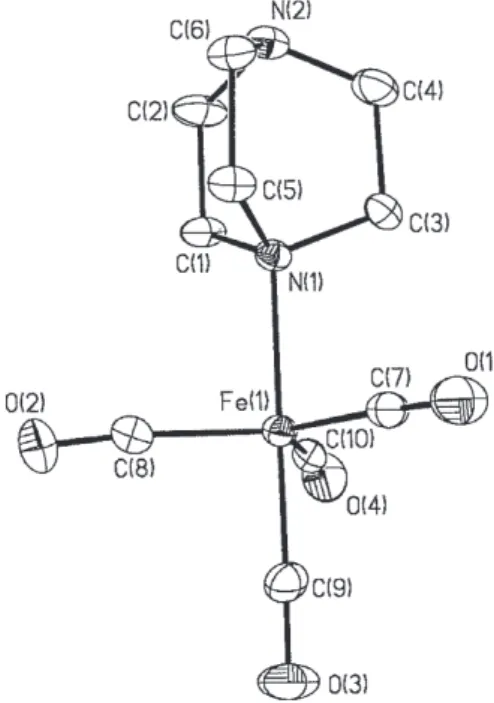
Figure 1. Ortep diagram at 30% probability of 1. Selected bond distances (Å) and angles (o): Fe(1)-C(9) 1.774(6); Fe(1)-C(7)
4 4 4
CDCl3, 25 oC): δ 166.6 (m). 1H NMR (400.13 MHz, CD 3CN, 25 oC): δ 3.02 (virt. t, 6 Hz separation, CH
3).
13C{1H} NMR
(100.62 MHz, CDCl3, 25 oC): δ 37.1 (overlapping dd, 2J(CP) = 3J(CP) = 3.52 Hz, CH
3), 217.8 (overlapping dd
2J(CP) =
5J(CP) = 10.77 Hz, CO). 13C NMR (100.62 MHz, CDCl 3, 25 oC) δ 37.1 (q of overlapping dd, 1J(CH) 138.14 Hz, 2J(CP) = 3J(CP) = 3.52 Hz, CH
3), 217.8 (overlapping dd, 2J(CP) =
5J(CP) 10.77 Hz, CO). IR ν max/cm
-1 2059s, 1992s and 1959vs
(CO) (Nujol). Calcd for C14H18N6O8P2Fe2: C, 29.37%; H, 3.15%; N, 14.64%. Found: C, 29.40%; H, 3.23% N, 14.70%.
Structure determination of 1
Data collection. A yellow crystal was selected under
oil under ambient conditions and attached to the tip of a glass capillary. The crystal was mounted in a stream of cold nitrogen at 173(2)K and centered in the X-ray beam by using a video camera. Crystal evaluation and data collection were performed on a Bruker CCD-1000 diffractometer with Mo Kα radiation with a diffractometer to crystal distance of 4.9 cm. The initial cell constants were obtained from three series of ω scans at different starting angles. Each series consisted of 20 frames collected at intervals of 0.3o in a 6o range about ω with an exposure time of 10 seconds per frame. A total of 64 reflections were obtained. The reflections were successfully indexed by an automated indexing routine built into the SMART program. The final cell constants were calculated from a set of 6257 strong reflections from the actual data collection. The data were collected using the hemisphere data collection routine. The reciprocal space was surveyed to the extent of a full sphere to a resolution of 0.80 Å. A total of 20682 data were harvested by collecting three sets of frames with 0.3o scans in ω with an exposure time 30 sec per frame. These highly redundant data sets were corrected for Lorentz and polarization effects. The absorption correction was based on fitting a function to the empirical transmission surface as sampled by multiple equivalent measurements.16
Structure solution and refinement. The systematic
absences in the diffraction data were uniquely consistent
for the space group P21/c which yielded chemically
reasonable and computationally stable refinement results.17 A successful solution by the direct methods provided most hydrogen atoms from the E-map. The remaining non-hydrogen atoms were located in an alternating series of least-squares cycles and difference Fourier maps. All non-hydrogen atoms were refined with anisotropic displacement coefficients. All hydrogen atoms were included in the structure factor calculation at idealized positions and were
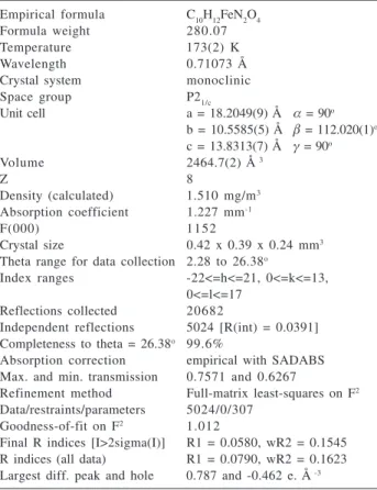
allowed to ride on the neighboring atoms with relative isotropic displacement coefficients. There are two independent molecules of the Fe complex in the asymmetric unit. The final least-squares refinement of 572 parameters 6071 data resulted in residuals R (based on F2 for I > 2σ) and wR (based on F2 for all data) of 0.0580 and 0.1545, respectively. The final difference Fourier map was featureless. Crystallographic data for 1 are summarized in Table 1.
Results and Discussion
The photochemical reaction of [Fe(CO)5] with DABCO in THF affords complex 1 in virtually quantitative yield after 4 h. For periods longer than this, formation of a brown precipitate is observed although interestingly, we observed no evidence for the formation of dinuclear 2 nor for the disubstituted complex [Fe(CO)3(DABCO)2] in this reaction, even when various ratios of reagents were used. We were able to obtain dinuclear 2 by irradiating 1 for 48 h in the presence of [Fe(CO)5]. However, we were not able to prepare [Fe(CO)3(DABCO)2] even when a mixture of 1 and excess DABCO was irradiated for 48 h. After this time, a large amount of paramagnetic material was formed. After filtering the THF solution through Florisil, only a mixture containing unreacted materials could be obtained. Table 1. Crystal and structure refinement data for 1
Empirical formula C10H12FeN2O4 Formula weight 280.07
Temperature 173(2) K
Wavelength 0.71073 Å
Crystal system monoclinic
Space group P21/c
Unit cell a = 18.2049(9) Å α = 90o
b = 10.5585(5) Å β = 112.020(1)o
c = 13.8313(7) Å γ = 90o
Volume 2464.7(2) Å 3
Z 8
Density (calculated) 1.510 mg/m3
Absorption coefficient 1.227 mm-1
F(000) 1152
Crystal size 0.42 x 0.39 x 0.24 mm3
Theta range for data collection 2.28 to 26.38o
Index ranges -22<=h<=21, 0<=k<=13, 0<=l<=17
Reflections collected 20682
Independent reflections 5024 [R(int) = 0.0391] Completeness to theta = 26.38o 99.6%
Absorption correction empirical with SADABS Max. and min. transmission 0.7571 and 0.6267
Refinement method Full-matrix least-squares on F2
Data/restraints/parameters 5024/0/307 Goodness-of-fit on F2 1.012
Complexes 1 and 2 are remarkably stable in the solid state and they can be manipulated in air for long periods of time. However they decompose very easily in solution, especially in hydrocarbons, producing a brown precipitate. Similar observations have been reported for other aminetetracarbonyl complexes.4-6
The 1H NMR spectrum of 1 in CD
3CN shows two virtual triplets, with band separations of 9.2 Hz corresponding to the two sets of chemically equivalent but magnetically
nonequivalent hydrogens on each CH2CH2 moiety in
DABCO. The virtually coupled triplet at δ 2.75 is assigned to the CH2 groups bonded to the uncoordinated nitrogen (CA) due to its proximity to the chemical shift of free DABCO (δ 2.65) whereas the virtually coupled triplet at δ
3.04 is attributed to the CH2 groups bonded to the
coordinated nitrogen (CB). Complex 1 shows a 1H NMR spectrum in which the chemical shifts and multiplet patterns are rather solvent dependent. In CDCl3, its 1H NMR spectrum exhibits two rather broad singlets at δ 2.83 and δ
3.06, and in CD2Cl2 broad singlets are also observed (δ
2.78 and δ 3.03). In benzene-d6 solution, however, the singlets are observed at δ 1.90 and δ 2.33. A somewhat smaller difference in proton chemical shifts is observed for free DABCO in CD3CN (δ 2.65) compared with
benzene-d6 (δ 2.47).
The 13C{1H} NMR spectrum of 1 exhibits two singlets
for the two chemically different CH2 groups in each
CH2CH2 moiety in DABCO, and their assignments were easily made from its heteronuclear decoupling (HMQC) NMR spectrum or by assuming that the chemical shift closer to that of free DABCO (δ 48.4) would correspond to the CH2 bonded to the uncoordinated nitrogen, i.e.δ 48.2 (CA) rather than δ 59.8 (CB). The carbonyl region revealed only one signal for the two types of CO groups, suggesting fluxionality which is rapid on the NMR time scale, a phenomenon commonly observed for substituted pentacarbonyl complexes,3,18-20 and also for [Fe(CO)
5] which is stereochemically nonrigid on the NMR time scale even at –170 oC.15,21 Our conclusion on this point for 1 is supported by its 13C{1H} NMR spectrum which maintains a singlet for the CO groups down to –80 oC. The process is generally assumed to proceed through Berry pseudorotation which, because bond rupture is not involved, spin-spin coupling is preserved.3,12,15,17 It had been believed at one time that exchanging one CO with a ligand different in its σ-donor and π-acceptor properties (e.g., pyridine) would inhibit pseudorotation by virtue of a strong σ-donor capability and its extremely weak π -acceptor property, which would create a preference for the ligand to bond in an axial position. However, the 13C NMR
spectrum of [Fe(CO)4py] showed no change from room
temperature to temperatures as low as –100 oC.3 The 13C NMR spectrum of 1, as expected, shows two triplets of triplets for each CH2 group (with 1J
CH and 3J
CH coupling constants in the expected range) and a singlet for the CO groups.
The IR spectrum of 1 exhibits four bands in the CO region (see Experimental) with the lowest reciprocal wave length displaying a very weak intensity. For a complex of
C3v symmetry only three bands are normally expected
unless splitting of the E mode occurs.22 The normally axial ligand placement in Fe(CO)4L complexes has been amply documented by X-ray diffraction studies,23-25 including [Fe(CO)4(PHPh2)], for example, for which splitting of the E mode has also been observed.21
Despite the tendency for tetracarbonyl iron(0) amine complexes to decompose in hydrocarbon solvents,4-6 a yellow single crystal of 1 suitable for X-ray study was obtained from an n-hexane solution of this complex. The molecular structure of 1 (Figure 1) is a trigonal bipyramid with the DABCO ligand in an axial position, as would be expected due to the lack of π-acceptor character of the ligand. The Fe-N bond length [2.096(4) Å] is longer than in [Fe(CO)4(pyridazine)] [2.013(5) Å],26 [Fe(CO)
4(py)] [2.046(5) Å],3 [Fe(CO)
4(pyrazine)] [2.031(2) Å] 3 and
[CpFeC5H4-4-CHCH-py-Fe(CO)4][2.041(4) Å]27 by more than three times the esd values. However, this is not unexpected since in complex 1 the donating nitrogen lone pair has sp3 character whereas the examples cited possess nitrogens with an sp2 lone pair in which the s character is augmented. To the best of our knowledge, 1 represents the first example of a complex of the type [Fe(CO)4(amine)] whose structure has been determined by X-ray crys-tallographic means in which the amine is aliphatic. The Fe-C(ax) bond distance [1.774(6) Å] found for complex 1 is very close to those in [Fe(CO)4(pyridazine)] [1.765(7) Å],26 [Fe(CO)4(py)] [1.772(7) Å],3 [Fe(CO)
4(pyrazine)] [1.774(4) Å]3 and [CpFeC
5H4CHCH-py-Fe(CO)4] [1.760(6) Å]. 27 The
Fe-C(eq) bond distances (avg. 1.807 Å) are in the normal range found for complexes of the type [Fe(CO)4L].3,26,27
The dinuclear complex 2 features a singlet at δ 3.11 in its 1H NMR spectrum, corresponding to the two chemically equivalent CH2 groups. Its 13C{1H} NMR spectrum consists of a singlet due to the CH2 groups (δ 60.1) and, similarly to
the 13C{1H} NMR spectrum of 1, only one signal
corresponding to the CO groups at (δ 218.2) is present. The 13C NMR spectrum of this compound displays a triplet
of triplets at δ 60.1 with a one-bond HC coupling
4 4 4
in the CO region (2047, 1967 and 1931 cm-1).
Unlike DABCO, P(NMeNMe)3P in the presence of
[Fe(CO)5] under photolysis conditions does not yield the
corresponding mononuclear complex {[Fe(CO)5]
[P(NMeNMe)3P]} even when the reaction is carried out with a 1:1 ratio of the reactants. The products isolated from the reactions using a 1:1 or 1:2 ligand-to-metal ratio are the same, namely, {[Fe(CO)5]2[P(NMeNMe)3P]} (3) in the latter case and excess ligand in the former. The 31P{1H} NMR spectrum of 3 shows a singlet at δ 166.6 whereas the 31P NMR spectrum consists of a multiplet containing nine bands centered at the same chemical shift, corresponding to the X part of an A9XX’A9’ virtually coupled second order spectrum. The 1H NMR spectrum of 3 exhibits a virtually coupled triplet (indicative of strong PP coupling) corresponding to the A part of an A9XX’A9’ spectrum at δ 3.02 with a band separation of 6 Hz. Its 13C{1H} NMR spectrum consists of two apparent triplets (δ 37.1 and δ 217.8). We assign the upfield triplet to the CH3 protons which are coupled equally to the two phosphorus atoms (2J
CP = 3J
CP = 3.52 Hz) giving rise to an overlapping pair of doublets. Similarly, the low-field triplet is attributed to accidentally equal coupling of the CO carbons to the two phosphorus nuclei (2J
CP = 5J
CP = 10.77 Hz). While it may seem odd that the five-bond CP coupling is approximately as large as its two-bond counterpart, it should be noted that there are three through-bond pathways for the latter coupling to occur. That spin-spin coupling in a diphosphorus cages of this type can indeed be substantial and is supported by the observation that the PP coupling in P(CH2NMe)3P is 27 Hz and is 116 Hz in S=P(CH2NMe)3P=S.28 Like the DABCO complexes 1 and 2, complex 3 is also fluxional on the NMR time scale, since only one 13C chemical shift (δ 217.8) is observed.
Acknowledgements
We thank the Donors of the PRF administered by the American Chemical Society for funding this work. RMM thanks CNPq-Brazil for a travel award and UFMG for the award of a sabbatical year.
Supplementary Material
Crystallographic data (excluding structure factors) for the structures in this paper have been deposited with the Cambridge Crystallographic Data Centre as supplementary publication no CCDC 176925. Copies of the data can be obtained, free of charge via ww.ccdc.cam.ac.uk/conts/ retrieving.html (or from the Cambridge Crystallographic Data Centre, CCDC, 12 Union Road, Cambridge CB2 1EZ, UK; fax: +44 1223 336033; or e-mail: [email protected]).
References
1. Whitmire, K. H. In Comprehensive Organometallic Chemistry II; Abel; E. A.; Stone, F. G. A.; Wilkinson, G., eds.; Elsevier
Science: Oxford, UK, 1995, vol. 7, p.1.
2. Shubert, E. H.; Sheline, R. K.; Inorg. Chem.1966, 5, 1071. 3. Cotton, F. A.; Troup, J. M.; J. Am. Chem. Soc.1974, 96, 3438. 4. Fanchinetti, G.; Fochi, G.; Funaioli, T.; Zanazzi, P. F.; J. Chem.
Soc., Chem. Commun.1987, 87.
5. Mealli, C.; Proserpio, D. M.; Fachinetti, G.; Funaioli, T.; Fochi, G.; Zanazzi, P. F.; Inorg. Chem.1989, 28, 1122.
6. Birencwaig, F.; Shamai, H.; Shvo, Y.; Tetrahedron Letters1979, 31, 2947.
7. Elzinga, J.; Hogeveen, H.; J. Chem. Soc., Chem. Commun. 1977, 705.
8. Hieber, W.; Werner, R.; Chem. Ber.1957, 90, 286. 9. Hieber, W.; Angew. Chem.1960, 72, 795.
10. Hieber, W.; Kahlen, R.; Chem. Ber.1958, 91, 2223. 11. Edgell, W. F.; Yang, M. T.; J. Am. Chem. Soc.1966, 88, 4839. 12. Shriver, D. F.; Whitmire, K. H., In Comprehensive Organome-tallic Chemistry, Wilkinson, G.; Stone, F. G. A.; Abel, E. W.
eds., Pergamon Press: Oxford, UK, 1982, vol. 4, p. 243. 13. Hieber, W.; Beutner, H.; Angew. Chem., Int. Ed. Engl. 1962,
1, 116.
14. Goetze, R.; Nöth, H.; Payne, D. S.; Chem. Ber.1972, 105, 2637. 15. Nöth, H. ; Ullmann, R.; Chem. Ber.1974, 107, 1019. 16. Blessing, R. H.; Acta Cryst.1995, A51, 33.
17. All software and sources of the scattering factors are con-tained in the SHELXTL (version 5.1) program library (G. Sheldrick, Bruker Analytical X-Ray Systems, Madison, WI). 18. Adams, R. D.; Cotton, F. A.; In Dynamic Nuclear Magnetic
Resonance Spectroscopy, Jackman, L. M.; Cotton,F. A., eds., Academic Press: New York, 1975, p. 489.
19. Udovich, C. A.; Clark, R. J.; Hass, H.; Inorg. Chem.1969, 8, 1066.
20. Langford, G. R.; Akhtar, M.; Ellis, P. D.; MacDiarmid; A. G.; Odom, J. D.; Inorg. Chem.1975, 98, 5728.
21. Jesson, J. P.; Meakin, P.; J. Am. Chem. Soc.1973, 95, 1344. 22. Darensbourg, D. J.; Nelson, III, H. H.; Hyde, C. L.; Inorg.
Chem.1974, 13, 2135.
23. Riley, P.; Davis, R. E; Inorg. Chem., 1980, 19, 159. 24. Pickardt, J.; Rosch, L.; Schumann, H.; J. Organomet. Chem.
1976, 107, 241.
25. Kilbourn, B. T.; Raeburn, U. A.; Thompson, D. T.; J. Chem. Soc. (A)1969, 1906.
26. Cotton, F. A.; Hanson, B. E.; Isr. J. Chem.1977, 15, 165. 27. Lee, I. S.; Lee, S. S.; Chung, Y. K.; Kim, D.; Song, N. W.;
Inorg. Chim. Acta, 1998, 279, 243.
28. P. M. Stricklen; J.G. Verkade, unpublished work.
Received: June 21, 2002