Chemical composition of the mushroom
Meripilus giganteus
Karst. and
bioactive properties of its methanolic extract
Dejan S. Stojkovi
c
a,1, Nata
sa Kova
cevi
c-Gruji
ci
c
b,1, Filipa S. Reis
c, Slobodan Davidovi
c
b,
Lillian Barros
c, Jelena Popovi
c
b, Isidora Petrovi
c
b, Aleksandar Pavi
c
b,
Jasmina Glamo
clija
a, Ana
Ciri
c
a, Milena Stevanovi
c
b,***, Isabel C.F.R. Ferreira
c,**,
Marina Sokovi
c
a,*aInstitute for Biological Research“Sinisa Stankovic”, Department of Plant Physiology, University of Belgrade, Bulevar Despota Stefana 142, 11000 Belgrade,
Serbia
bInstitute of Molecular Genetics and Genetic Engineering, University of Belgrade, Vojvode Stepe 444a, PO Box 23, 11010 Belgrade, Serbia cMountain Research Centre (CIMO), School of Agriculture, Polytechnic Institute of Bragança, Campus de Santa Apolonia, Apartado 1172, 5300-253
Bragança, Portugal
a r t i c l e
i n f o
Article history:
Received 24 November 2016 Received in revised form 4 January 2017 Accepted 16 January 2017 Available online 17 January 2017
Keywords:
Meripilus giganteusKarst. Antioxidant activity Antimicrobial activity Antitumor activity Apoptosis
a b s t r a c t
WildMeripilus giganteusKarst belongs to the order Polyporales, in which some members are known to possess a wide range of pharmacological properties.M. giganteusshowed to be rich in carbohydrates (74.49 g/100 g) and proteins (15.94 g/100 g), presenting low fat content (1.51 g/100 g). Chemical composition was determined by using chromatographic techniques. Also, various bioactive compounds were detected including all four tocopherol isoforms withd- andg-tocopherols being predominant (123.35 and 77.80mg/100 g, respectively); five organic acids (oxalic, malic, quinic, citric and fumaric acids) with predominant malic acid (3.17 g/100 g); and three phenolic acids and related compounds (p -hydroxybenzoic,p-coumaric and cinnamic acids; 1010, 2420 and 340mg/100 g, respectively).M. giganteus methanolic extract exhibited antioxidant activity tested byfive different assays with the strongest po-tential in TBARS assay (EC500.31 mg/mL); and antimicrobial activities (MIC/MBC 0.0125e5 mg/mL; MIC/ MFC 0.025e0.4 mg/mL). Furthermore, treatment of cervical carcinoma cell line (HeLa) led to reduction in cell's viability in MTT assay (IC500.41 mg/mL after 48 h), induced process of apoptosis and inhibited cell's migrationin vitro.The analysed extract was not toxic for zebrafish embryos (at 0.5 mg/mL), indicating its biosafety and potential application as a dietary supplement in chemoprevention.
©2017 Elsevier Ltd. All rights reserved.
1. Introduction
Besides their high nutritional value (Kalac, 2013), mushrooms possess a wide variety of beneficial effects to human health, with special emphasis on pharmacological properties such as antioxi-dant (Ferreira, Barros,&Abreu, 2009), antimicrobial (Alves et al., 2013) and antitumor (Popovic,Zivkovi c, Davidovic, Stevanovic,& Stojkovic, 2013) activities. Among the bioactive constituents in
medicinal and edible mushrooms, tocopherols, fatty acids, organic acids, polyphenols, polysacharides and proteins are the most frequently reported. Bioactivity of tocopherols includes antioxidant capacity (Ferreira et al., 2009) that confers ability to prevent dis-eases correlated with increased formation of free radicals and oxidative stress. Also, polyunsaturated fatty acids (PUFAs) seem to be endogenous mediators for cell signaling, being involved in the regulation of gene expression and other biological processes (Choque, Catheline, Rioux,&Legrand, 2014). Furthermore, phenolic compounds are known to exert multiple biological effects, including antioxidant, antitumor, antimutagenic and antibacterial properties (Kancheva&Kasaikina, 2013).
Reactions leading to the production of free radicals namely, reactive oxigen species (ROS), reactive nitrosative species (RNS) and reactive sulphur species (RSS) have been linked to many severe *Corresponding author.
**Corresponding author.
***Corresponding author.
E-mail addresses:milenastevanovic@imgge.bg.ac.rs(M. Stevanovic),iferreira@ ipb.pt(I.C.F.R. Ferreira),mris@ibiss.bg.ac.rs(M. Sokovic).
1 These authors contributed equally to this work.
Contents lists available atScienceDirect
LWT - Food Science and Technology
j o u r n a l h o m e p a g e :w w w . e l s e v i e r . c o m / l o c a t e / l w t
http://dx.doi.org/10.1016/j.lwt.2017.01.045
diseases, such as cancer. Therefore, searching for novel sources of antioxidants from natural origin, being able to neutralize free radicals, is one of the major concerns in thefield of cancer che-moprevention. In addition, more than 15% of malignancies world-wide have been related to an infectious cause (Mager, 2006). Although there are effective ways and reliable drugs to treat mi-crobial infections, some bacteria and fungi develop mechanisms to overcome the effects of commercial antimicrobial agents, mostly due to their misuse. Proper treatment of microbial infections is leading to lower cancer occurrence and this should influence the well-being of human population (Assaf et al., 2013). Additionally, mycotherapy of cancer is emerging as a rapidly growing and promising discipline that involves the study of cancer chemo-preventive and anticancer properties of mushrooms extracts and their bioactive compounds (Popovic et al., 2013).
Meripilus giganteusKarst. is a wild growing basidiomycete with an edible fruiting body, belonging to the family Meripilaceae. It is a parasitic species on trees, being also characterized as saprobic. It is frequently found onQuercusandFagusspecies, but it can also be found on some hardwoods and coniferous species.M. giganteusis mainly distributed in northern hemisphere, in Europe, Turkey, Iran and northern Asia (Schmidt, 2006). So far, reports on chemical composition ofM. giganteusgrowing wild in Serbia, as well as on bioactive properties of its methanolic extract are very scarce (Karaman, Jovin, Malbasa, Matavuly, &Popovic, 2010; Karaman, Kaisarevic, Somborski, Kebert, & Matavulj, 2009). The present study aims to characterize the chemical composition of M. giganteus and to evaluate its biological properties namely, antioxidant potential, antimicrobial activity and the effect on can-cer cell's viability, apoptosis and migration in vitro. Finally, its toxicity was evaluated on zebrafish.
2. Materials and methods
2.1. Samples
The samples of wild growing Meripilus giganteusKarst. were collected in Bojcinska forest, Belgrade, Serbia, in autumn 2012, and authenticated by Dr. Jasmina Glamoclija (Institute for Biological Research). A voucher specimen was deposited at the Fungal Collection Unit of the Mycological Laboratory, Department for Plant Physiology, Institute for Biological Research “Sinisa Stankovic”, Belgrade, Serbia, under the number Mg 181 2012. The samples were lyophilized (LH Leybold, Lyovac GT2, Frenkendorf), reduced to afine dried powder (20 mesh) and stored in a desiccator, protected from light, until further analysis.
2.2. Standards and reagents
Acetonitrile 99.9%,n-hexane 95% and ethyl acetate 99.8% were of HPLC grade from Fisher Scientific (Lisbon, Portugal). The fatty acids methyl ester (FAME) reference standard mixture 37 (standard 47885-U) was purchased from Sigma (St. Louis, MO, USA), as also were other individual fatty acid isomers, trolox (6-hydroxy-2,5,7,8-tetramethylchroman-2-carboxylic acid), tocopherol and sugar standards. Phenolic compound standards were from Extrasynthese (Genay, France). Racemic tocol, 50 mg/mL, was purchased from Matreya (Pleasant Gap, PA, USA). 2,2-Diphenyl-1- picrylhydrazyl (DPPH) was obtained from Alfa Aesar (Ward Hill, MA, USA). Dul-becco's modified Eagle's medium (DMEM), fetal bovine serum (FBS) and MEM non-essential amino acids (NEAA) were obtained from Invitrogen (NY, USA). 3-(4,5-Dimethylthiazol-2yl)-2,5-diphenyltetrazolium bromide (MTT), paraformaldehyde (PFA) and dimethylsulfoxide (DMSO) were from Merck (KGaA, Germany). 40,6-Diamidino-2-phenylindole dihydrochloride (DAPI) and
propidium iodide (PI) were obtained from Sigma (St. Louis, MO, USA) and doxorubicin from Pfizer Inc (NY, USA). APOPTEST™-FITC kit was obtained from Dako (Agilent Technologies Inc., Denmark). MuellereHinton agar (MH) and malt agar (MA) were obtained from
the Institute of Immunology and Virology, Torlak (Belgrade, Serbia). Water was treated in a Milli-Q water purification system (TGI Pure Water Systems, Greenville, SC, USA).
2.3. Preparation of the M. giganteus methanolic extract
Samples (~5 g) were extracted by stirring with 150 mL of methanol (25 C at 150 rpm) for 1 h and subsequently
filtered through Whatman No. 4 paper. The residue was then extracted with an additional portion of methanol. The combined methanolic extracts were evaporated under reduced pressure (rotary evapo-rator Büchi R-210; Flawil, Switzerland) to dryness (Heleno, Barros, Sousa, Martins,&Ferreira, 2010). The extract was redissolved in i) methanol (final concentration 20 mg/mL) for antioxidant activity assays, ii) 30% EtOH (final concentration 1.5 mg/mL) for antimi-crobial activity assays and for cytotoxic activity evaluation (80 mg/ mLfinal concentration). Thefinal solutions were further diluted to different concentrations to be submitted to distinct bioactivity evaluation in vitro assays. The results were expressed in i) EC50 values (sample concentration providing 50% of antioxidant activity or 0.5 of absorbance in the reducing power assay) for antioxidant activity; ii) MIC (Minimum inhibitory concentration) and MBC/MFC (Minimum bactericidal concentration/Minimum fungicidal con-centration) values forin vitroantimicrobial activity iii) IC50value (sample concentration providing 50% inhibition of cell growth in MTT test) for cytotoxicity assay.
2.4. Chemical characterization of the M. giganteus methanolic extract - bioactive compounds
Tocopherols were determined following a procedure previously described by the authors (Heleno et al., 2010) using HPLC-fluorescence. The compounds were identified by chromatographic comparisons with authentic standards. Quantification was based on thefluorescence signal response of each standard, using the IS (tocol) method and by using calibration curves obtained from commercial standards of each compound:
a
-tocopherol (y¼1.295x;R2¼0.991);b
-tocopherol (y¼0.396x;R2¼0.992);g
-tocopherol (y ¼0.567x; R2 ¼0.991);d
-tocopherol (y¼ 0.678x; R2¼0.992). The results were expressed inm
g per 100 g of dry weight (dw). Organic acids were determined by ultrafast liquid chromatography coupled to a photodiode array detector (UFLC-PAD), following a procedure previously described by the authors (Barros, Pereira,&Ferreira, 2013). The organic acids found were quantified by comparison of the area of their peaks recorded at 215 nm with calibration curves obtained from commercial stan-dards of each compound: oxalic acid (y ¼ 1 107xþ96178; R2¼0.999); quinic acid (y¼601768xþ8853.2; R2¼1); malic acid (y ¼ 952269xþ17803; R2 ¼ 1) and fumaric acid (y¼172760xþ52193;R2¼0.999. The results were expressed in g per 100 g of dry mushroom (dw). Phenolic compounds were determined by the same methodology using 280 nm and 370 nm as preferred wavelengths, according to a procedure previously described by the authors (Barros, Duenas, Ferreira, Baptista,~ & Santos-Buelga, 2009). The phenolic compounds were character-ized according to their UV and mass spectra and retention times, and comparison with authentic standards when available. For the quantitative analysis of phenolic compounds, a calibration curve was obtained by injection of known concentrations (5e80m
g/mL)(y ¼ 51195xþ1 106x; R2 ¼ 0.992); cinnamic acid (y¼86366xþ88451;R2¼0.999). The results were expressed as
m
g per 100 g of dry weight (dw).2.5. Antioxidant activity
DPPH radical-scavenging activity was evaluated by using an ELX800 microplate reader (Bio-Tek Instruments, Inc; Winooski, VT, USA), and calculated as a percentage of DPPH discolouration using the formula: [(ADPPH-AS)/ADPPH]100, where ASis the absorbance of the solution containing the sample at 515 nm, and ADPPHis the absorbance of the DPPH solution. Reducing power was evaluated by two different assays: Ferricyanide/Prussian blue assay, which de-termines the capacity to convert Fe3þ into Fe2þ, measuring the absorbance at 690 nm in the microplate reader mentioned above; Folin-Ciocalteu assay: the colour development was measured at 765 nm (Analytikjena spectrophotometer; Jena, Germany). Gallic acid was used to obtain the standard curve and the reduction of Folin-Ciocalteu reagent by the samples was expressed as mg of gallic acid equivalents (GAE) per g of extract (Heleno et al., 2010). Inhibition of
b
-carotene bleaching was evaluated though theb
-carotene/linoleate assay; the neutralization of linoleate free radi-cals avoidsb
-carotene bleaching, measured at 470 nm and calcu-lated by the formula:b
-carotene absorbance after 2 h of assay/ initial absorbance)100 (Heleno et al., 2010). Lipid peroxidation inhibition in porcine (Sus scrofa) brain homogenates was evaluated by the decreasing in thiobarbituric acid reactive substances (TBARS); the colour intensity of the malondialdehyde-thiobarbituric acid (MDA-TBA) was measured by its absorbance at 532 nm; the inhibition ratio (%) was calculated using the following formula: [(A - B)/A]100%, where A and B were the absorbances of the control and the sample solution, respectively (Heleno et al., 2010). Trolox was used as a positive control in all of the antioxi-dant assays.2.6. Antimicrobial properties
The following Gram-negative bacteria were used: Escherichia coli(ATCC 35210),Pseudomonas aeruginosa(ATCC 27853), Salmo-nella typhimurium(ATCC 13311),Enterobacter cloacae(ATCC 35030) and the following Gram-positive bacteria: Staphylococcus aureus (ATCC 6538), Bacillus cereus (clinical isolate), Micrococcus flavus (ATCC 10240), andListeria monocytogenes(NCTC 7973).
For the antifungal bioassays, microfungi were used:Aspergillus fumigatus (1022),Aspergillus ochraceus (ATCC 12066), Aspergillus versicolor (ATCC 11730),Aspergillus niger(ATCC 6275),Penicillium funiculosum (ATCC 36839),Penicillium ochrochloron (ATCC 9112), Trichoderma viride (IAM 5061), and Penicillium aurantiogriseum (food isolate). The organisms were obtained from the Mycological Laboratory, Department of Plant Physiology, Institute for Biological Research“Sinisa Stankovic”, Belgrade, Serbia.
In order to investigate the antimicrobial activity of the M. giganteus methanolic extract, a modified microdilution tech-nique was used (CLSI, 2009). Minimum inhibitory concentration (MIC) determinations were performed by a serial dilution tech-nique using 96-well microtiter plates. The minimum bactericidal concentrations (MBCs) and minimum fungicidal concentrations (MFCs) were determined by serial subcultivation of a 2
m
L sample into microtiter plates containing 100m
L of broth per well and further incubation for 48 h at 37C or 72 h at 28C. The lowest concentration with no visible growth was defined as MBC/MFC, respectively, indicating 99.5% killing of the original inoculum. Streptomycin (streptomycin sulfate (ICN-Galenika, Belgrade, Serbia) and ampicillin (Panfarma, Belgrade, Serbia) were used as positive controls in antibacterial assay, while commercialfungicides, bifonazole (Srbolek, Belgrade, Serbia) and ketoconazole (Zorkapharma, Sabac, Serbia), were used as positive controls in antifungal tests (1 mg/ml in sterile physiological saline). 30% EtOH was used as a negative control.
2.7. Evaluation of cytotoxicity by MTT assay
Human cervical cancer cell line HeLa and human lung fi bro-blasts MRC-5 were cultured in DMEM supplemented with 1% NEAA and 10% FBS at 37C in a humidi
fied incubator with 5% CO2. Cell viability was determined by the colorimetric MTT assay. Briefly, 24 h before treatment 4103HeLa cells/well and 104MRC-5 cells/ well were seeded in 96-well plates. Cells were then treated for 24 and 48 h with various concentrations ofM. giganteusmethanolic extract (prepared by dilution from the inicial stock solution, 80 mg/ mL) or doxorubicin as a positive reference compound. Relative cell viability was obtained by measuring the absorbance at 550 nm in a Multiscan RC microplate reader (Thermo Labsystems, Helsinki, Finland). Concentrations that caused 50% (IC50) or 80% (IC80) of inhibition of cells growth after 24 and 48 h of treatment were calculated based on their survival rate compared with control cells treated with vehicle only (30% ethanol).
2.8. Double DAPI and PI staining of live cells
Control cells and cells treated with IC50and IC80concentrations of extract were grown for 24 and 48 h on coverslips in 6-well plates. After washing three times in phosphate buffered saline (PBS), the cells were incubated in PBS containing 1
m
g/mL of DAPI and 5m
g/mL of PI for 10 min at RT. After staining, cells were washed three times in PBS andfixed with 4% PFA for 15 min. Cells were examined under the Zeiss Axiovert inverted fluorescent microscope (Carl Zeiss Foundation, Oberkochen, Germany) equipped with AxioVision4.8 software, using 365/550 nm excitation; thefluorescence emission was measured at 445 nm for DAPI and 605 nm for PI with a 20 objective.2.9. Detection of apoptosis by a double staining method with Annexin V-FITC and PI
Apoptosis assays were conducted using the APOPTEST™-FITC kit according to the manufacturer's instructions. Briefly, control cells (treated with vehicle only) and cells treated with IC50 con-centration of extract for 48 h were washed twice with cold PBS, resuspended in 1 Binding Buffer at a concentration of ~ 1106cells/mL and Annexin V-FITC and PI were added at thefinal concentrations of 25 ng/mL and 2.5
m
g/mL, respectively. The cells were gently mixed, incubated for 10 min in the dark at RT, and immediately analyzed (within 1 h) by PartecCyFlow®Space (Partec GmbH, Münster, Germany). Theflow cytometer collected 100,000 events and analysis was performed using Flomax Software © Version 2.9.
2.10. Wound-scratch migration assay
2.11. In vivo zebrafish toxicity assay
Assay is described in detail inSupplementary material.
2.12. Statistical analysis
Three samples were used and all the assays were carried out in triplicate. The results are expressed as mean values± standard deviation (SD) or standard error mean (SEM). The results were analyzed using one-way analysis of variance (ANOVA) followed by Tukey's HSD test with
a
¼0.05 or Student'st-test. This analysis was carried out using SPSS 22.0 program.3. Results and discussion
3.1. Chemical composition of M. giganteus methanolic extract-bioactive compounds
Bioactive compounds (tocopherols, organic and phenolic acids) identified inM. giganteusare presented inTable 1. All tocopherol isoforms were detected, but
d
- andg
-tocopherols were the pre-dominant isoforms (Table 1andFig. 1A). On the other hand,b
- anda
-tocopherols were found at lower levels (Table 1). The most suitable sources of antioxidants, such as tocopherols, are provided by our diet, more than by antioxidant supplements (pills or tablets) (Bjelakovic, Nikolova, &Gluud, 2014). M. giganteus represents a food source regarding intake of antioxidant tocopherols and to the best of our knowledge, tocopherols composition in this species was not previously reported in literature.Quantitative values of organic acids identified inM. giganteus are given inTable 1and its profile is represented inFig. 1B. Malic acid, a dicarboxylic acid occurring naturally in all fruits, many vegetables and mushrooms, was the predominant organic acid, which was also observed for other mushroom species (Barros et al., 2013). Oxalic, citric, quinic and fumaric acids were also identified and quantified. Health-beneficial effects and bioactive properties of some organic acids are well-known and include antimicrobial, antioxidant and acidifying properties. Our results are in accordance to those published for other mushroom species (Barros et al., 2013). Phenolic acids profile was provided inFig. 1Cp-Coumaric acid was the most abundant phenolic component identified in M. giganteus, while p-hydroxybenzoic and cinnamic acids were found at lower levels (Table 1). A previous study performed by Karaman et al. (2010)reported gallic (675
m
g/g) and protocatechuic (279m
g/g) acids as the only phenolic compounds inM. giganteus, whilep-coumaric acid was not detected. The observed differences might be attributed to different extraction methodologies or different maturity stages of fruiting bodies, among other factors.Nutritional value, sugars composition and fatty acids profile are presented inSupplementary material.
3.2. In vitro analysis of bioactive properties
In order to analyze bioactive properties ofM. giganteus meth-anolic extract, we have investigated antioxidant activity,
Table 1
Bioactive composition ofM. giganteuson dry weight basis (mean±SD).
Phenolic acids (mg/100 g) Tocopherols (mg/100 g) Organic acids (g/100 g)
p-Hydroxybenzoic acid 1010±110 a-tocopherol 3.58±0.39 Oxalic acid 1.32±0.02
p-Coumaric acid 2420±590 b-tocopherol 9.05±0.13 Quinic acid 0.48±0.03 Cinnamic acid 340±40 g-tocopherol 77.80±0.66 Malic acid 3.17±0.01 Total phenolic acids* 3430±470 d-tocopherol 123.35±40.11 Citric acid 0.77±0.01 Total tocopherols 213.78±40.52 Fumaric acid 0.16±0.00 Total organic acids 5.90±0.03
antimicrobial properties, and the effect on cancer cell's viability, apoptosis and migration in vitro. Finally, toxicity testing of M. giganteusmethanolic extract was performed on zebrafish em-bryos and these results are presented inSupplementary material.
3.2.1. The antioxidant activity
Since natural matrices with antioxidant activity important for the discovery of novel cancer chemopreventive and chemothera-peutic agents, we have analyzed the antioxidant activity of M. giganteus methanolic extract byfive different in vitroassays
(Table 2).
Total phenolics content in the methanolic extract was 58.20 mg GAE/g of extract which is higher than the one reported byKaraman et al. (2010) (5.11 mg of chlorogenic acid equivalents/g of dry weight ofM. giganteus). The obtained difference might be related to the different extraction methods used. Lipid peroxidation inhibi-tion, evaluated by the decreasing in TBARS formainhibi-tion, gave the best result with EC50value of 0.31 mg/mL. For Ferricyanide/Prussian blue assay, EC50value was 0.83 mg/mL. On the other hand, DPPH scavenging activity and inhibition of
b
-carotene bleaching gave slightly higher results with EC50values of 2.08 mg/mL and 2.14 mg/ mL, respectively. DPPH scavenging activity of M. giganteus 70% methanolic extract was previously evaluated and the EC50value was 0.155 mg/mL (Karaman et al., 2010). In the present study, the EC50 DPPH scavenging activity value was significantly higher compared to the mentioned report and this difference might be explained by the different extraction conditions and drying pro-cesses applied to mushrooms (e.g.drying in the oven at 50C). As far as we know, this is thefirst study reporting antioxidant activity of pure methanolic extract ofM. giganteus. The observed antioxi-dant activity may be related to the presence of phenolic acids and tocopherols, as generalized for other mushroom species (Ferreira et al., 2009).3.2.2. Antimicrobial properties
Antibacterial and antifungal activity of theM. giganteusextract was assessed by microdilution methods, and the results are pre-sented as MIC and MBC/MFC inTable 3. The most sensitive bacterial species to M. giganteus methanolic extract were S. aureus and
Table 2
Antioxidant properties of theM. giganteusmethanolic extract (mean±SD). Antioxidant activity tests Extract
Folin- Ciocalteuassay (mg GAE/g extract)
58.20±5.10 Ferricyanide/Prussian blue assay (EC50;mg/mL) 0.83±0.01
DPPH scavenging activity (EC50;mg/mL)
2.08±0.06
b-Carotene/linoleate assay (EC50;mg/mL)
2.14±0.12 TBARS assay
(EC50;mg/mL)
0.31±0.02
Concerning the Folin-Ciocalteu assay, higher values mean higher reducing power; for the other assays, the results are presented in EC50values, what means that higher
values correspond to lower reducing power or antioxidant potential. EC50: Extract
concentration corresponding to 50% of antioxidant activity or 0.5 of absorbance for the Ferricyanide/Prussian blue assay. Trolox EC50values: 41mg/mL (Ferricyanide/
Prussian blue assay), 42mg/mL (DPPH scavenging activity), 18mg/mL (b-Carotene/ linoleate assay) and 23mg/mL (TBARS assay).
Table 3
In vitroantimicrobial activity ofM. giganteusextract.
Bacteria M. giganteus
MIC/MBC (mg/mL)
Streptomycin MIC/MBC (mg/mL)
Ampicillin MIC/MBC (mg/mL)
Staphylococcus aureus 0.0125±0.0008a
0.035±0.002a
0.04±0.002b
0.09±0.003b
0.25±0.000c
0.37±0.0100c
Bacillus cereus 0.0125±0.0008a
0.035±0.002a
0.09±0.003b
0.17±0.0100b
0.25±0.0100c
0.37±0.007c
Micrococcusflavus 0.60±0.03c
1.25±0.080b
0.17±0.010a
0.34±0.010a
0.25±0.0200b
0.37±0.000a
Listeria monocytogenes 0.30±0.000b
0.60±0.010c
0.17±0.010a
0.34±0.010a
0.37±0.010c
0.49±0.010b
Pseudomonas aeruginosa 0.60±0.020b
1.25±0.020b
0.17±0.010a
0.34±0.000a
0.74±0.000c
1.24±0.010b
Salmonella typhimurium 0.025±0.002a
0.035±0.002a 0.17 ±0.010b
0.34±0.010b 0.37 ±0.010c
0.49±0.020c
Escherichia coli 0.15±0.007a
0.60±0.000a
0.17±0.007b
0.34±0.010b
0.25±0.000c
0.49±0.020c
Enterobacter cloacae 2.50±0.070b
5.00±0.000c
0.26±0.000a
0.52±0.007a
0.37±0.010a
0.74±0.010b
Fungi M. giganteus
MIC/MFC (mg/mL)
Bifonazole MIC/MFC (mg/mL)
Ketoconazole MIC/MFC (mg/mL)
Aspergillus fumigatus 0.10±0.010a
0.20±0.000a
0.15±0.000b
0.20±0.020a
0.20±0.010c
0.50±0.020b
Aspergillus versicolor 0.025±0.000a
0.050±0.002a
0.10±0.010b
0.20±0.000b
0.20±0.020c
0.50±0.010c
Aspergillus ochraceus 0.20±0.010a
0.40±0.010a
0.15±0.007a
0.20±0.020a
1.50±0.070b
2.00±0.100b
Aspergillus niger 0.30±0.010c
0.40±0.000a
0.15±0.000a
0.20±0.010b
0.20±0.010b
0.50±0.020c
Trichoderma viride 0.05±0.002a
0.20±0.010a
0.15±0.007a
0.20±0.000a
1.00±0.070b
1.00±0.100b
Penicillium funiculosum 0.025±0.000a
0.400±0.010a 0.20 ±0.010b
0.25±0.000b 0.20 ±0.010b
0.50±0.010c
Penicillium ochrochloron 0.20±0.000a
0.40±0.020a
0.20±0.010a
0.25±0.007b
2.50±0.070b
3.50±0.070c
Penicillium aurantiogriseum 0.20±0.010a
0.40±0.000a
0.10±0.010b
0.20±0.010b
0.20±0.000b
0.30±0.020c
B. cereuswith the same MICs (0.0125 mg/mL) and MBCs (0.035 mg/ mL). E. cloacae strain was the most resistant to the effect of M. giganteusextract with MIC of 2.50 mg/mL and MBC of 5.00 mg/ mL. It should be noted that the most sensitive bacterial strains were
Gram positive. Although,M.flavus(Gram positive) andP. aeruginosa (Gram negative) had the same inhibitory and bactericidal values (0.60 mg/mL and 1.25 mg/mL), it might be suggested that the antimicrobial activity is dependent on bacterial species used. The
Table 4
Cytotoxic activity ofM. giganteusmethanolic extract (mean±SD) on HeLa and MRC-5 cell lines.
Extract (mg/mL) Doxorubicin (mg/mL)
24 h 48 h 24 h 48 h
HeLa IC50 0.72±0.27b 0.41±0.08b 0.0069±0.0006a 0.0027±0.0003a
IC80 1.32±0.23b 0.80±0.04b 0.0091±0.0010a 0.0039±0.0007a
MRC-5 IC50 >1.6 >1.6 0.0059±0.0012 0.0025±0.0004
IC80 na na 0.0091±0.0022 0.0046±0.0005
The results are expressed as IC50and IC80values corresponding to the extract/compound concentration, which inhibited 50% and 80% of cell growth after 24 and 48 h of
treatment, respectively. Doxorubicin was used as a positive reference compound. Different letters (a, b) mean significant differences between extract and doxorubicin after 24 h and 48 h of treatment, respectively (p0.05). Na-not acquired.
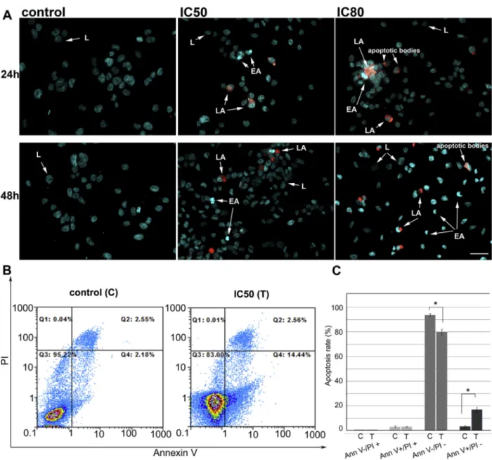
Fig. 2.M. giganteusmethanolic extract induces apoptosis of HeLa cells.A)Representative composite images showing morphological changes of HeLa cells detected with dual DAPI/ PI staining. Cells were treated with IC50and IC80concentrations ofM. giganteusextract for 24 and 48 h and imaged byfluorescence microscope. Arrows indicate (L) viable cells with
normal nuclei, (EA) live cells with apoptotic nuclei showing chromatin condensation, (LA) dead cells in late apoptosis/necrosis. Scale bare50mm.B)Flow cytometry analysis of
Annexin-FITC staining and propidium iodide accumulation after treatment of HeLa cells withM. giganteusmethanolic extract. Cells were treated either with vehicle (C) or IC50
activity of the extract decreased in order: S. aureus¼B. cereus>S. typhimurium>E. coli>L. monocytogenes> M.flavus¼P. aeruginosa>E. cloacae.Karaman et al. (2010), even though applying different method, also foundS. aureusstrain as the most sensitive to the antimicrobial action ofM. giganteus.
Regarding antifungal activity, A. versicolor and P. funiculosum were the most susceptible micromycetes with MICs of 0.025 mg/mL and MFCs of 0.050 mg/mL.A. nigerwas the most resistant strain with MIC of 0.30 mg/mL and MFC of 0.40 mg/mL. Antifungal activity of the tested sample decreased in order: A. versicolor ¼ P. funiculosum > T. viride > A. fumigatus > A. ochraceus ¼P. ochrochloron¼P. aurantiogriseum >A. niger. In general, fungi were more sensitive than bacteria to the effect of M. giganteus extract. To the best of our knowledge, no previous studies reported antifungal activity ofM. giganteus. Standard anti-biotics (streptomycin and ampicillin) and antimycotics (bifonazole and ketoconazole) were used as positive controls, but comparison with M. giganteus extract should be avoided, since the extract presents a mixture of compounds in which the concentration of each bioactive compound is much lower than the MIC. Antimicro-bial activity could be related to the presence of the phenolic acids
identified in the extract that also show antimicrobial properties against bacteria and fungi (Heleno et al., 2013).
3.2.3. Evaluation of cytotoxic properties on HeLa cells
The effect ofM. giganteusmethanolic extract on the proliferation and viability of HeLa cells was assessed using MTT assay. A dose-dependent growth inhibition was observed in HeLa cell line after treatment with the extract for 24 h and 48 h. Extract concentrations required for 50% inhibition of growth (IC50) were 0.72 mg/mL and 0.41 mg/mL for 24 and 48 h of treatment, respectively (Table 4). The concentrations required for 80% of growth inhibition (IC80) were 1.32 mg/mL and 0.80 mg/mL for 24 h and 48 h, respectively. These results are consistent with two previously published showing that M. giganteusmethanolic extract displays cytotoxicity against hu-man breast carcinoma cells MCF-7 (with similar range of IC50 concentrations) and against murine Lewis lung carcinoma cell line 3LL (Karaman et al., 2009; Tomasi, Lohezic-Le Devehat, Sauleau, Bezivin,&Boustie, 2004). Methanolic extract ofM.giganteusdid not cause 50% inhibition of growth (IC50) of normalfibroblast cell line MRC-5, even at the highest concentration tested (1.6 mg/mL, Table 4).
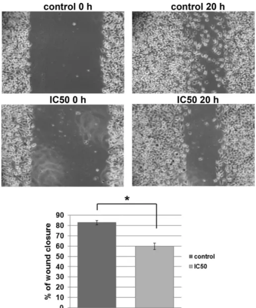
Fig. 3.Effect ofM. giganteusextract on migration of HeLa cells (wound-scratch migration assay). Cells migration was quantified 20 h after scratching in constant presence of
These results suggest the presence of bioactive compounds that impair cell growth in the analyzed extract. The identified phenolic acids could be, at least partly, responsible for this property. p -Coumaric acid, most abundant phenolic component ofM. giganteus methanolic extract, was previously shown to inhibit proliferation and induce apoptosis of colorectal adenocarcinoma cell lines (HCT-15 and HT-29) (Jaganathan, Supriyanto,&Mandal, 2013). Cytotoxic activity on several malignant cell lines was also demonstrated for cinnamic acid and its derivatives (Sova et al., 2013).
3.2.4. Induction of apoptosis of HeLa cells
First we have observed morphological changes in HeLa cells nuclei after treatment with M. giganteus extract using double staining with DAPI and PI. Cells treated withM. giganteusextract for 24 or 48 h exhibited typical features of apoptosis such as nuclear condensation, fragmentation and formation of apoptotic bodies (Fig. 2A). Cells in early apoptosis have highly condensed chromatin that is characterized by more intense bluefluorescence compared to the nuclei of healthy cells, while cells in late apoptosis/necrosis have lost the integrity of their plasma membranes and their condensed/fragmented chromatin is characterized by bright red fluorescence. Observed morphological changes suggest that mechanism underlying inhibition of cell growth byM. giganteus extract relies on induction of apoptosis. This mode of action has been demonstrated previously for many mushroom extracts and their bioactive principles (Popovic et al., 2013).
These results were confirmed byflow cytometry (Fig. 2B and C). Obtained results suggested that M. giganteusmethanolic extract induced early apoptosis in HeLa cells (Annexinþ/PI ). Based on three independent experiments the percentage of cells in early apoptosis raised to 17% compared to control cells treated with vehicle only (Fig. 2C). At the same time, the low percentage of cells with necrosis (Annexin /PIþ) and late apoptosis (Annexinþ/PIþ) remained unchanged. Taken together, these results suggest that M. giganteusextract induced apoptosis of HeLa cells.
3.2.5. The effect on migratory potential of HeLa cells
Since the ability of cancer cells to migrate is closely associated with their capacity to colonize distant organs, we tested migratory potential of HeLa cells, upon treatment withM. giganteusextract, using wound scratch assays. We detected that treated HeLa cells were slower in closing the scratched area than control cells (Fig. 3), showing thatM. giganteusextract exhibits not only cytotoxic, but also antimigratory effect on HeLa cells.
Antimigratory potential of mushroom's extracts has been re-ported for other classes likeFomitopsis pinicolawhich belongs to the Basidiomycota fungal class that could inhibit the migration of colon cancer cell line SW-480 (Wang et al., 2014), orGanoderma lucidum that exerts strong antimigratory effect on ovarian cancer cell lines (Zhao et al., 2011). This is thefirst report presenting antimigratory potential ofM. giganteusextract on cancer cellsin vitro.
4. Conclusions
Chemical composition analysis of M. giganteus methanolic extract revealed the presence of bioactive compounds (all tocoph-erol isoforms, five organic and three phenolic acids) along with carbohydrates, fatty acids and proteins. Trehalose was identified as the predominant free sugar. Polyunsaturated fatty acids predomi-nated over monounsaturated and saturated fatty acids, highlighting linoleic acid as the most dominant one.M. giganteusis a natural source of agents exhibiting antioxidant, antimicrobial and anti-tumor activities highlighting this species as a valuable dietary supplement in chemoprevention. Importantly,M. giganteusextract was not toxic for zebrafish embryos indicating its biosafety and
potential for further applications in food or pharmaceutical industry.
Acknowledgements
This work was supported by the Ministry of Education, Science and Technological Development of the Republic of Serbia (grant numbers 173032, 173051 and 47025). Authors are grateful to the Foundation for Science and Technology (FCT, Portugal) forfinancial support to CIMO (Pest-OE/AGR/UI0690/2015) and L. Barros grant (SFRH/BPD/107855/2015). We would like to thank dr Jelena Dinic for help in performing DAPI/PI staining.
Appendix A. Supplementary data
Supplementary data related to this article can be found athttp:// dx.doi.org/10.1016/j.lwt.2017.01.045.
References
Alves, M. J., Ferreira, I. C. F. R., Dias, J., Teixeira, V., Martins, A., & Pintado, M. (2013). A review on antifungal activity of mushroom (basidiomycetes) extracts and isolated compounds.Current Topics in Medicinal Chemistry, 13, 2648e2659. Assaf, A. M., Haddadin, R. N., Aldouri, N. A., Alabbassi, R., Mashallah, S.,
Mohammad, M., et al. (2013). Anti-cancer, inflammatory and anti-microbial activities of plant extracts used against hematological tumors in traditional medicine of Jordan.Journal of Ethnopharmacology, 145, 728e736. Barros, L., Duenas, M., Ferreira, I. C. F. R., Baptista, P., & Santos-Buelga, C. (2009).~
Phenolic acids determination by HPLC-DAD-ESI/MS in sixteen different Portu-guese wild mushrooms species.Food and Chemical Toxicology, 47, 1076e1079. Barros, L., Pereira, C., & Ferreira, I. C. F. R. (2013). Optimized analysis of organic acids
in edible mushrooms from Portugal by ultra fast liquid chromatography and photodiode array detection.Food Analytical Methods, 6, 309e316.
Bjelakovic, G., Nikolova, D., & Gluud, C. (2014). Antioxidant supplements and mortality.Current Opinion in Clinical Nutrition and Metabolic Care, 17, 40e44. Choque, B., Catheline, D., Rioux, V., & Legrand, P. (2014). Linoleic acid: Between
doubts and certainties.Biochimie, 96, 14e21.
CLSI. Clinical and Laboratory Standards Institute. (2009).Methods for dilution anti-microbial susceptibility tests for bacteria that grow aerobically. Approved standard (8th ed.). Wayne, PA: CLSI publication M07-A8. Clinical and Laboratory Stan-dards Institute.
Ferreira, I. C. F. R., Barros, L., & Abreu, R. M. V. (2009). Antioxidants in wild mush-rooms.Current Medicinal Chemistry, 16, 1543e1560.
Heleno, S. A., Barros, L., Sousa, M. J., Martins, A., & Ferreira, I. C. F. R. (2010). To-copherols composition of Portuguese wild mushrooms with antioxidant ca-pacity.Food Chemistry, 119, 1443e1450.
Heleno, S. A., Ferreira, I. C. F. R., Calhelha, R. C., Esteves, A. P.,Ciric, A., Glamoclija, J., et al. (2013). Antimicrobial and demelanizing activity ofGanoderma lucidum extract,p-hydroxybenzoic and cinnamic acids and their synthetic acetylated glucuronide methyl esters.Food and Chemical Toxicology, 58, 95e100. Jaganathan, S. K., Supriyanto, E., & Mandal, M. (2013). Events associated with
apoptotic effect ofp-Coumaric acid in HCT-15 colon cancer cells.World Journal of Gastroenterology, 19, 7726e7734.
Kalac, P. (2013). A review of chemical composition and nutritional value of wild-growing and cultivated mushrooms.Journal of the Science of Food and Agricul-ture, 93, 209e218.
Kancheva, V. D., & Kasaikina, O. T. (2013). Bio-antioxidantsea chemical base of their antioxidant activity and beneficial effect on human health.Current Me-dicinal Chemistry, 20, 4784e4805.
Karaman, M., Jovin, E., Malbasa, R., Matavuly, M., & Popovic, M. (2010). Medicinal and edible lignicolous fungi as natural sources of antioxidative and antibacterial agents.Phytotherapy Research, 24, 1473e1481.
Karaman, M., Kaisarevic, S., Somborski, J., Kebert, M., & Matavulj, M. (2009). Bio-logical activities of the lignicolous fungusMeripilus giganteus(Pers.: Pers) Karst. Archive of Biological Sciences, 61, 853e861.
Mager, D. L. (2006). Bacteria and cancer: Cause, coincidence or cure? A review. Journal of Translational Medicine, 4, 14.
Popovic, V., Zivkovic, J., Davidovic, S., Stevanovic, M., & Stojkovic, D. (2013). Mycotherapy of cancer: An update on cytotoxic and antitumor activities of mushrooms, bioactive principles and molecular mechanisms of their action. Current Topics in Medicinal Chemistry, 13, 2791e2806.
Schmidt, O. (2006).Wood and tree fungi: biology, damage, protection, and use. Berlin: Springer.
Sova, M., Zizak, Z., Stankovic, J. A., Prijatelj, M., Turk, S., Juranic, Z. D., et al. (2013). Cinnamic acid derivatives induce cell cycle arrest in carcinoma cell lines. Medical Chemistry, 9, 633e641.
Wang, Y., Cheng, X., Wang, P., Wang, L., Fan, J., Wang, X., et al. (2014). Investigating migration inhibition and apoptotic effects of Fomitopsis pinicola chloroform extract on human colorectal cancer SW-480 Cells.PLoS One, 9, e101303.