Arq Bras Endocrinol Metab vol 47 nº 6 Dezembro 2003 744
ABSTRACT
Periodic paralysis is an uncommon complication of primary aldostero-nism in the non-Asian population. We describe the case of a Brazilian woman who presented to the emergency room with proximal symmetric tetraparesis that was later diagnosed as primary aldosteronism. This case report shows that primary aldosteronism should be included in the differ-ential diagnosis of periodic paralysis, especially among hypertensive patients.(Arq Bras Endocrinol Metab 2003;47/6:744-747)
Keywords: Primary aldosteronism; Symmetric tetraparesis; Periodic paral-ysis; Metabolic myopathy
RESUMO
Paralisia Periódica: Uma Complicação Incomum do Hiperaldosteronis-mo Primário.
A paralisia periódica é uma complicação rara do aldosteronismo primário na população não asiática. Nós relatamos o caso de uma mu-lher brasileira que se apresentou à unidade de emergência com tetra-paresia proximal simétrica e recebeu posteriormente o diagnóstico de aldosteronismo primário. Este relato de caso mostra que o aldosteronis-mo primário deve ser incluído no diagnóstico diferencial da paralisia periódica, especialmente entre pacientes hipertensos. (Arq Bras Endocrinol Metab 2003;47/6:744-747)
Descritores: Aldosteronismo primário; Miopatia metabólica; Paralisia per-iódica; Tetraparesia simétrica
T
H IS CASE REPO RT D ESCRIBESa patient with an uncommon andpoten-tially severe manifestation of primary aldosteronism that is the proxi-mal symmetric tetraparesis due to a metabolic myopathy secondary to hypokalemia.
CASE PRESENTATION
A 36-year-old female born and living in Belo H orizonte, Minas Gerais, Brazil, presented to the emergency room of the Santa Casa H ospital in Belo H orizonte in February 2000 complaining of paresthesia in her upper and lower limbs for about a week, that worsened in the last 24 hours. She also had proximal muscle weakness and could barely climb stairs or per-form simple movements like combing her hair. She had been diagnosed with arterial hypertension two weeks before admission and was taking hydrochlorotiazide 25mg/ day. She denied other symptoms or previous hospitalizations and informed smoking five cigarettes/ day and mild alco-hol consumption.
apresentação de casos
Peri odi c Paralysi s: A n U ncommon
Compli cati on of Pri mar y A ldosteroni sm
R i cardo M. Costa de Frei tas
A lexandre Sampai o Moura
Enferm aria de Clínica Médica (R MCF) e U nidade de Pronto A tendim ento (A SM), Santa Casa de Belo H orizonte, MG.
Arq Bras Endocrinol Metab vol 47 nº 6 Dezembro 2003
Periodic Paralysis in H yperaldosteronism
Freitas & Moura
745
H er physical examination showed no abnormal-ities except for a systolic heart murmur II/ VI. Blood pressure was 150/ 100mmH g, the pulse rate 80bpm and the respiratory rate 8mpm.
Serum potassium levels were 2.0mEq/ L, total cre-atine-kinase (total CK) was 36,540U / L (normal range [NR]: 26-165), AST was 1,522U / L (NR: 8-39), ALT was 461U / L (NR: 8-31), and aldolase was 330U I/ L (NR: 3-7). Serum measurements of urea, creatinine, bilir-rubins, alkaline phosphatase, glucose, magnesium, calci-um, chloride and sodicalci-um, as well as the complete blood cell count (CBC) were within normal limits.
Arterial blood gases showed metabolic alkalosis with partial compensatory respiratory acidosis (pH 7.47; pCO2 39mmH g; pO2 90mmH g; H CO329.3mEq/ L;
base excess +5.9 and satO2 97.5%). U rinalysis was
nor-mal including measurements of 24h-urine excretion of sodium and potassium, and creatinine clearance. The ECG revealed a sinusal rhythm and presence of U waves. Echocardiogram and chest X-rays were normal.
Electromyography of upper limbs was normal and deltoid muscle biopsy showed necrotic areas and myofibrils degeneration without inflammatory evi-dence. These findings were consistent with the diag-nosis of a primary myopathy (figure 1a).
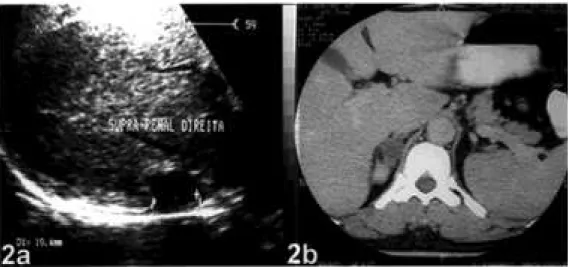
The levels of plasma and 24h-urine aldosterone were increased with values of 23.3ng/ dL (N R: 4-14ng/ dL) and 26.2ug/ 24h (N R: 5-14ug/ 24h), respectively, and plasma renin activity was reduced to 0.1ng/ mL/ h (N R: 0.3-1.6ng/ mL/ h). An abdominal ultrasonography (U S) showed a nodular, hypoe-chogenic image measuring 19mm with well-defined borders in the topography of the right adrenal gland (figure 2a). An adrenal CT revealed the presence of a small mass with soft tissue density with regular and well-defined borders, measuring about 2.0x1.1cm, with no contrast enhancement, located in the right adrenal. The left adrenal was normal (figure 2b).
After admission, the patient received IV potas-sium replacement and serum potaspotas-sium levels stabi-lized between 2.0 and 3.5mEq/ L. The symptoms relieved and the patient became asymptomatic after 3 days. O ral potassium replacement was substituted for the IV replacement and spironolactone 100mg/ day was added. The total CK, aldolase and transaminases levels decreased progressively and became normal in 3 weeks. The patient was discharged asymptomatic with normal potassium levels and mild arterial hypertension using captopril 25mg TID , oral KCl 10mgTID , and spironolactone 100mg/ day. After an 8-wk use of spi-ronolactone, potassium levels remained normal and the patient underwent a right adrenalectomy.
Patho-logical findings revealed a 1.8cm nodule. Microscopi-cal evaluation showed predominance of lipid-laden cortical cells with no areas of anaplasia, necrosis and neither capsule nor vessels invasion (figure 1b). The patient recovered well and both potassium and blood pressure levels stabilized within the normal ranges after progressive tapering of the medication in use.
DISCUSSION
Primary aldosteronism is seldom the cause of sys-temic arterial hypertension being responsible for about 2% of the total of cases in unselected patients (1). The aldosterone-producing adrenal adenoma (APA) is the main cause of primary aldosteronism (1) and should always be ruled out in hypokalemic hypertensive patients. The clinical manifestations of primary aldosteronism include arterial hypertension, muscle weakness, paresthesia, headache, polyuria and polydipsia (1,2). T his case report describes an uncommon initial presentation of primary aldostero-nism that is the symmetric proximal tetraparesis. The symptom probably resulted from severe hypokalemia t h at was po ssib ly accen t u at ed b y t h e u se o f hydrochlorotiazide. Muscular weakness was totally reversed following interruption of the anti-hyperten-sive drugs and potassium replacement.
Most cases of the syndrome of hypokalemic paralysis is due to familial or primary hypokalemic peri-odic paralysis and sporadic cases are related with other conditions such as hyperthyroidism, renal disorders, barium poisoning, gastrointestinal potassium losses and endocrinopathies. The clinical investigation is helpful in identifying the cause of hypokalemic paralysis (3).
the diagnosis of primary aldosteronism. N o correlation was found between the severity of hypokalemia and the size of the adenoma or the level of urinary aldos-terone excretion.
In Taiwan, H uang et al (5) showed that 21 (49%) out of 43 cases of primary aldosteronism pre-sented with muscular paralysis as the initial symptom. They also showed that the serum potassium level of those with muscular paralysis was significantly lower than of those without it.
The differential diagnosis of primary aldostero-nism includes APA, bilateral adrenal hyperplasia and less frequently glucocorticoid-suppressible hyperaldostero-nism and adrenal carcinoma. Measurement of plasma 18-hydroxycorticosterone and assessment of changes in aldosterone levels after corticotropin administration or after changing from supine to erect position might be helpful in defining the specific etiology (6).
Imaging studies should be performed after the initial biochemical analysis. O ver 20% of adrenal ade-nomas are less than 1cm in diameter and difficult to detect (7). Although U S may reveal adrenal masses, CT scanning is the method of choice for the initial characterization of an adrenal mass with an overall sen-sitivity greater than 90% (2). A non-enhanced exami-nation should be performed followed by a contrast–enhanced study if necessary (8). MR imaging has been considered a specific test for detection of adrenal adenomas, with sensitivity of 70%, specificity of 100% and accuracy of 85%, comparable to that reported with CT (9).
Adrenal cortical scintigraphy is rarely needed, given the improvements in CT technology, but it can be useful to determine whether the abnormality is uni-lateral or biuni-lateral (10). Iodine-131 6-beta-iodomethyl-19-norcholesterol (N P-59) can be used to
Periodic Paralysis in H yperaldosteronism
Freitas & Moura
746 Arq Bras Endocrinol Metab vol 47 nº 6 Dezembro 2003
Figure 2. Abdominal US (2a) and CT scan (2b) of the right adrenal gland identifying the aldos-terone-producing tumor.
detect aldosteronomas and other hyperfunctioning cortical tumors. It is still considered investigational by the U .S. Food and D rug Administration despite many years of safe use (8).
In summary, periodic paralysis seems to be a rare event among non-Asian patients with primary aldosteronism. H owever, this possibility must be raised in patients with hypertension associated to hypokalemia and those with severe hypertension (1,6). Appropriate biochemical and imaging studies need to be carried out to avoid misdiagnosis (11) and other causes of myopathies should be ruled out.
REFERENCES
1. Ganguly A. Primary aldosteronism. N Engl J Med 1998;339:1828-34.
2. Bravo EL. Primary aldosteronism.Endocrinol Metab Clin North Am 1994;23:271-82.
3. Ahlawat SK, Sachdev A. Hypokalaemic paralysis. Post-grad Med J 1999;75/882:193-7.
4. Ma JTC, Wang C, Lam KSL, et al. Fifty cases of primary hyperaldosteronism in Hong Kong Chinese with a high frequency of periodic paralysis. Evaluation of tech-niques for tumour localization. QJM New Series 61 1986;235:1021-37.
5. Huang YY, Hsu BR, Tsai JS. Paralytic myopathy - a lead-ing clinical presentation for primary aldosteronism in Tai-wan.J Clin Endocrinol Metab 1996;81:4038-41.
6. Stewart PM. Mineralocorticoid hypertension. Lancet 1999;353:1341-6.
7. Dunnick NR, Leight GS, Robidoux MA, et al. CT in the diagnosis of primary aldosteronism: sensitivity in 29 patients. Am J Roentgenol 1993;160:321-4.
8. Mayo-Smith WW, Boland GW, Noto RB, Lee MJ. State-of-the-art adrenal imaging. Radiographics 2001 ;21:995-1012.
9. Sohaib SA, Peppercorn PD, Allan C, et al. Primary hyper-aldosteronism (Conn Syndrome): MR imaging findings.
Radiology 2000;214:527-31.
10. Ikeda DM, Francis IR, Glazer GM, et al. The detection of adrenal tumors and hyperplasia in patients with primary aldosteronism: comparison of scintigraphy, CT and MR imaging.Am J Roentgenol 1989;153:301-6.
11. Gordon RD, Stowasser M, Rutherford JC. Primary aldos-teronism: are we diagnosing and operating on too few patients?World J Surg 2001;25:941-7.
Endereço para correspondência:
Ricardo Miguel Costa de Freitas Rua Santo Antônio do Monte, 386 30330-220 Belo Horizonte, MG
Periodic Paralysis in H yperaldosteronism
Freitas & Moura
