ABSTRACT
The association of differentiated thyroid cancer and a functioning no-dule is very low. We report on a case of papillary carcinoma in an autonomously functioning thyroid nodule in a 39 year-old female patient. The nodule extended to the whole right lobe and 131I scintigraphy has detected a “hot” nodule and a partial suppression of 131I uptake in the left lobe. Serum TSH levels (RIA) were undetectable (<1.0µUI/mL), but total T3 (190ng/dL) and T4 (8.5µg/dL) were normal. The patient under-went a partial thyroidectomy and an adenomatous nodule was found with a small central nucleus (7mm) hosting a papillary carcinoma. Whole body scans detected only residual thyroid uptake and the patient was subsequently treated with 100mCi of 131I. The patient has been on replacement therapy with 150µg of L-thyroxine and free of tumor recur-rence for 12 years after surgery. In conclusion: the present report confirms other published cases in which the presence of a “hot” thyroid nodule does not exclude the concomitance of a well-differentiated thyroid car-cinoma.(Arq Bras Endocrinol Metab 2003;47/6:739-743)
Keywords: Carcinoma; Papillary; Graves’ disease; Hot nodule; Thyroid carcinoma; Thyroid nodule
RESUMO
Carcinoma Papilar da Tiróide em Nódulo Funcionante Autônomo. A associação de câncer diferenciado da tiróide com nódulo funcio-nante é muito baixa. Apresentamos uma mulher de 39 anos com carci-noma papilar em nódulo autônomo funcionante da tiróide. O nódulo ocupava todo o lobo direito e o estudo cintilográfico com I131detectou um nódulo quente e supressão parcial da captação de I131pelo lobo esquerdo. Os níveis de TSH (RIE) eram indetectáveis (<1,0µUI/mL), mas os de T3 (190ng/dl) e T4 total (8,5µg/dl) normais. Submetida à tiroidectomia parcial, encontrou-se nódulo adenomatoso com um pequeno núcleo central (7mm) com carcinoma papilar. A pesquisa de corpo inteiro mostrou somente captação residual da tiróide e a paciente foi tratada com uma dose de 100mCi de I131. A paciente está recebendo terapia substitutiva com 150µg de L-tiroxina e 12 anos após a cirurgia não apre-senta qualquer sinal de recorrência tumoral. Em conclusão: a paciente descrita confirma outros casos publicados de que a presença de um nódulo “quente” da tiróide não exclui a possibilidade da ocorrência de um carcinoma bem diferenciado. (Arq Bras Endocrinol Metab 2003;47/6:739-743)
Descritores: Carcinoma papilar; Doença de Graves; Nódulo quente; Carcinoma da tiróide; Nódulo da tiróide
T
H E ASSO CIATIO N O F TH YRO ID carcinoma and hyperthyroidism is presently considered more frequent than it was in the past (1). The most frequent association of thyroid carcinoma is with “hot” nodules, withJosé U li sses M. Calegaro
Mari a Stella Oli vei ra D i as
Sung H oon Bae
Si oeme da Si lva Moraes
Eni o de Frei tas Gomes
Lui z A ugusto Casulari
N uclear Medicine U nit (JU MC,SH B), H ospital de Base do Distrito Federal; Endocrinology (MSOD,EFG) and Pathology (SSM) U nits, H ospital de Taguatinga; Escola Superior em
Ciências da Saúde (LA C), H ospital de Base do Distrito Federal, Brasília, DF.
or without manifestations of hyperthyroidism (2-5), whereas the association with Graves’ disease is consi-dered to be rare (6,7).
The majority of thyroid carcinomas associated with hyperthyroidism or with a hyperfunctioning nodule is of the papillary type, followed by follicular carcinoma (2-5,8); less frequently, H ürtle cell (5), anaplastic (8), and medullary carcinoma (9) have also been described.
The diagnosis of thyroid cancer before surgery through biopsy and cytology is not frequent (4), due to the following: a) the association of thyroid cancer and hyperthyroidism is considered to be very rare; b) thyroid carcinoma originates inside the hyperfunctioning no-dule, but it can be located someplace else in the gland (1,6,9). The belief that hyperthyroidism or hyperfunc-tioning nodule would dismiss the possibility of thyroid cancer, and the fact that a thyroid carcinoma can occur just or near the hyperfunctioning nodule, or someplace else in the gland (1), are what makes the pre-surgical diagnosis by biopsy and cytology less frequent (4).
H erein we report the case of a woman with a papillary thyroid carcinoma presenting in an autonomously hyperfunctioning nodule, an uncom-mon combination until 1990, when only 17 cases of this association were reported (1). H owever, in the past 12 years several other cases have been published (3,4,6,7,10-12).
CASE REPORT
A 39 year-old white woman was referred for obesity treatment in 1988. She was nullipara, with regular menses, and had no clinical evidence of hyperthyroidism.
Although the patient was from an endemic goitrous area, there was no record of thyroid diseases in her family his-tory. She did not have a record of radiotherapy on her neck area. H er height and weight were 152cm and 74.5kg (BMI: 31.1). O n physical examination, a large nodule was palpable in the right thyroid lobe.
131I-scintigraphy demonstrated an area of high
iodine uptake occupying all right lobe, with reduced uptake in the left lobe (figure 1). After a T3 suppres-sion test (75µg daily for 8 days), the thyroid scinti-gram remained unchanged: basal and post-suppressive thyroid 131I-uptakes were 47.8% and 32.7%,
respec-tively. T3 (190ng/ dL; N R: 80-210ng/ dL) and T4 (8.5µg/ dL; N R: 4.5-12µg/ dL) were normal by IRM A, but T SH levels were undetectable (<1.0µU I/ mL), as were thyroglobulin and microso-mal antibodies. Routine laboratory data were unre-markable. The patient was diagnosed with a sub-clini-cal autonomous functioning thyroid nodule and observation was recommended.
Three years later the nodule had increased sig-nificantly, extending to the entire right lobe. 131
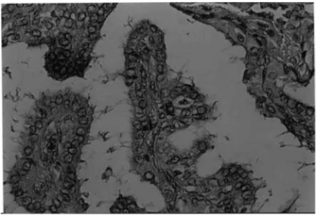
I-scintigraphy demonstrated the same large hyperfunc-tioning area. Two ultrasound (U S)-guided fine needle biopsy aspirations (FN BA) disclosed follicular cells without atypia, and a thyroid surgery was performed. The right thyroid lobe weighed 15g and measured 4.5 x 4.5 x 2.5cm. O n cutting section a 0.7cm white nod-ule of increased elastic consistence was observed; his-tological examination revealed an adenomatous no-dule with a small papillary carcinoma focus (7mm) (figure 2) and vessel invasion. Total thyroidectomy was then performed and the rest of the gland was his-tologically normal.
Figure 1.Thyroid131I-scintigram shows a “hot” nodule in the
right lobe and decreased131I-uptake in the left lobe.
Because a subsequent 131I whole-body scan
showed some uptake in the cervical area, a 100mCi dose of 131I was administered. The patient was
there-after maintained on suppressive L-thyroxine therapy with daily doses of 150µg. O n follow-up TSH (<0.05µU I/ mL) and thyroglobulin (0.5ng/ ml; N R: <38.2) were kept suppressed. Subsequent 131I-whole
body scans were consistently negative.
O ne-year later, conjunctival hyperemia and mild bilateral exophthalmia were observed. O rbital computerized tomography disclosed increased density of the retro-orbital fat, mainly in the right side, with a small thickening of the retro-lateral muscle. The patient has been in good health and free of symptoms for the past 12 years.
DISCUSSION
The association of autonomous nodular goiter and dif-ferentiated thyroid carcinoma seems more frequent than it was supposed, having been increasingly report-ed lately in surgical series: 3.1% (8), 6% (2), 16% (3), 17.8% (5), and 17.9% (13). In children this association is described in 11.3% of the cases (14).
A carcinoma associated with hyperthyroidism is rarely diagnosed before surgery (4) and this is largely due to the fact that the majority of the patients had an occult microcarcinoma defined as a tumor with less than 1cm (5). Fine-needle aspiration biopsy (FN AB) coupled with cytological examination is the most effec-tive procedure for the diagnosis of thyroid cancer (15). H owever, this method is limited when small amount of cells is obtained. The diagnosis of occult thyroid carci-noma was missed in our patient after two FN AB.
Another problem in diagnosing cancer associat-ed to hyperfunctioning nodules by FN AB is that it may not reside inside the adenoma, as in the present case, but can also be someplace else in the gland (1,13). For instance, hyperthyroidism and thyroid car-cinoma were two separate illnesses in 45% of patients (13). Thus, it is important to perform U S-guided FN AB to increase the chances of diagnosing a thyroid cancer, including occult ones with <1cm that could reside in different areas of the gland (15).
As in the present case, hyperthyroidism is most commonly associated with a papillary cancer (2-5,8), and the cytological diagnosis is straightforward provided enough material is obtained by FNAB (15). H owever, in a few patients the cytological diagnosis of follicular cancer is challenging, since capsular or blood vessel inva-sion, as required for diagnosis, may not be evident.
H yperfunctioning thyroid nodule is a benign condition that progress slowly to symptomatic hyper-thyroidism, since most patients are diagnosed after 50 years of age (16). The most common association of hyperfunctioning nodules is papillary cancer (2-5,8), and age (>50 years) seems to be one of the most important prognostic factors in papillary cancer (16). U sually, a careful observation through periodic evalu-ations is indicated for the asymptomatic or sub-clinical forms (16). When clinical hyperthyroidism takes place or TSH becomes suppressed, surgery or radioiodine therapy is indicated; recently, percutaneous intranodu-lar ethanol injections have been also used (17-19).
O ur patient was initially treated with a partial thyroidectomy, due to a large hyperfunctioning right lobe nodule. This type of treatment is effective in pro-viding immediate relief of symptoms (3,16,19,20). Later, when the presence of an occult carcinoma inside the thyroid nodule was verified, total thyroidectomy complemented by ablative 131I-therapy (100mCi) was
carried out. Some investigators recommend that occult thyroid carcinomas (<10mm in diameter, as in our patient), should be treated by lobectomy plus T4 sup-pressive therapy (8,21); they contend that the preva-lence of an occult thyroid carcinoma in a normal popu-lation is 5-10%, whereas the prevalence of clinically evi-dent thyroid cancer is only 0.05%; thus, during a lifes-pan only 1-2% of occult carcinomas may progress to an overt tumor (21). O n the other hand, Furlan et al. (22) demonstrated similar incidences of metastatic nodal ease, distant metastases, recurrent neck metastatic dis-ease, and multicentricity in bearers of occult thyroid cancer and obvious thyroid carcinoma. These authors thereby indicate near-total thyroidectomy and plus radioiodine ablation, as done in our patient. Besides, the blood vessel invasion seen in our patient’s occult carci-noma is an unfavorable histological feature.
Radioiodine therapy for the hyperfunctioning nodule may fail in improving thyroid function (3), and may be associated with a high incidence of hypothy-roidism (16). Radiation doses higher than those used to treat Graves’ disease may be necessary, due to the greater resistance of adenomas (16). H owever, 131
I-therapy seems as effective in treating well-differentiat-ed metastatic thyroid cancer (23), as in treating occult thyroid carcinoma associated with a hyperfunctioning nodule, as in the present case.
hepatocellular carcinoma (24). It is also possible that PIEI could destroy the differentiated carcinoma inside the hyperfunctioning nodule. We are not aware of any patient bearing a hyperfunctioning nodule and treated with PIEI who has developed metastases of an occult carcinoma in the long run. Recently, it has been shown in patients with papillary thyroid carcinoma with metastases limited to cervical nodes that PIEI was successful in the long-term control of metastatic adenopathy (25). This observation justifies the use of PIEI to treat a hyperfunctioning thyroid nodule har-boring an occult thyroid carcinoma (as in our case) without the need of a prior biopsy and/ or surgery.
Finally, we could not rule out the possibility that our patient had Graves’ disease. She had an elevated and only slightly T3-suppressive 131I-uptake. O n
fol-low-up a moderate mostly unilateral exophthalmia occurred and findings of an orbital CT were suggestive of Graves’ disease. The concurrence of toxic adenoma and Graves’ disease is rare (6,7). In addition, the possi-ble association of thyroid carcinoma and Graves’ dis-ease would have made this case even more unusual, since only a few cases have been reported to date (3,4,7,26-29). In a series of 202 patients with hyper-thyroidism who underwent thyroidectomy, thyroid cancer was diagnosed in 5.3% of patients with Graves’ disease (5). In another series of 273 patients with thy-roid carcinoma, 1.5% had Graves’ disease (13).
In conclusion, the present report confirms other published cases in whom the presence of a “hot” nodule on thyroid scintigraphy does not exclude the possible concomitance of a well-differentiated thyroid carcinoma.
ACKNOWLEDGEMENTS
We are indebted to Luiz Gustavo D omingues Casulari da Motta for technical assistance.
REFERENCES
1. De Rosa G, Testa A, Maurizi M, Satta MA, Aimoni C, Artu-so A, et al. Thyroid carcinoma mimicking a toxic adeno-ma.Eur J Nucl Med 1990;17:179-84.
2. Smith M, McHenry C, Jarosz H, Lawrence AM, Paloyan E. Carcinoma of the thyroid in patients with autonomous nodules.Am Surg 1988;54:448-9.
3. David E, Rosen IB, Bain J, James J, Kirsh JC. Management of the hot thyroid nodule. Am J Surg 1995;170:481-3. 4. Ragni F, Pinelli D, Facchini M, Ghedi M, Piccini I, Pasini M,
et al. Thyroid carcinoma in hyperthyroid syndromes. G Chir 1996;17:158-65.
5. Zanella E, Rulli F, Sianesi M, Sciacchitano S, Danese D, Pontecorvi A, et al. Hyperthyroidism with concurrent thy-roid cancer. Ann Ital Chir 2001;72:293-7.
6. Michigishi T, Mizukami Y, Shuke N, Satake R, Noguchi M, Aburano T, et al. An autonomously functioning thyroid carcinoma with euthyroid Graves’ disease. J Nucl Med 1992;33:2024-6.
7. Valenti TML, Macchia E, Pisa R, Bucalo ML, Russo V, Col-letti I, et al. Toxic adenoma and papillary thyroid carci-noma in a patient with Graves’ disease. J Endocrinol Invest 1999;22:701-4.
8. Chou FF, Sheen-Chen SM, Chen YS, Chen MJ. Hyperthy-roidism and concurrent thyroid cancer. Int Surg 1993;78:343-6.
9. Calegaro JUM, Almeida MS, Spadeto JR, Moraes VC, Pinheiro ES. Carcinoma medular da tireóide associado a bócio nodular autônomo: relato de caso. Rev Bras Cancerol 1994;40:43-7.
10. Intenzo CM, Park CH, Cohen SN. Thyroid carcinoma pre-senting as an autonomous thyroid nodule. Clin Nucl Med 1990;15:313-4.
11. Vieira Filho JPB, Cervantes O, Takahashi MH, Kayath MJ, Silva RC. Índia xavante com bócio nodular tóxico asso-ciado a carcinoma folicular. Arq Bras Endocrinol Metab 1992;36:137-9.
12. Appetecchia M, Ducci M. Hyperfunctioning differentiat-ed thyroid carcinoma. J Endocrinol Invest 1998 ;21:189-92.
13. Rösler H, Wimpfheimer C, Ruchti C, Kinser J, Teuscher J. Hyperthyroidism in thyroid cancer. Retrospective study of 53 cases. Nuklearmedizin 1984;23:293-300.
14. Croom RD, Thomas CG, Reddick RL, Tawi MT. Autonomously functioning thyroid nodules in childhood and adolescence. Surgery 1987;102:1101-8.
15. Yokozawa T. Câncer da tireóide detectado pela punção aspirativa por agulha fina guiada pelo ultra-som.Arq Bras Endocrinol Metab 1998;42:296-8.
16. Mazzaferri EL. Management of a solitary thyroid nodule.
N Engl J Med 1993;328:553-9.
17. Livraghi T, Paracchi A, Ferrari C, Bergonzi M, Garavaglia G, Raineri P, et al. Treatment of autonomous thyroid nodules with percutaneous ethanol injection: prelimi-nary results. Work in progress. Radiology 1990;175:827-9. 18. Lippi F, Ferrari C, Manetti L, Rago T, Santini F, Monzani F, et al. Treatment of solitary autonomous thyroid nodules by percutaneous ethanol injections: results of an Italian multicenter study. J Clin Endocrinol Metab 1996; 81:3261-4.
19. Kunori T, Shinya H, Satomi T, Abe M, Kawaguchi S, Honda H, et al. Management of nodular goiters and their oper-ative indications. Surg Today 2000;30:722-6.
20. Montenegro FLM, Mettig PG, Araujo Filho VJF, Brandão LG, Cordeiro AC, Ferraz AR. Considerações sobre o tratamento cirúrgico do bócio nodular tóxico autônomo.Arq Bras Endocrinol Metab 1997;41:168-72. 21. Pelizzo MR, Piotto A, Rubello D, Casara D, Fassina A,
Bus-nardo B. High prevalence of occult papillary thyroid car-cinoma in a surgical series for benign thyroid disease.
22. Furlan JC, Bedard Y, Rosen IB. Biologic basis for the treat-ment of microscopic, occult well-differentiated thyroid cancer.Surgery 2001;130:1050-4.
23. Calegaro JUM, Calegaro NQM, Duarte LV, Araujo MRA, Miziara MD, Gomes EF, et al. Tratamento do carcinoma diferenciado de tireóide com cirurgia e radioiodo-131.
Rev Bras Cancerol 1996;42:209-17.
24. Livraghi T, Salmi A, Bolondi L, Marin G, Arienti V, Monti F, et al. Small hepatocellular carcinoma: percutaneous alcohol injection: results in 23 patients. Radiology 1988;168:313-7.
25. Lewis BD, Hay ID, Charbouneau JW, McIver B, Reading CC, Goellner JR. Percutaneous ethanol injection for treatment of cervical lymph node metastases in patients with papillary thyroid carcinoma. Am J Roentgenol 2002;178:699-704.
26. Shapiro SJ, Friedman NB, Perzik SL, Catz B. Incidence of thyroid carcinoma in Graves’ disease. Cancer 1970;26:1261-70.
27. Farbota LM, Calandra DM, Lawrence AM, Paloyan E. Thyroid carcinoma in Graves’ disease. Surgery 1985;98:1148-53.
28. Behar R, Arganini M, Wu TC, McCormik M, Straus FH, DeGroot LJ, et al. Surgery 1986;100:1121-6.
29. Belfiore A, Garofalo MR, Giuffrida D, Runello F, Filetti S, Fiumara A, et al. Increased aggressiveness of thyroid cancer in patients with Graves’ disease. J Clin Endocrinol Metab 1990;70:830-5.
Endereço para correspondência:
José Ulisses Manzzini Calegaro Unidade de Medicina Nuclear, Hospital de Base do Distrito Federal 70335-900 Brasília, DF