Incidence of pressure ulcers in cardiopulmonary
intensive care unit patients*
Ticiane Carolina Gonçalves Faustino Campanili1, Vera Lúcia Conceição de Gouveia Santos2, Kelly Cristina Strazzieri-Pulido3, Priscilla de Brito Mendes Thomaz1, Paula Cristina Nogueira2
Incidência de úlceras por pressão em pacientes de Unidade de Terapia Intensiva Cardiopneumológica
La incidencia de las úlceras por presión en los pacientes de unidad de cuidados intensivos cardiopneumológica
* Extracted from the dissertation “Incidência de úlcera por pressão e de lesão por fricção em pacientes de Unidade de Terapia Intensiva Cardiopneumológica”, Escola de Enfermagem, Universidade de São Paulo, 2014.
1 Universidade de São Paulo, Faculdade de Medicina, Hospital das Clínicas, Instituto do Coração, São Paulo, SP, Brazil.
2 Universidade de São Paulo, Escola de Enfermagem, Departamento de Enfermagem Medico-Cirúrgica, São Paulo, SP, Brazil. 3 Universidade de São Paulo, Escola de Enfermagem, Programa de Pós-Graduação em Enfermagem na Saúde do Adulto, São Paulo, SP, Brazil.
Received: 04/10/2015 Approved: 07/14/2015
Original article DOI: 10.1590/S0080-623420150000700002
aBStract
Objective: Identify and analyze the incidence coeicients of pressure ulcers (PU) and the risk factors for PU development in critical patients with cardiopulmonary diseases. Method: A prospective cohort study conducted in the cardiopulmonary intensive care unit (ICU) of a large hospital in the city of São Paulo, from November 2013 to February 2014. In total, 370 patients over 18 years old who did not present PU at admission and who had been in the ICU for less than 24 hours were studied. Data were analyzed by univariate and multivariate analyses (Classiication And Regression Tree - CART). Results: he incidence coeicients of PU were: 11.0% for total participants, 8.0% for male and 3.0% for female patients (p=0.018); 10.0% for white patients and 6.5% for patients 60 years or older. he main risk factors were length of stay in the ICU for 9.5 days or more, age 42.5 years or older, and being of the white race. Conclusion: his study was related to the epidemiology of PU in critical patients with cardiopulmonary diseases, facilitating the planning of speciic preventive care for these patients.
DeScriPtOrS
Pressure Ulcer; Incidence; Intensive Care Units; Inpatients; Nursing Care; Epidemiologic Studies.
Correspondence Addressed to:
Ticiane Carolina Gonçalves Faustino Campanili Rua Tagipuru, 1060, Apto. 83, A1 – Barra Funda CEP 01156-000 – São Paulo, SP, Brasil [email protected]
INTRODUCTION
Skin injuries are a great challenge to health profes-sionals who provide care to critical patients, considering that, in clinical practice, the occurrence of pressure ulcers (PU) have become more frequent, with an increase in treatment costs, longer hospitalization periods, discomfort and negative impact on the quality of service provided and the quality of life of patients(1-3).
Skin care programs for the prevention of injuries, based on international guidelines(1,4), have contributed to a reduced number of skin injuries in intensive care units (ICUs), although not all risk factors for these in-juries are known, due to the speciicities of each patient, each associated disease(5) and each scenario. Particularly in cardio pulmonology, due to improved diagnostic and therapeutic methods, repair and corrective surgeries have been indicated late in the process or for patients with serious conditions. his has caused a greater number of risky situations, as these procedures are compromised by comorbidities and other pre-existing diseases(6). he real dimensions of skin injury problems in cardiopulmonary ICU patients has raised the interest of professionals who provide care to these patients.
PU is among skin injuries frequently found in critical patients, highlighted due to its multifactorial etiology(1,7). Several factors are associated with the development of PU, including, speciically for critical patients, extrinsic compres-sion associated with advanced age, nutritional deiciency, humidity, immobility and bed rest, reduced tissue perfusion, use of vasoactive drugs, sedation, and comorbidities like dia-betes mellitus and vascular disease(8-9).
Reduced sensory perception caused by anesthetic agents and longer periods of immobility due to the im-possibility of patient mobilization during the operation(8) facilitate the development of tissue hypoperfusion areas after cardiopulmonary surgeries. Patients whose sensory perception is impaired, that is, who are not able to detect sensations that indicate the need to change position, are among the most susceptible to PU development(6,8-9).
In surgery patients, PU usually appears in the period that includes the surgery up to ive days after the exposure to the compression applied in the surgery room, although here it is often unnoticed or described as hyperemia on-ly,(8-9). he cardiovascular system tolerance to a surgery procedure is inluenced by pharmacological agents that keep venous return and proper systemic vascular resistance. However, the circulatory and respiratory systems may be afected during the surgery, causing hypotension and con-sequent alteration to tissue perfusion, requiring vasoactive drugs to improve blood circulation and preserve cerebral and myocardial tissue irrigation(5,8-9).
In normal conditions, the capillary pressure in skin artery termination is around 32 mmHg, while in venous artery termination, it is about 12 mmHg. Some situations may afect skin perfusion during the patient period in an ICU(1,8-9), such as: reduced blood pressure due to cardiovas-cular alteration, systemic inlammatory response syndrome
(SIRS), septic shock, hemorrhagic shock and shock due to drug use; external pressure kept over 32 mmHg for an extended period on rigid tables or hospital beds without a proper support surface; hemodynamic stability with el-evated external pressure under the body or hemodynamic stability with continuous external pressure. In any of these situations, critical patients have interrupted blood low to the area under pressure, afecting tissue oxygenation and nutrition in that area, with the possibility of ischemia, hy-poxia, tissue acidosis, edema and tissue necrosis(8).
Tissue tolerance to pressure and ischemia depends on tissue nature and it is inluenced by the ability of the skin and supporting structures to redistribute the pressure ap-plied(6,8-9). Critical determinants for the development of PU have been widely discussed by experts for over 30 years and, every day, new factors are discovered regarding the speciicity of each patient and each disease(9).
Considering that health institutes seek to improve the service quality and patient safety, since the 1980s in Brazil, the occurrence of new cases of PU have been used as indica-tors of health service quality, especially in nursing, requiring the evaluation of results from prevention protocol imple-mentation processes. More recently, in April 2013, the Min-istry of Health created the National Patient Safety Program (PNSP), through Ministerial Directive MS/GM 529/2013, and one of the objectives of this program is to monitor PU incidence to minimize it in hospitalized patients(10).
In Brazil, the incidence of PU in critical patients has been explored in few units, especially in the state of São Paulo. National studies show a great variation either in PU incidence coeicients found in critical patients (10.0% to 62.5%)(11-12) or in the evaluation periods (1 to 15 months)(11, 12). International studies show PU incidence in ICU between 3.2% and 39.0%( 13-17).
Considering that critical patients are highly vulnerable to the development of skin injuries and that these injuries involve speciic risk factors, depending on the underlying disease and associated conditions, this study was conduct-ed to identify and analyze the incidence coeicients of PU and the risk factors for PU development in cardiopulmo-nary intensive care unit patients.
METHOD
his is an epidemiological prospective cohort study conducted in the cardiopulmonary ICU of a large public hospital in the city of São Paulo. he study sample had 370 patients admitted to the ICU from November 26, 2013 to February 26, 2014, who met the following inclusion crite-ria: the patients had to be at least 18 years old, have no PU at ICU admission, be in the ICU for less than 24 hours, and present ≤18 Braden scale.
use and time of extracorporeal circulation - ECC, use of blood products, use and concentration of vasoactive drugs at admission, type and number of artifacts (venous and arterial catheters, drains), hospitalization period, date of PU onset, and ICU discharge or death. Besides this in-strument, this study also used: Braden scale, Glasgow Co-ma Scale, Ramsay scale and Acute Physiology and Chronic Health Evolution (APACHE II).
Sociodemographic data, comorbidities, habits and surgery data were collected at admission, while the other clinical data, were collected on a daily basis during the ICU period.
he results were expressed in mean and standard de-viation values for continuous variables, and absolute and relative frequencies for categorical variables. he asso-ciations of PU incidence with demographic, clinical and surgical variables used Fisher exact test for categorical variables, the Student’s test for numerical variables that pre-sented normal distribution and Wilcoxon-Mann-Whitney test for variables with other distribution. After univariate analyses, a decision tree was developed with the Classiica-tion And Regression Tree (CART) algorithm to identify risk
factors for PU development. his statistical analysis tech-nique was selected for its easy result interpretation through charts (tree). Another advantage of the CART is that it does not depend on hypothesis tests. Due to a low preva-lence of the studied efects, logistic regression, frequently used in cohort studies, could lead to estimation problems in this study. he analyses were conducted using R 3.1.0, with 5.0% signiicance level.
he study project was approved by the Research Eth-ics Committees of the institution and hospital (process nº 20780713.4.0000.5392) and followed the ethical standards.
RESULTS
he sample of this study consisted of 360 patients, mean age 57.2 years old (SD=16.0), ranging from 18 to 96 years; with predominance of male (202; 55.0%) and white (307; 8.0%) patients.
Regarding their clinical characteristics, the sample pre-sented mean BMI of 26.0 kg/m² (SD=4.1) ( overweight,) ranging from 17 to 39; and comorbidities, such as: DM (100; 27.0%); SAH (221; 60.0%); DL (112; 30.3%); cig-arette smoking (51; 14.0%); EVA (24; 6.5%) and AF (40; 11.0%); mean APACHE II score of 22.2 (SD=5.0), rang-ing from 2 to 32; mean Braden score of 10.0 (SD=3.0); mean Ramsay score of 5.1 (SD=2.0), ranging from 2 to 6; mean Glasgow scores of 6.0 (SD=5.0) at admission and 15.0 (SD=1.2) at discharge, ranging from 3 to 15; mean time in ICU of 5.5 days (SD=6.0), ranging from 1 to 63 days; use of blood transfusion in 98 patients (26.5%) and vasoactive drugs in 285 (77.0%) participants of the study. Due to the critical condition of patients, artifacts had to be inserted in the skin for cardiac and pulmonary moni-toring, drug administration and luid drainage (venous and arterial catheters, chest drains, temporary epicardial pacing and vesical catheters), leading to mean 5.4 (SD=2.2) arti-fact use by patients, ranging from 0 to 11 artiarti-fact use by patients at admission.
Among the participants, 120 patients (32.4%) were submitted to myocardial revascularization (MR) and 74 (20.0%) changed heart valves; extracorporeal circulation (ECC) was used in 210 patients (57.0%), with mean time of 100.0 minutes (SD=40.3) in surgical procedures, which lasted the mean time of 278.3 minutes (SD=100.0). Of 370 patients of this study sample, 350 (95.0%) were discharged from the ICU; 17 (5.0%) died and 3 (1.0%) remained in the ICU after this study was concluded.
In total, 40 PUs were detected in 40 patients, resulting in a global incidence coeicient of 11.0%: 8.0% in male patients and 3.0% in female patients (p=0.018); and 10.0% in white patients. Regarding age, 6.5% incidence was ob-served in people 60 years or older (Table 1).
Also with regard to age, a statistically signiicant dif-ference (p=0.014) was observed between the mean values for patients with PU (63 years; SD=13.4) and without PU (56.5 years; SD=16.0).
Of 40 PUs found in this study, 16 (40.0%) were classi-ied as category I, 16 (40.0%) as category II, and 8 (20.0%) as suspected deep tissue injury, afecting the sacral region. he PUs were identiied from day 2 to day 23 of the ICU period; with day 3 being the day with the greatest number of PUs diagnosed (eight injuries in the sacral region and two in the calcaneus.
Among the clinical quantitative variables, the groups were significantly different, except for ICU period, longer for patients with PU (14.0; SD=12.0) (p<0.001) and, among the surgical variables, longer ECC time in the group of patients with PU (120.0; SD=68.2) (p=0.030) (Table 2).
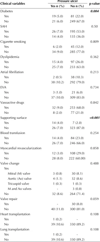
Variables DM and use of supporting surface pre-sented a statistically signiicant diference between the groups (p=0.004; p<0.001, respectively) with and with-out PU (Table 3).
According to the CART analysis, three factors were observed that better identiied the group with PU, that is,
Table 1 - Incidence of PU according to demographic variables - São Paulo, São Paulo, Brazil, 2013-2014.
Demographic variables Pressure ulcer p-value
Yes n (%) no n (%)
Age 0.400
18 to 29 years - 26 (7.0)
30 to 39 years 3 (1.0) 27 (7.1)
40 to 49 years 5 (1.3) 47 (13.0)
50 to 59 years 8 (2.1) 62 (17.0)
60 years or older 24 (6.5) 168 (45.0)
Gender 0.018
Female 11 (3.0) 157 (42.0)
Male 29 (8.0) 173 (47.0)
Ethnicity 0.206
White 36 (10.0) 271 (73.1)
Black - 27 (7.2)
Yellow 1 (0.2) 6 (1.5)
the risk factors for the development of PU: ICU period of 9.5 days or more, age of 42.5 years or older and being of white race (Figure 1).
DISCUSSION
In the last decade, international studies in ICU have shown PU incidence coeicients of PU between 3.2% and 39.0%( 13-17). In post-op cardiac surgery patients(18), an inci-dence of 16.4% was found. In a retrospective study(19) with 2,695 adult surgical patients, conducted in an American quaternary general hospital, an incidence of 11.0% was found, conirming the indings of this study.
In Brazil, the incidence found in ICUs ranges from 10.0(11) to 62.5%( 12). Ina study conducted in four distinct ICUs (two neurological units, one cardiac unit and one general unit) of two private hospitals in the city of São Paulo, the authorsobtained a practically identical result (11.1%)(20) to the results from this study. he lowest index found in Brazil (10, 0%) – also similar to this study – was identiied in a neurosurgical ICU(11), while in another study(12), conducted in an ICU of a large tertiary hospital located in the state of São Paulo, the highest incidence was 62.5%.
In a study(21) about the incidence of PU in a university hospital that used the same methodology as this study, the authors found 41.0% coeicient among critical patients before the implementation of a prevention protocol. More recently, in 2012, one of the same authors found 23.1% in-cidence after the implementation of a prevention protocol, which is still very high, despite a signiicant reduction(4).
Triggering or risk factors are associated with the de-velopment of PU(22-24). In a retrospective study using 222 medical charts of patients with diferent diseases who were submitted to surgeries lasting more than two hours, the authors related the development of PU to factors that af-fected tissue perfusion, like hypertension and diabetes(25). In a comprehensive review of the literature on the use of vasopressors in critical patients and the development of PU(26), the use of a vasopressor (noradrenaline) along with age and extended stay in the ICU were found to be risk factors for the development of PU. In another study(19), the risk factor was the use of blood products used during the
surgery. Other publications(26-28) highlight advanced age, diabetes mellitus and arterial hypertension, including body weight and chronic diseases (vasculopathies, neuropathies and anemia) as risk factors. Two of the factors highlighted (ICU period and age) were conirmed in this study.
In a recent systematic review (54 studies) conducted to identify independent risk factors for PU(27), the authors concluded that there are primary and secondary factors in-volved in the development of PU: mobility/activity,
perfu-Table 2 - Clinical and surgical quantitative variables of patients with and without PU - São Paulo, São Paulo, Brazil, 2013-2014.
clinical variables Pressure ulcer p-value
Yes - Mean (SD) no - Mean (SD)
BMI 25.5 (4.1) 26.0 (4.2) 0.591
Hospitalization time until ICU
admission (days) 9.2 (12.0) 9.5 (35.0) 0.363
ICU period (days) 14.0 (12.0) 4.5 (3.0) <0.001
Number of artifacts (admission) 6.0 (2.5) 5.4 (2.1) 0.551
APACHE II 22.3 (5.0) 22.2 (5.0) 0.689
Ramsay 5.1 (2.0) 5.0 (2.0) 0.901
Braden 10.0 (3.0) 10.0 (3.0) 0.761
ECC time (min.) 120.0 (68.2) 92.1 (35.1) 0.030
Surgery time (min.) 293.0 (133.3) 277.0 (95.5) 0.387
Table 3 - Clinical and surgical categorical variables of patients with and without PU - São Paulo, São Paulo, Brazil, 2013-2014.
clinical variables Pressure ulcer p-value
Yes n (%) no n (%)
Diabetes 0.004
Yes 19 (5.0) 81 (22.0)
No 21 (6.0) 249 (67.0)
SAH 0.50
Yes 26 (7.0) 195 (53.0)
No 14 (4.0) 135 (36.0)
Cigarette smoking 0.809
Yes 6 (2.0) 45 (12.0)
No 34 (9.0) 285 (77.0)
Dyslipidemia 0.362
Yes 15 (4.0) 97 (26.0)
No 25 (7.0) 233 (63.0)
Atrial ibrillation 0.213
Yes 2 (0.5) 38 (10.3)
No 38 (10.2) 292 (79.0)
EVA 0.734
Yes 3 (1.0) 21 (6.0)
No 37 (10.0) 309 (83.0)
Vasoactive drugs 0.842
Yes 32 (9.0) 253 (68.0)
No 8 (2.0) 77 (21.0)
Supporting surface <0.001
Yes 14 (4.0) 7 (2.0)
No 26 (7.0) 323 (87.0)
Blood transfusion 0.254
Yes 14 (4.0) 84 (23.0)
No 26 (7.0) 246 (66.0)
Myocardial revascularization 0.858
Yes 12 (3.0) 108 (29.0)
No 28 (8.0) 222 (60.00)
Valve change 0.488
Yes
Mitral (M) valve 3 (0.8) 30 (8.1)
Aortic (Ao) valve 4 (1.1) 32 (8.6)
Tricuspid valve 1 (0.3) 1 (0.3)
M and Ao valves - 3 (0.8)
No 32 (8.6) 264 (71.4)
Valve repair 0.059
Yes - 30 (8.0)
No 40 (11.0) 300 (81.0)
Heart transplantation 0.108
Yes 1 (0.2)
-No 39 (10.6) 330 (89.2)
Lung transplantation 0.108
Yes 1 (0.2)
sion (including diabetes, vascular disease, arterial pressure, cigarette smoking, edema, and reactive hyperemia were identiied as primary factors; and skin hydration, age, met-abolic alterations, nutrition, and general health condition as secondary factors.
Today, nursing professionals are more and more en-couraged to identify risk factors early and explore them scientiically. he literature shows that more risk factors have to be recognized and analyzed in speciic populations, so that care plans can be better developed(20-23).
he risk factors found in this study are also conirmed partly by the results of other studies, which identiied age above 69 years, ICU period above eight days(28); being of the white race as risk factors.
Age is a widely discussed factor, alone or combined with other variables like cardiovascular diseases, lifestyle and ICU period, as it involves physiological alterations re-lated to the aging process. Loss of muscle mass, reduced inlammatory response, reduced albumin levels and subcu-taneous tissue associated with reduced dermal-epidermal junction cause a higher risk of skin integrity rupture, as the skin does not have the proper ability to redistribute the mechanical load applied, presenting an important item for risk classiication(28-31). he Braden scale, although com-monly used and studied, at national and international lev-els, does not include age(1). Experts claim risk scales should be developed according to the speciic risk factors of each population, based on multivariate analyses. In clinical prac-tice, risk assessment scales that include age as one of the factors involved in PU development should be used, such as the Cubin-Jackson scale(1).
Regarding the ICU period, in a prospective study with 140 patients conducted in Turkey, the time spent by the patients in ICU was found to be a risk factor for the development of PU (p<0.001), always associated with mobility and level of consciousness of patients(31), which is similar to the indings of this study. In this study, the
patients were conined to bed and had reduced mobility due to the great number of artifacts inserted in the skin, use of sedatives and the patient’s fear of feeling pain and discomfort when moving.
A longer period in the cardiopulmonary ICU is neces-sitated due to to the recovery of the physiological condi-tion of patients submitted to invasive procedures, and the use of technologies that allow organ reconditioning and control of associated diseases. he population with car-diopulmonary alterations seeks health services late due to alternative clinical treatments, making surgeries the last option. he patients who developed PU in this study pre-sented impaired skin integrity, predominantly on the third day of hospitalization, conirming the indings of another study(29) that describes onset of 98.0% of PUs in the irst three days of post-operative hospitalization. In two other studies conducted with surgical(30) and critical(20) patients, the authors found out that PUs were diagnosed on the irst two days of ICU hospitalization and that preventive mea-sures should be used during and after surgical procedures. In these studies, the sacral region was also the most afect-ed area, with category II injuries, conirming the results of this study and those found in the literature. he literature shows category II PUs are more frequent in patients with systemic alterations that afect tissue perfusion, such as cardiorespiratory diseases and diabetes mellitus, while cat-egory I PUs are found especially in patients in the imme-diate post-operative period(1,14,28). he preferential region is explained by the dorsal decubitus patient position for long periods, hemodynamic instability and use of artifacts that do not allow or do not favor a change in position in the irst 24 hours after surgery in the ICU. Also regarding cardiac surgery, immediate sternal instability does not en-courage nursing staf to early patient mobilization. hese patients have their sternum sutured with steel threads that allow more efective movements on a bed only after extu-bation, which happens, on average, six to 24 hours after
Node 2 (n=330)
0 0.2 0.4 0.6 0.8
1 Node 4 (n=8)
0 0.2 0.4 0.6 0.8
1 Node 6 (n=8)
0 0.2 0.4 0.6 0.8 1
No
Yes
No
Yes
No
Yes
No
Yes
Node 7 (n=24)
0 0.2 0.4 0.6 0.8 1 5
Race 1
ICU period
<9.5 ≥9.5
≥42.5
<42.5
Indian, Black, Pardo White 3
Age
ICU admission, when the patient can make more active movements, get up and sit on an armchair(1, 28).
In two studies(28, 30), race was an independent predictor of PU development; however, these indings were contra-dictory, as in one of these studies(28), white race was asso-ciated with increased risk for PU (as in this study) and, in the other study(30), black race was similarly associated. he authors of both studies conclude that race and gender are not signiicant independent factors for the development of PU, but they are part of a complex interaction of triggering factors for this injury.
In clinical practice, an association of intrinsic and extrinsic factors for the development of PU in critical pa-tients is a real fact, and they may be avoided by the health team’s ability of early identiication of these predisposing risk factors or factors associated with the development of this type of injury.
he quality of health service has been widely discussed at national and international levels, due to the high cost of service maintenance, scarcity of available resources and the global aging of the population. It should be noted that the quality of service provided has also been evaluated with
re-gard to skin injury, identifying the services that prevent it, and not the services that best treat it. By recognizing that successful prevention of PU is largely dependent on the knowledge and skills of health professionals, it is mandatory to understand individual and institutional factors that in-luence this knowledge and the subsequent use of scientiic evidence. Systematized strategies of PU prevention may be planned and used in institutions, to increasingly reduce and improve the incidence coeicients in speciic populations(1, 28) and in critical post-op cardiopulmonary patients.
CONCLUSION
he results of this study showed the global PU inci-dence coeicient was 11.0%, with predominance of male patients, white race and patients 60 years or older. When analyzing the risk factors or predictors of PU development, the results were; period in the ICU of 9.5 days or more, age of 42.5 years or older, and being of the white race.
his study contributes to knowledge related to the epi-demiology of PU in critical patients with cardiopulmonary diseases, facilitating the planning of speciic preventive care for these patients.
reSUMO
Objetivo: Identiicar e analisar os coeicientes de incidência de úlceras por pressão (UP) e os fatores de risco para o seu desenvolvimento em pacientes críticos com doenças cardiopneumológicas. Método: Estudo de coorte, prospectivo realizado na Unidade de Terapia Intensiva (UTI) Cardiopneumológica de um hospital de grande porte na cidade de São Paulo, durante os meses de novembro de 2013 a fevereiro de 2014. Participaram do estudo 370 pacientes maiores de 18 anos, que não apresentavam UP na admissão e que estavam na UTI há menos de 24 horas. Os dados foram analisados por meio de análises univariadas e multivariada (Classiication And Regression Tree - CART). Resultados: Os coeicientes de incidência de UP foram: 11,0% para o total, distribuindo-se em 8,0% entre os homens e 3,0% para as mulheres (p=0,018); 10,0% na raça branca e 6,5% em pessoas com idade igual e superior a 60 anos. Os principais fatores de risco encontrados foram tempo de permanência na UTI igual ou superior a 9,5 dias, idade igual ou superior a 42,5 anos e raça branca. Conclusão: O estudo contribui para os conhecimentos relacionados à epidemiologia das UP em pacientes críticos com doenças cardiopneumológicas, favorecendo o planejamento de cuidados preventivos especíicos para essa clientela.
DeScritOreS
Úlcera por Pressão; Incidência; Unidades de Terapia Intensiva; Pacientes Internados; Cuidados de Enfermagem; Estudos Epidemiológicos.
reSUMen
Objetivo: Identiicar y analizar la incidencia de las úlceras por presión (UP) y los factores de riesgo para su desarrollo en los pacientes críticos con enfermedades cardiopneumológicas. Método: Estudio prospectivo de cohorte realizado en la Unidad de Cuidados Intensivos (UCI) Cardiopneumológica de un gran hospital en São Paulo, durante los meses de noviembre 2013 hasta febrero de 2014. El estudio incluyó a 370 pacientes mayores de 18 años que no mostraron PU en la admisión a la UCI y que no eran menos de 24 horas. Los datos fueron analizados mediante el análisis univariado y multivariado (regresión y clasiicación Tree - CART). Resultados: La incidência de UP fue el 11,0% in general; de 8,0% en los hombres y de 3,0% en las mujeres (p=0,018); 10,0% en los caucásicos y 6,5% en personas de 60 años o más. Los principales factores de riesgo fueron la duración de la estancia en la UCI inferior a 9,5 días; edades entre 42,5 años y blancos. Conclusión: El estudio contribuye al conocimiento sobre la epidemiología de la UP en pacientes críticamente enfermos con enfermedades cardiopneumológicas ( incidência de 11%), favoreciendo la planiicación de la atención preventiva especíica para esta clientela.
DeScriPtOreS
Úlcera por Presión; Incidencia; Unidades de Cuidados Intensivos; Pacientes Internos; Atención de Enfermería; Estudios Epidemiológicos.
ReFeReNCes
1. National Pressure Ulcer Advisory Panel; European Pressure Ulcer Advisory Panel and Pan Paciic Pressure Injury Alliance. Prevention and
treatment of pressure ulcers: quick reference [Internet]. Washington, DC: EPUAP; 2009 [cited 2015 Apr 04]. Available from: http://www. epuap.org/guidelines/Final_Quick_Treatment.pdf
2. Cox J. Predictors of pressure ulcers in adult critical care patients. Am J Crit Care. 2011;20(5):364-75.
4. Rogenski NMB, Kurcgant P. The incidence of pressure ulcers after the implementation of a prevention protocol. Rev Latino Am Enfermagem [Internet]. 2012 [cited 2015 Apr 04];20(2):333-9. Available from: http://www.scielo.br/scielo.php?script=sci_arttext&pid=S0104-11692012000200016&lng=en&nrm=iso&tlng=en
5. Shanin ES, Dassen T, Halfens RJ. Pressure ulcer prevalence and incidence in intensive care patients: a literature review. Nurs CritCare. 2008;13(22):71-9.
6. Rodrigues Junior GR, Amaral JLG. Inluence of sedation on morbidity and mortality in the intensive care unit. São Paulo Med J.
2004;122(1):8-11.
7. Wound Ostomy and Continence Nurses Society (WOCN). Guideline for prevention and management of pressure ulcers. Mount Laurel; 2010.
8. Lewicki LJ, Mion L, Splane KG, Samstag D, Secic M. Patient risk factors for pressure ulcers during cardiac surgery. AORN J. 1997;65(5):933-42.
9. Sewchuk D, Padula C,Osborne B. Prevention and early detection of pressure ulcers in patients undergoing cardiac surgery. J Wound Ostomy Continence Nurs. 2008;35(1):66-75.
10. Brasil. Ministério da Saúde. Portaria MS/GM n. 529, de 1º de abril de 2013. Institui o Programa Nacional de Segurança do Paciente
(PNSP). Diário Oicial da União, Brasília, 2 abr. 2013. Seção 1, p.43-4.
11. Diccini S, Camaduro C, Ilda LIS. Incidência de úlcera por pressão em pacientes neurocirúrgicos de hospital universitário. Acta Paul
Enferm. 2009;22(2):205-9.
12. Fernandes LM, Caliri MHL. Using the braden and glasgow scales to predict pressure ulcer risk in patients hospitalized at intensive care units.Rev Latino Am Enfermagem [Internet]. 2008 [citado 2015 abr. 15];16(6):973-8. Available from: http://www.scielo.br/scielo. php?script=sci_arttext&pid=S0104-11692008000600006&lng=pt&nrm=iso&tlng=en
13. Shahin ESM, Dassen T, Halfens RJG.Incidence, prevention and treatment of pressure ulcers in intensive care patients: a longitudinal study. Int J Nurs Stud. 2009;46(4):413-21.
14. Sayar S, Turgut S, Doðan H, Ekici A, Yurtsever S, Demirkan F, et al. Incidence of pressure ulcers in intensive care unit patients at risk
according to the Waterlow scale and factors inluencing the development of pressure ulcers. J Clin Nurs. 2009;18(5):776-7.
15. Manzano F, Navarro MJ, Roldán D, Moral MA, Leyva I, Guerrero C, et al.; Granada UPP Group. Pressure ulcer incidence and risk factors in ventilated intensive care patients. J Crit Care. 2010;25(3):469-76.
16. De Laat EH, Pickkers P, Schoonhoven L, Verbeek AL, Feuth T, Van Achterberg T. Guideline implementation results in a decrease of pressure ulcer incidence in critically ill patients. Crit Care Med. 2007;35(3):815-20.
17. Nassaji M, Askari Z, Ghorbani R. Cigarette smoking and risk of pressure ulcer in adult intensive care unit patients. Int J Nurs Pract.
2014;20(4):418-23.
18. Chen HL, Shen WQ, Xu YH, Zhang Q, Wu J. Perioperative corticosteroid administration as a risk factor for pressure ulcers in cardiovascular
surgical patients: a retrospective study. Int Wound J. 2013 Dec 10. [Epub ahead of print].
19. O’Brien DD, Shanks AM, Talsma A, Brenner PS, Ramachandran SK. Intraoperative risk factors associated with postoperative pressure ulcers in critically ill patients: a retrospective observational study. Crit Care Med. 2014;42(1):40-7.
20. Serpa LF, Santos VLCG, Oliveira AS, Caetano VC, Donadon SR. Incidência de úlceras por pressão em pacientes críticos. Rev Estima.
2011;9(3):21-6.
21. Rogenski NMB, Santos VLCG. Estudo sobre a incidência de úlceras por pressão em um hospital universitário. Rev Latino Am Enfermagem
2005;13(4):474-80.
22. Bavaresco T, Medeiros RH, Lucena AF. Implantação da Escala de Braden em uma Unidade de Terapia Intensiva de um hospital universitário.
Rev Gaúcha Enferm. 2011;32(4):703-10.
23. Pereira LC, Luz MHBA, Santana WS, Bezerra SMG, Figueiredo MLF. Incidência de úlceras por pressão em uma Unidade de Terapia Intensiva de um hospital público. Rev Enferm UFPI On Line [Internet]. 2013 [cited 2015 Apr 04];2(4):21-7. Disponível em: http://www. ojs.ufpi.br/index.php/reufpi/article/view/1325/pdf
24. Ventura JA, Moura LTR, Carvalho MFAA. Braden scale and incidence of pressure ulcers in an Intensive Care Unit. Rev Enferm UFPE On Line [Internet]. 2014 [cited 2015 Apr 04];8(7):2047-53. Available from: http://www.revista.ufpe.br/revistaenfermagem/index.php/revista/ article/view/5295/pdf_5507
25. Lumbley JL, Ali SA, Tchokouani LS. Retrospective review of predisposing factors for intraoperative pressure ulcer development. J Clin Anesth. 2014;26(5):368-374.
26. Cox J. Pressure ulcer development and vasopressor agents in adult critical care patients: a literature review. Ostomy Wound Manage. 2013;59(4):50-4,56-60.
27. Coleman C, Gorecki C, Nelson E, Closs SJ, Deloor T, Halfens R, et al. Patient risk factors for pressure ulcer development: systematic
review. Int J Nurs Stud. 2013;50(7):974-1003.
28. Corniello AL, Moyse T, Bates J, Karafa M, Hollis C, Albert NM. Predictors of pressure ulcer development in patients with vascular disease. J Vasc Nurs. 2014;32(2):55-62.
29. Karadag M, Gümüskaya N. The incidence of pressure ulcers in surgical patients: a sample hospital in Turkey. J Clin Nurs. 2006;15(4):413-21.
30. Schoonhoven L, Deloor T, Grypdonck M. Incidence of pressure ulcers due to surgery. J Clin Nurs. 2002;11(4):479-87.
31. Alderden J, Whitney JD, Taylor SM, Zaratkiewicz S. Risk Proile characteristics associated with outcomes of hospital-acquired pressure