Andréia Rodrigues Gonçalves AyresI
Gulnar Azevedo e SilvaII
I Programa de Pós-graduação em Saúde Coletiva. Instituto de Medicina Social (IMS). Universidade Estadual do Rio de Janeiro (UERJ). Rio de Janeiro, RJ, Brasil II IMS-UERJ. Rio de Janeiro, RJ, Brasil
Correspondence:
Andréia Rodrigues Gonçalves Ayres R. Aramã, 9 – Bento Ribeiro 21550-350 Rio de Janeiro, RJ, Brasil E-mail: [email protected] Received: 9/30/2009
Approved: 2/22/2010
Article available from: www.scielo.br/rsp
Cervical HPV infection in
Brazil: systematic review
ABSTRACT
OBJECTIVE: To assess the prevalence of human papillomavirus (HPV)
infection in women in Brazil.
METHODS: A systematic literature review was conducted with an active
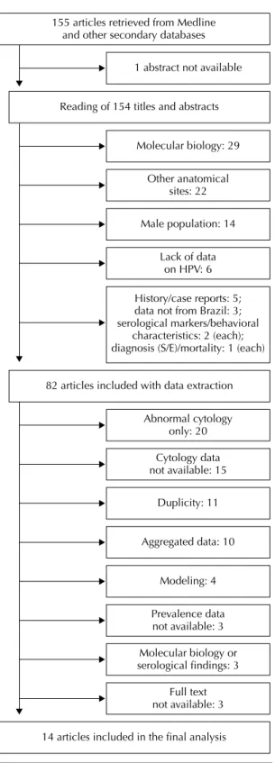
search in PubMed and Virtual Health Library databases using the terms “human papillomavirus,” “HPV,” “prevalence,” and “Brazil”. Of 155 articles retrieved, 82 were selected after reading their title and abstract. After a thorough examination, 14 articles were included in the study.
RESULTS: The 14 articles selected were published between 1989 and 2008
and comprised studies from four Brazilian macroregions (Southeast – 43%; South – 21.4%; Northeast – 21.4%; and North – 7.1%). Nine were cross-sectional studies. Eight articles used polymerase chain reaction and seven used hybrid capture for HPV detection. The study samples ranged from 49 to 2,329 women. The overall prevalence of HPV cervical infection was between 13.7% and 54.3%; and women with cytologically normal results had 10% to 24.5% prevalence of HPV cervical infection. Four articles described the most common HPV types.
CONCLUSIONS: The cytology techniques available use different
classifi cations leading to different HPV prevalence estimates. However, considering the studies individually according to the detection technique used, the HPV prevalence has increased. HPV16 was the most prevalent type among women, regardless of the cytology result. The concentration of studies in the Southeast region, especially in metropolitan regions, evidences that further investigations are needed to improve information coverage of Brazilian women.
DESCRIPTORS: Papillomavirus Infections, epidemiology. Uterine
Cervical. Neoplasms, prevention & control. Scientifi c and Technical Publications. Review.
INTRODUCTION
Cervical cancer is the sixth most common cancer in the general population and the second most common among women.39 In Brazil it is estimated 20,000 new
cases of cervical cancer per year, an estimated rate of 20 per 100,000.a Mortality
rates from cervical cancer show a steady trend with signifi cant reductions in capital cities.60,b There is epidemiological evidence showing that human
papil-lomavirus (HPV) infection is necessary but not suffi cient for the development of cervical cancer.5,35 Low screening coverage and changes in exposure to risk
factors for HPV infection have been described in the analysis of the epidemiology of cervical cancer.6,34
Studies on the prevalence of HPV infection published in Brazil mostly analyzed data from women who sought health services for screening or treatment. Many of them showed exclusively data of women with abnormal cervical cytology results. HPV detection methods and terminology used for reporting results have improved, which may infl uence the assessment of exposure to HPV and cytological diagnosis. Moreover, the fi ndings have not been analyzed together, making it diffi cult to understand the distribution of this infection based on the literature.
The lack of consistent results on the magnitude of cervical cancer creates limitations to the planning of surveillance and control actions. A critical review of studies of Brazilian women on the estimated preva-lence of HPV infection may provide epidemiological knowledge necessary for strengthening and redirecting policies for cervical cancer control. The objective of the present study was to assess the prevalence of HPV infection among Brazilian women, especially in women with normal cytology results, as this estimate would be close to the prevalence of exposure to HPV in the general population.
METHODS
A systematic review of studies on HPV infection in Brazilian women was conducted. There were reviewed publications indexed in the Medical Literature Analysis and Retrieval System (Medline), accessed through PubMed database, the Latin American and Caribbean Health Sciences (LILACS), Cochrane Library, and Scientifi c Electronic Library Online (SciELO). No period of publication was pre-defi ned. Two independent reviewers used a free search strategy using the terms “human papillomavirus,” “prevalence,” and “Brazil” and the Boolean operators “NOT HIV” and “NOT pregnant.” This search was conducted in April 2009. All articles were retrieved in both PubMed and Virtual Health Library.
There were retrieved 155 references that were assessed based on their titles and abstracts and sorted by date of publication (Figure). Most (133) were from PubMed/ MEDLINE. There were selected 82 articles that met the inclusion criteria: they must be conducted with Brazilian women and include cervical cytology results, HPV detection methods, the overall prevalence of HPV in women with cervical cytology results and the prevalence by HPV type in these women.
There were excluded from the review studies on women
c JabRef Reference Manager. [cited 2008 Jun 17] Available from: http://jabref.sourceforge.net/
only with abnormal cervical cytology, immunocom-promised or women who underwent hysterectomy or other surgical procedures with excision of the entire uterus or part of it including the cervix; and studies only reporting laboratory test results with no reference to the population of women studied.
A thorough examination of the articles retrieved was performed and an additional 22 articles were found in the references of these articles. The examination of the entire content of the texts led to the identifi cation of other exclusion criteria, previously not identifi ed, and the exclusion of more articles. The articles were then submitted to a process of data extraction and quality assessment by two independent reviewers.
In the case of publications with data from the same study, we chose to include the article that was most recent, with the largest sample and study period, and more complete data. A total of 14 articles were selected for the study.
All articles retrieved were stored using the open source application Jabref Reference Manager, v. 2.5.c
Data on methodology and study results from the articles were stored in a separate instrument. The authors of three of the articles selected were contacted for complete information.
For quality assessment it was used an instrument consisting of 24 items, adapted from 22 criteria proposed by the STROBE Statement based on epide-miological investigation principles.64 This assessment
aimed to identify the relevance of articles for the prepa-ration of a panel where information could be easily identifi ed and captured to guide recommendations for future studies.
Eighteen criteria items were used for assessing the quality of the studies and six for assessing availability of data for extraction. Each question was given either a score of 0 or 1 (no or yes). The highest score of articles reporting hybrid capture (HC) for HPV detection was 23, and for those reporting polymerase chain reaction (PCR) was 24 as item 12 was only applicable to the latter. Agreement of results was measured by the intra-class correlation coeffi cient (ICC) proposed by Shrout58
(1998) and estimated using SPSS, version 17.0.
RESULTS
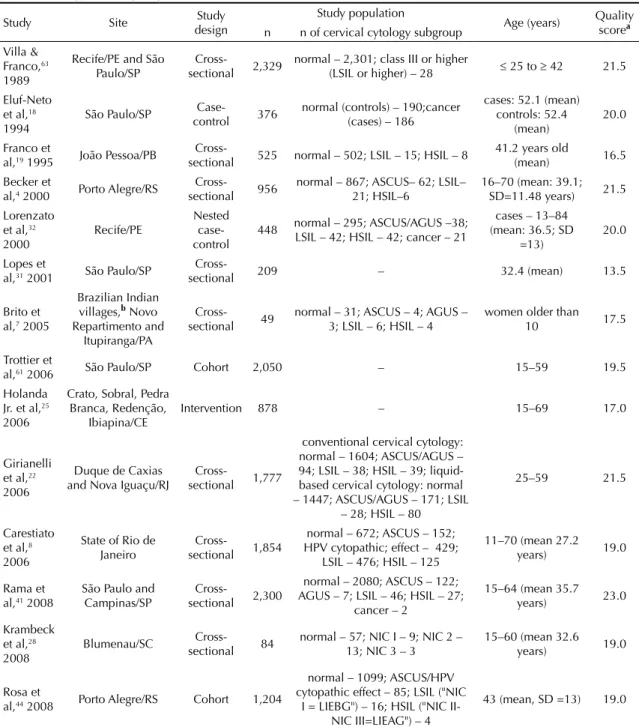
Table 1 shows the distribution of the articles included in the review. Three of them were published between 1989 and 1995;63,18,19 and the remaining were published
from the year 2000.4,7,8,22,25,28,31,32,41,44,61 Three articles
Most studies were conducted in the Southeast region, followed by the South and Northeast (three articles each), and the North (one article). One article is about a study conducted in two regions, Northeast and Southeast.
As for study design, nine were cross-sectional, two were cohort, two were case-control and one was an
experimental study. Four articles reported the preva-lence of HPV in women with any cervical cytology test results, and ten reported the prevalence in women by cytology results, including women with normal cytology.
The samples studied ranged from 49 to 2,329 women.7,63
Eight articles included up to 1,000 women and six
Table 1. Description and quality assessment of the studies selected.
Study Site Study
design
Study population
Age (years) Quality scorea n n of cervical cytology subgroup
Villa & Franco,63 1989
Recife/PE and São Paulo/SP
Cross-sectional 2,329
normal – 2,301; class III or higher
(LSIL or higher) – 28 ≤ 25 to ≥ 42 21.5
Eluf-Neto et al,18 1994
São Paulo/SP
Case-control 376
normal (controls) – 190;cancer (cases) – 186
cases: 52.1 (mean) controls: 52.4
(mean)
20.0
Franco et
al,19 1995 João Pessoa/PB
Cross-sectional 525 normal – 502; LSIL – 15; HSIL – 8
41.2 years old
(mean) 16.5 Becker et
al,4 2000 Porto Alegre/RS
Cross-sectional 956
normal – 867; ASCUS– 62; LSIL– 21; HSIL–6
16–70 (mean: 39.1; SD=11.48 years) 21.5 Lorenzato
et al,32 2000
Recife/PE
Nested case-control
448 normal – 295; ASCUS/AGUS –38; LSIL – 42; HSIL – 42; cancer – 21
cases – 13–84 (mean: 36.5; SD
=13)
20.0
Lopes et
al,31 2001 São Paulo/SP
Cross-sectional 209 – 32.4 (mean) 13.5
Brito et al,7 2005
Brazilian Indian villages,b Novo Repartimento and
Itupiranga/PA
Cross-sectional 49
normal – 31; ASCUS – 4; AGUS – 3; LSIL – 6; HSIL – 4
women older than 10 17.5
Trottier et
al,61 2006 São Paulo/SP Cohort 2,050 – 15–59 19.5
Holanda Jr. et al,25 2006
Crato, Sobral, Pedra Branca, Redenção,
Ibiapina/CE
Intervention 878 – 15–69 17.0
Girianelli et al,22 2006
Duque de Caxias and Nova Iguaçu/RJ
Cross-sectional 1,777
conventional cervical cytology: normal – 1604; ASCUS/AGUS – 94; LSIL – 38; HSIL – 39; liquid-based cervical cytology: normal – 1447; ASCUS/AGUS – 171; LSIL
– 28; HSIL – 80
25–59 21.5
Carestiato et al,8 2006
State of Rio de Janeiro
Cross-sectional 1,854
normal – 672; ASCUS – 152; HPV cytopathic; effect – 429;
LSIL – 476; HSIL – 125
11–70 (mean 27.2 years) 19.0
Rama et al,41 2008
São Paulo and Campinas/SP
Cross-sectional 2,300
normal – 2080; ASCUS – 122; AGUS – 7; LSIL – 46; HSIL – 27;
cancer – 2
15–64 (mean 35.7 years) 23.0
Krambeck et al,28 2008
Blumenau/SC Cross-sectional 84
normal – 57; NIC I – 9; NIC 2 – 13; NIC 3 – 3
15–60 (mean 32.6 years) 19.0
Rosa et
al,44 2008 Porto Alegre/RS Cohort 1,204
normal – 1099; ASCUS/HPV cytopathic effect – 85; LSIL ("NIC
I = LIEBG") – 16; HSIL ("NIC II-NIC III=LIEAG") – 4
43 (mean, SD =13) 19.0
aThe quality assessment scoring was based on a 1-23 scale for hybrid capture studies and 1–24 for polymerase chain reaction
(PCR) studies.
b Parakanã, Paranatinga, Maroxewara, Paranowaona, Itaigoa and Inaxinganga
included more than 1,000 women. Eleven articles described that women were stratifi ed according to their cervical cytology results while no references in this regard were made in the remaining articles.
The minimum age was “older than ten years” and the maximum age was 84 years.7,32 Four articles reported
only the mean age of the women studied. In 12 articles women were recruited from health services and two articles were population-based studies.
The mean score in the quality assessment was 19.18. All articles met at least 15 out of the 18 epidemio-logical research criteria and at least four criteria for data extraction.
There was a signifi cant agreement between scores assigned by reviewers, ICC = 0.81 (95% CI: 0.408, 0.939, p = 0.003).
With respect to the techniques for cervical cytology most articles reported the use of conventional cervical
cytology. Three articles reported using liquid-based and conventional cytology in the same study and sample of women. One article reported the use of “cytology” but the technique applied could not be identifi ed. As for terminology used for reporting test results, the Bethesda System for Reporting Cervical Cytology was mentioned in ten articles, the Bethesda System including the Richart Classifi cation in two articles, Papanicolaou Classifi cation in one article and one article did not mention any classifi cation.
As for HPV detection method, eight articles reported the use of PCR and seven HC. Two articles described both techniques and one article reported in situ hybridization, not currently employed.
Tables 2–4 present the HPV prevalence estimates obtained from the articles studied. Table 2 shows the studies that reported PCR for HPV detection. Six of them reported the use of MY09/11 primer set, one used the GP5/GP6 prime and another one used a generic probe.
Table 2. Characteristics of studies with the use of polymerase chain reaction (PCR) for HPV DNA detection and prevalence of HPV infection among Brazilian women.
Study Site Primer used for typing
Study population HPV prevalence
General population
Women with normal cervical
cytology
General population
Women with normal cervical
cytology Eluf-Neto et al18 1994 São Paulo, SP GP5/GP6 - 194 - 17.0% Franco et al19 1995 João Pessoa, PB MY09/MY11 525 - 18.3% -Becker et al4 2000 Porto Alegre, RS generic probe 956 867 23.0% 20.5%
Lorenzato et al32 2000a Recife, PE MY09/MY11 - 295 - 14.3% Trottier et al61 2006 São Paulo, SP MY09/MY11 2,050 - 16.8% -Krambeck et al28 2008 Blumenau, SC MY09/MY11 84 57 28.6% 18.0% Rosa et al44 2008 Porto Alegre, RS MY09/MY11 1,204 1,099 24.6% 24.5% a Only tested high-risk HPV
SP: São Paulo; PB: Paraíba; RS: Rio Grande do Sul; PE: Pernambuco; SC: Santa Catarina.
Table 3. Characteristics of studies with the use of hybrid capture for HPV DNA detection and prevalence of HPV infection among Brazilian women.
Study Site
Study population HPV prevalence
General population
Women with normal cervical cytology
General population
Women with normal cervical cytology Becker et al4 2000 Porto Alegre, RS 956 867 14.0% 11.6%
Lopes et al31 2001 São Paulo, SP 209 - 21.1% -Girianelli et al22
2006
Nova Iguaçu e Duque de
Caxias, RJ 1,777 1,604 13.7%
10.4% (AR 7.2%; BR 3.2%) Holanda Jr. et al25
2006
Crato, Sobral, Pedra Branca,
Redenção e Ibiapina, CE 878 - 33.9%
-Carestiato et al8
2006 Niterói, RJ 1,854 672 54.3%
12.6% (AR 7.4%; BR 1.8%;
The overall prevalence of HPV infection among women, not stratifi ed by cervical cytology results, ranged from 16.8% (Trottier et al61, 2006) to 28.6%
(Krambeck et al28, 2008). Eluf-Neto et al18 (1994) and
Lorenzato et al32 (2000) articles were not included in
the overall HPV prevalence as they were case-control studies. However, data from controls are presented in Table 2 that shows the studies on women without any lesions. In the articles that included women with normal cytology tested with PCR, the number of women ranged from 5728 to 1,099.19
Two articles showed the overall prevalence of HPV types detected by PCR (Table 4). Franco et al19 (1995)
showed that the overall prevalence of low-risk HPV infection was between 0.2% (HPV54 and HPV68) and 2.3% (HPV11). The same study showed an overall prevalence of high-risk HPV infection between 0.2% (HPV68) and 5.3% (HPV16); 4.0% were not detected.
In Rosa et al study44 (2008), the overall high-risk HPV
prevalence was 3.3% (HPV18) to 18.6% (HPV16).
Two studies estimated the prevalence of HPV types by cytology results, including women with normal cytology (Table 4). Eluf-Neto et al18 (1994) reported a
50.0% prevalence of high-risk HPV types (HPV18) in women with low-grade squamous intraepithelial lesion (LSIL), of low malignant potential and high potential for cure. Women with normal cytology had 0.5% prevalence of low-risk HPV infection (HPV6); 6.3% prevalence of multiple HPV types; 10.0% prevalence of non-detected HPV types; and 0.5% (HPV18) to 5.3% (HPV16) prevalence of high-risk HPV. Krambeck et al28
(2008) reported among women with normal cytology 2.0% (HPV6 and HPV54) and 4.0% (HPV72) preva-lence of low-risk HPV. The prevapreva-lence of HPV CP4773 was 2.0% regardless of risk classifi cation. In women with LSIL the prevalence of low-risk HPV was 4.5%
Table 4. Overall prevalence of HPV types detected by polymerase chain reaction (PCR) and by cytology results.
HPV type detected Overall prevalence
a Prevalence by cervical cytology
Franco et al19 Rosa et al14 Eluf-Neto et al18,b Krambeck et al28,c
Cervical cytology result All All Normal Normal LSIL HSIL Low-risk
HPV 6 2.1% - 0.5% 2.0% -
-HPV 11 2.3% - - - 4.5%
-HPV 54 0.2% - - 2.0% -
-HPV 62 - - - - 4.5%
-HPV 72 - - - 4.0% -
-HPV 81 - - - - 4.5%
-High-risk
HPV 16 5.3% 18.6% 5.3% 2.0% 9.0% 67.0%
HPV 18 2.1% 3.3% 0.5% - -
-HPV 31 0.6% 15.8% - - -
-HPV 33 2.5% - - 2.0% -
-HPV 35 0.8% - - - -
-HPV 45 0.6% - - - 4.5%
-HPV 52 0.8% - - - 4.5%
-HPV 53 1.0% - - 4.0% -
-HPV 56 0.4% - - - -
-HPV 58 1.3% - - - -
-HPV 66 1.3% - - - 4.5%
-HPV 68 0.2% - - - -
-HPV CP4773d - - - 2.0% -
-Not detected 4.0% - 10.0% - 9.0%
-Multiple types - - 6.3% - -
-a women with all cervical cytology results were aggregated b GP5/GP6 primer set
c MY09/MY11 primer set
d not classifi ed according to cervical cancer risk
(HPV 11, HPV 62 and HPV 81), and the same was seen for high-risk HPV types (HPV45, HPV52 and HPV66); 9.0% of HPV types were not detected. In women with high-grade squamous intraepithelial lesion (HSIL)
including moderate to severe dysplastic, precancerous lesions, and carcinoma in situ, the prevalence of HPV was 67.0% (HPV16).
The studies that reported the use of HC for HPV detection are shown in Table 3. The prevalence of HPV infection in all women was between 13.7%22 and
54.3%.8 The number of women with normal cytology
ranged from 6728 to 2.080.41 One study31 reporting
the use of HC described only the overall prevalence and no information was given on the prevalence in women with normal cytology. In those women tested with HC, the prevalence of HPV infection ranged from 10.4%22 to 14.3%.41 Two articles reported HPV groups
detected in women with normal cytology, stratifying them into high- and low-risk. Girianelli et al22 (2006)
and Holanda et al25 (2006) reported the prevalences
of HPV in physician and self-collected samples. Two studies reported the prevalence of HPV by groups: women with normal cytology had prevalence between 1.8% and 3.2% for low-risk HPV and 7.2% and 7.4% for high-risk HPV.8,22
Only one study7 described HPV detection by both
techniques (PCR and HC); however, the prevalences were not reported by technique used. The overall prevalence was 42.8% and the prevalence in women with normal cytology (n=30) was 29.0%. This study is highly relevant since it shows the prevalence in a very specifi c population of Brazilian Indian women.7
DISCUSSION
The overall prevalence of cervical HPV infection ranged from 13.7% to 54.3%. The prevalence of HPV infection among women with normal cytology ranged between 10.4% and 24.5%. Meta-analyses conducted in other settings or that included data from South America countries including Brazil showed prevalence rates in women with normal cytology between 10.0% and 15.9%.2,16
The articles selected showed differences regarding: technique used in cervical cytology; terminology used for reporting results; and methods used for HPV detec-tion or typing.
The articles reported HPV DNA detection and typing of high and low-risk types in women with normal cytology. Studies including women with normal cytology results have gained importance because of improved accuracy of estimates from population-based studies. Studies that include only women with abnormal cytology who were referred to health services because of previous abnormal fi ndings or clinical symptoms tend to overestimate the prevalence of HPV infection in the general population. Studies that include women recruited in family plan-ning or women’s health care units tend to report low or intermediate prevalences.
Figure 1. Flowchart of the selection process of studies on the prevalence of human papilloma virus infection among Brazilian women.
155 articles retrieved from Medline and other secondary databases
1 abstract not available
Reading of 154 titles and abstracts
82 articles included with data extraction Molecular biology: 29
Other anatomical sites: 22
Male population: 14
Lack of data on HPV: 6
Abnormal cytology only: 20
Cytology data not available: 15
Duplicity: 11
Aggregated data: 10
Modeling: 4
Prevalence data not available: 3
Molecular biology or serological findings: 3
Full text not available: 3
14 articles included in the final analysis History/case reports: 5; data not from Brazil: 3; serological markers/behavioral
Thus, prevalence estimates obtained in women recruited at health care services can be over- or underestimated,65
and understanding the dynamics of HPV infection in women with normal cytology is expected to minimize selection bias.16 Knowing the distribution of HPV
infection is essential for the development of new HPV tests and for the assessment of the impact of vaccines in different scenarios.62
The analysis of the distribution of studies over time showed an increasing number of articles published since 2000. It is parallel to the growth of epidemiological research on HPV infection following the establishment of a causal relationship between HPV and cervical cancer. In addition, advances in technologies and methods for HPV detection and typing have improved the estimates.
The cytology techniques available use different classifi -cations leading to different HPV prevalence estimates, which prevents a direct comparison between results. The prevalent HPV type varies depending on cervical cytology results, either normal or not. HPV prevalence also varies according to cytological abnormalities found (type-specifi c; by cytology results; or both).2,14
Also, prevalences vary with the use of certain primers for HPV detection that may be more sensitive to some viral types than others.13 Therefore, the sampling issue
should be considered from a statistical and inferential perspective. Noncompliance of these aspects may introduce bias in the estimates.40
The increased use of liquid-based cytology and HC in recent years led to lower prevalence estimates compared to those reported in studies using conventional cytology and PCR. These inconsistencies, which sometimes are seen within a group of women, support the fi ndings of other studies reporting different sensitivity and positive predictive value of cytology techniques, methods of sample collection (physician or self-collected samples), and HPV detection methods by comparing them and often with colposcopy plus biopsy, using the histopatho-logical result as reference.1,26,68
The present review found that the prevalences esti-mated using PCR are generally higher than those using HC, although no differences in variability between estimates obtained with PCR and HC are seen (ANOVA, p>0.2861). Articles reporting the use of HC are more common in recent years and show lower estimates.4,8,22,25,31,41 They are studies with larger
samples (956 to 2,300 women) than those using PCR (84 to 2,050 women). Only Trottier et al61 (2006) and
Rosa et al44 (2008) studied samples with more than
1,000 women, which may have affected the preci-sion of their estimates. Thus, the use of PCR seems to produce increased HPV prevalences. For example, Becker et al4 (2000) reported 23.0% prevalence with
PCR and 14.0% with HC in the same sample of
956 women. HC seems to produce more consistent estimates with less variability over time. However, considering the studies individually according to the detection technique used, higher HPV prevalences were described in more recent studies. Among the articles that reported the use of PCR, the fi rst one (a 1994 study18) showed a prevalence of 17% and the last
one (a 2008 study44), of 24.5%. Among those reporting
the use of HC, the prevalences were 11.6% and 14.3% in 20004 and 2008,42 respectively.
In the present review estimates of HPV prevalence by type and cytology results are in part coherent with the fi ndings in the literature. A meta-analysis67 carried out
with women across all continents found that HPV16 was the most prevalent type among women, regard-less of the cytology result. However, estimates of HPV prevalence among women with normal cytology described in these studies are not consistent concerning the level of importance of HPV18. Studies using HC reported a prevalence of high-risk HPV about two times higher than that of low-risk HPV. As for low-risk types, we found low prevalence of HPV6 in women with normal cytology in contrast to data reported by other study67 that detected it only in women with
LSIL. Although eight studies used PCR and were able to detect HPV types in all women studied, only two of them reported HPV types found in women with normal cytology. This fact stresses the need for further investigating the prevalence of HPV types by Brazilian states and macroregions and exploring differ-ences in estimates that may due to regional population characteristics.
The analysis of the prevalence and types of HPV by age was prevented by the fact that different age groups were studied and data was not available for each group.
There was a concentration of studies in women in the Southeast region, followed by South, then Northeast and North, and no reports were found for the Central-west region. We identifi ed two large groups of longi-tudinal studies comprising several substudies using the same sample: the Latin American Screening Study (LAMS)17,23,24,29,30,38,42,50,51,59 and the Ludwig-McGill
Study.20,21,27,33,45-48,53-57 Both were prospective cohort
studies for expanding the knowledge about incidence, persistence, and regression of HPV infection.
Although there were retrieved research studies on the subject matter of interest conducted in the state of Rio de Janeiro,9-11 Minas Gerais,3,36,37 and the Federal
District,12,15 these articles did not provide prevalence
estimates including women with normal cytology and thus were not included in the present review.
coverage of Brazilian women. Brazil has Because of Brazil’s continental dimension and socioeconomic and cultural diversity, it is reasonable to assume that Brazilian women have different risks for factors asso-ciated with HPV infection. These differences should be considered in the development of cervical cancer actions and decisions must be taken based on regional contexts and the resolving capacity of health services. Increased screening coverage with assurance of appro-priate follow-up treatment will certainly promote a more favorable scenario.52,66
The articles included in this review do not allow to performing a meta-analysis since the period in which they were published coincided with great improvements in molecular biology techniques. It per se may lead to the calculation of a summary measure based on studies developed in the wake of technology advances in terms of diagnosis. Thus, a meta-analysis would provide results that are diffi cult to interpret and inaccurate. A systematic review including 22 studies was conducted in the United States in 2005 and pointed out similar arguments for not performing a meta-analysis.43
With respect to the limitations of this review, it is possible that other articles of interest were not identi-fi ed with the search strategy. However, we believe this is unlikely to have occurred considering that the main databases were searched and less important articles were also retrieved.49
1. Alves VAF, Bibbo M, Schmidt FCL, Milanezi F, Longatto Filho A. Comparison of manual and automated methods of liquid-based cytology. A morphologic study. Acta Cytologica. 2004;48(2):187-93.
2. Bao YP, Li N, Smith JS, Qiao YL, ACCPAB members. Human papillomavirus type distribution in women from Asia: a meta-analysis. Int J Gynecol Cancer. 2008;18(1):71-9. DOI:10.1111/j.1525-1438.2007.00959.x
3. Barcelos AC, Adad SJ, Michelin MA, Murta EF. Atypical squamous cells of undetermined signifi cance: analysis of microbiology, cytological criteria and clinical conduct. Tumori. 2006;92(3):213-8.
4. Becker E Jr, Edelweiss MI, Nonnenmacher B, Bozzetti MC. Prevalence and epidemiologic correlates of atypical squamous cells of undetermined signifi cance in women at low risk for cervical cancer. Diagn Cytopathol. 2000;24(4):276-82. DOI:10.1002/dc.1059
5. Bosch FX, Lorincz A, Muñoz N, Meijer CJ, Shah KV. The causal relation between human papillomavirus and cervical cancer. J Clin Pathol. 2002;55(4):244-65.
6. Bosch FX, Burchell AN, Schiffman M, Giuliano AR, de Sanjose S, Bruni L, et al. Epidemiology and natural history of human papillomavirus infections and type-specifi c implications in cervical neoplasia. Vaccine. 2008;26 Suppl10:K1-16. DOI:10.1016/j. vaccine.2008.05.064
7. Brito EB, Silva ID, Stávale JN, Taromaru E, Menezess RC, Martins SJ. Amerindian women of the
Brazilian Amazon and STD. Eur J Gynaecol Oncol. 2006;27(3):279-81.
8. Carestiato FN, Silva KC, Dimetz T, Oliveira LH, Cavalcanti SM. Prevalence of human papillomavirus infection in the genital tract determined by hybrid capture assay. Braz J Infect Dis. 2006;10(5):331-6. DOI:10.1590/S1413-8670200600050000
9. Cavalcanti SMB, Frugulhetti ICPP, Passos MRL, Fonseca MEF, Oliveira LHS. Prevalence of human papilomavírus DNA in female cervical lesions from Rio de Janeiro, Brazil. Mem Inst Oswaldo Cruz. 1994;89(4):575-80. DOI:10.1590/S0074-02761994000400013
10. Cavalcanti SMB, Deus FCC, Zardo LG, Frugulhetti ICPP, Oliveira LHS. Human papillomavirus infection and cervical cancer in Brazil: a retrospective study. Mem Inst Oswaldo Cruz. 1996;91(4):433-40. DOI:10.1590/S0074-02761996000400009
11. Cavalcanti SM, Zardo LG, PassosMR, Oliveira LH. Epidemiological aspects of human papillomavirus infection and cervical cancer in Brazil. J Infect. 2000;40(1):80-7. DOI:10.1053/jinf.1999.0596
12. Cerqueira DM, Camara GN, Cruz MR, Silva EO, Brígido MM, Carvalho LG, et al. Variants of human papillomavirus types 53, 58 and 66 identifi ed in Central Brazil. Virus Genes. 2003;26(1):83-7. DOI:10.1023/A:1022386323921
13. Clifford GM, Gallus S, Herrero R, Muñoz N, Snijders PJF, Vaccarella S, et al. Worldwide distribution of human papillomavirus types in cytologically normal women in the International Agency for Research on Cancer HPV prevalence surveys: a pooled analysis.
Lancet. 2005;366(9490):991-8. DOI:10.1023/ A:1022386323921
14. Clifford GM, Smith JS, Plummer M, Muñoz N, Franceschi S. Human papillomavirus types in invasive cervical cancer worldwide: a meta-analysis. Br J Cancer. 2003;88(1):63-73. DOI:10.1038/sj.bjc.6600688
15. Cruz MR, Cerqueira DM, Cruz WB, Camara GNL, Brígido MM, Silva EO, et al. Prevalence of human papillomavirus type 16 variants in the Federal District, Central Brazil. Mem Inst Oswaldo Cruz. 2004;99(3):281-2. DOI:10.1590/S0074-02762004000300007
16. de Sanjosé S, Diaz M, Castellsagué X, Clifford G, Bruni L, Muñoz N, et al. Worldwide prevalence and genotype distribution of cervical human papillomavirus DNA in women with normal cytology: a meta-analysis. Lancet Infect Dis. 2007;7(7):453-9. DOI:10.1016/S1473-3099(07)70158-5
17. Derchain SFM, Rabelo-Santos SH, Sarian LO, Zeferino LC, Zambeli ERO, Westin MCA, et al. Human papillomavírus DNA detection and histological fi ndings in women referred for atypical glandular cells or adenocarcinoma in situ in their Pap smears. Gynecol Oncol. 2004;95(3):618-23. DOI:10.1016/j. ygyno.2004.08.033
18. Eluf-Neto J, Booth M, Muñoz N, Bosch FX, Meijer CJ, Walboomers JM. Human papillomavirus and invasive cancer in Brazil. Br J Cancer. 1994;69(1):114-9.
19. Franco EL, Villa LL, Ruiz A, Costa MC. Transmission of cervical human papillomavirus infection by sexual activity: differences between low and high oncogenic risk types. J Infect Dis. 1995;172(3):756-63.
20. Franco E, Villa L, Rohan T, Ferenczy A, Petzl-Erler M, Matlashewski G. Design and methods of the Ludwig-McGill longitudinal study of the natural history of human papillomavirus infection and cervical neoplasia in Brazil. Rev Panam Salud Publica. 1999;6(4):223-33.
21. Franco EL, Villa LL, Sobrinho JP, Prado JM, Rousseau MC, Désy M, et al. Epidemiology of acquisition and clearance of cervical human papillomavirus infection in women from a high risk area for cervical cancer. J Infect Dis. 1999;180(5):1415-23. DOI:10.1086/315086
22. Girianelli VR, Thuler LCS, Szklo M, Donato A, Zardo LMG, Lozana JA, et al. Comparison of human papillomavirus DNA tests, liquid-based cytology and conventional cytology for the early detection of cervix uteri cancer. Eur J Cancer Prevention. 2006;15(6):504-10. DOI:2006;15(6):504-10.1086/315086
23. Gontijo RC, Derchain SFM, Montemor EBL, Sarian LOZ, Serra MMP, Zeferino LC, et al. Citologia oncológica, captura de híbridos II e inspeção visual no rastreamento de lesões cervicais. Cad Saude Publica. 2005;21(1):141-9. DOI:10.1590/S0102-311X2005000100016
24. Gontijo RC, Derchain SF, Roteli-Martins C, Bragança JF, Sarian LO, Morais SS, et al. Human papillomavirus (HPV) infections as risk factor for cytological and histological abnormalities in baseline PAP smear-negative women followed-up for 2 years in the LAMS Study. Eur J Obstet Gynecol Reprod Biol. 2007;133(2):239-46. DOI:10.1016/j. ejogrb.2006.05.012
25. Holanda F Jr, Castelo A, Veras TM, de Almeida FML, Lins MZ, Dores GB. Primary screening for cervical cancer through self sampling. Int J Gynaecol Obstetr. 2006;95(2):179-84. DOI:10.1016/j.ijgo.2006.07.012
26. Karwalajtys T, Howard M, Sellors JW, Kaczorowski J. Vaginal self sampling versus physician cervical sampling for HPV among younger and older woman. Sex Transm Infect. 2006;82(4):337-9. DOI:10.1136/ sti.2005.019430
27. Kim JJ, Kuntz KM, Stout NK, Mahmud S, Villa LL, Franco EL, et al. Multiparameter calibration of a natural history model of cervical cancer. Am J Epidemiol. 2007;166(2):137-50. DOI:10.1093/aje/kwm086
28. Krambeck WM, Cadidé RM, Dalmarco EM, de Cordova CM. HPV detection and genotyping as an earlier approach in cervical cancer screening of the female genital tract. Clin Exp Obstet Gynecol. 2008;35(3):175-8.
29. Longatto-Filho A, Eržen M, Branca M, Roteli-Martins C, Naud P, Derchain SF, et al. Human papillomavirus testing as an optional screening tool in low-resource settings of Latin America: experience from the Latin American Screening Study. Int J Gynecol Cancer. 2006;16(3):955-62. DOI:10.1111/j.1525-1438.2006.00582.x
30. Longatto-Filho A, Roteli-Martins C, Hammes L, Etlinger D, Pereira SM, Eržen M, et al. Self-sampling for human papillomavirus (HPV) testing as cervical cancer screening option. Experience from the LAMS study. Eur J Gynaecol Oncol 2008;29(4):327-32.
31. Lopes F, Latorre MRDO, Pignatari ACC, Buchalla CM. Prevalência de HIV, papilomavírus humano e sífi lis na Penitenciária Feminina da Capital, São Paulo, 1997-1998. Cad Saude Publica. 2001;17(6):1473-80. DOI:10.1590/S0102-311X2001000600031
32. Lorenzato F, Ho L, Terry G, Singer A, Santos LC, de Lucena Batista R, et al. The use of human papillomavirus in detection of cervical neoplasia in Recife (Brazil). Int J Gynecol Cancer. 2000;10(2):143-50. DOI:10.1046/j.1525-1438.2000.00007.x
33. Maciag PC, Schlecht NF, Souza PS, Rohan TE, Franco EL, Villa LL. Polymorphisms of the human leukocyte antigen DRB1 and DQB1 genes and the natural history of human papillomavirus infection. J Infect Dis. 2002;186(2):164-72. DOI:10.1086/341080
34. Martins LFL, Thuler LCS, Valente JG. Cobertura do exame de Papanicolaou no Brasil e seus fatores determinantes: uma revisão sistemática da literatura. Rev Bras Ginecol Obstet. 2005;27(8):485-92. DOI:10.1590/S0100-72032005000800009
35. Muñoz N, Bosch FX, de Sanjosé S, Herrero R, Castellsagué X, Shah KV, et al. Epidemiologic classifi cation of human papillomavirus types associated with cervical cancer. N Engl J Med. 2003;348(6):518-27. DOI:10.1056/NEJMoa021641
36. Murta EFC, Lombardi W, Borges LS, Souza MAH, Adad SJ. Freqüência de infecção pelo papilomavírus humano em mulheres com ectopia cervical. Rev Bras Ginecol Obstet. 1999;21(8):447-9.
37. Murta EF, de Souza MA, Araújo Júnior E, Adad SJ. Incidence of Gardnerella vaginalis, Candida sp and
human papillomavirus in cytological smears. Sao Paulo Med J. 2000;118(4):105-8. DOI:10.1590/S1516-31802000000400006
38. Nonogaki S, Wakamatsu A, Longatto Filho A, Pereira SM, Utagawa ML, Ferreira Alves VA, et al. Hybrid capture II and polimerase chain reaction for identifying HPV infections in samples collected in a new collection medium: a comparison. Acta Cytol. 2004;48(4):514-20.
39. Parkin DM, Bray F, Ferlay J, Pisani P. Global cancer statistics, 2002. CA Cancer J Clin. 2005;55(2):74-108. DOI:10.3322/canjclin.55.2.74
40. Pearce N. Effect measures in prevalence studies. Environ Health Perspect. 2004;112(10):1047-50.
41. Rama CH, Roteli-Martins CM, Derchain SFM, Longatto-Filho A, Gontijo RC, Sarian LOZ, et al. Prevalência do HPV em mulheres rastreadas para o câncer cervical. Rev Saude Publica. 2008;42(1):123-30. DOI:10.1590/S0034-89102008000100016
42. Rama C, Roteli-Martins C, Derchain S, Longatto-Filho A, Gontijo R, Sarian L, et al. Rastreamento anterior para câncer do colo uterino em mulheres com alterações citológicas ou histológicas. Rev Saude Publica. 2008;42(3):411-9. DOI:10.1590/S0034-89102008000300004
43. Revzina NV, Diclemente RJ. Prevalence and incidence of human papillomavirus infection in women in the USA: a systematic review. Int J STD AIDS. 2005;16(8):528-37. DOI:10.1258/0956462054679214
44. Rosa MI, Fachel JM, Rosa DD, Medeiros LR, Igansi CN, Bozzetti MC. Persistence and clearance of human papillomavirus infection: a prospective cohort study. Am J Obstet Gynecol. 2008;199(6):617.e1-7.
45. Rousseau MC, Franco EL, Villa LL, Sobrinho JP, Termini L, Prado JM, et al. A cumulative case-control study of risk factor profi les for oncogenic and non-oncogenic cervical human papillomavirus infections. Cancer Epidemiol Biomarkers Prev. 2000;9(5):469-76.
46. Rousseau MC, Pereira JS, Prado JC, Villa LL, Rohan TE, Franco EL. Cervical coinfection with human papillomavirus (HPV) types as a predictor of acquisition and persistence of HPV infection. J Infect Dis. 2001;184(12):1508-17. DOI:10.1086/324579.
47. Rousseau MC, Abrahamowicz M, Villa LL, Costa MC, Rohan TE, Franco EL. Predictors of cervical coinfection with multiple human papillomavirus types. Cancer Epidemiol Biomarkers Prev. 2003;12(10):1029-37
48. Rousseau MC, Villa LL, Costa MC, Abrahamowicz M, Rohan TE, Franco EL. Occurrence of cervical infection with multiple human papillomavirus types is associated with age and cytologic abnormalities. Sex Transm Dis. 2003;30(7):581-7. DOI:10.1097/00007435-200307000-00010
49. Sampson M, Barrowman NJ, Moher D, Klassen TP, Pham B, Platt R, et al. Should meta-analysis search Embase in addition to Medline? J Clin Epidemiol. 2003;56(10):943-55. DOI:10.1016/S0895-4356(03)00110-0
(VILI), cervical cytology and HPV testing as cervical screening tools in Latin America. J Med. Screen. 2005;12(3):142-9. DOI:10.1258/0969141054855328
51. Sarian LO, Hammes LS, Longatto-Filho A, Guarisi R, Derchain SF, Roteli-Martins C, et al. Increased risk of oncogenic human papillomavirus infections and incident high-grade cervical intraepithelial neoplasia among smokers: experience from the Latin American screening study. Sex Transm Dis. 2009;26(4):241-8. DOI:10.1097/OLQ.0b013e3181935a7d
52. Schiffman M, Castle PE, Jeronimo J, Rodriguez AC, Wacholder S. Human papillomavirus and cervical cancer. Lancet. 2007;370(9590):890-907. DOI:10.1016/S0140-6736(07)61416-0
53. Schlecht NF, Kulaga S, Robitaille J, Ferreira S, Santos M, Miyamura RA, et al. Persistent human papillomavirus infection as a predictor of cervical intraepithelial neoplasia. JAMA. 2001;286(24):3106-14. DOI:10.1001/jama.286.24.3106
54. Schlecht NF, Platt RW, Duarte-Franco E, Costa MC, Sobrinho JP, Prado JC, et al. Human papillomavirus infection and time to progression and regression of cervical intraepithelial neoplasia. J Natl Cancer Inst. 2003;95(17):1336-43.
55. Schlecht NF, Platt RW, Negassa A, Duarte-Franco E, Rohan TE, Ferenczy A, et al. Modeling the time dependence of the association between human papillomavirus infection and cervical cancer precursor lesions. Am J Epidemiol. 2003;158(9):878-86. DOI:10.1001/jama.286.24.3106
56. Schlecht NF, Trevisan A, Baggio ML, Galan L, Duarte-Franco E, Greenberg MD, et al. Lack of agreement between cervicography and cytology and the effect of human papillomavirus infection and viral load. J Lower Genit Tract Dis. 2006;10(4):229-37. DOI:10.1097/01. lgt.0000225892.03613.2c
57. Schlecht NF, Trevisan A, Duarte-Franco E, Rohan TE, Ferenczy A, Villa LL, et al. Viral load as a predictor of the risk of cervical intraepithelial neoplasia. Int J Cancer. 2003;103(4):519-24. DOI:10.1002/ijc.10846
58. Shrout P. Measurement reliability and agreement in psychiatry. Stat Methods Med Res.1998;7(3):301-17. DOI:10.1191/096228098672090967
59. Syrjänen K, Naud P, Derchain S, Roteli-Martins C, Longatto-Filho A, Tatti S, et al. Comparing PAP
smear cytology, aided visual inspection, screening colposcopy, cervicography and HPV testing as optional screening tools in Latin America. Study and baseline data of the LAMS Study. Anticancer Res. 2005;25(5):3469-80.
60. Thuler LCS, Mendonça GA. Estadiamento inicial dos casos de câncer de mama e colo do útero em mulheres brasileiras. Rev Bras Ginecol Obstet. 2005;27(11):656-60. DOI:10.1191/096228098672090967
61. Trottier H, Mahmud S, Costa MC, Sobrinho JP, Duarte-Franco E, Rohan TE, et al. Human papillomavirus infections with multiple types and risk of cervical neoplasia. Cancer Epidemiol Biomarkers Prev. 2006;15(7):1274-80. DOI:10.1158/1055-9965.EPI-06-0129
62. Vaccarella S, Herrero R, Dai M, Snijders PJF, Meijer CJLM, Thomas JO, et al. Reproductive factors, oral contraceptive use, and human papillomavirus infection: pooled analysis of the IARC HPV prevalence surveys. Cancer Epidemiol Biomarkers Prev. 2006;15(11):2148-53. DOI:10.1158/1055-9965.EPI-06-0556
63. Villa LL, Franco EL. Epidemiologic correlates of cervical neoplasia and risk of human papillomavirus infection in asymptomatic women in Brazil. J Natl Cancer Inst. 1989;81(5):332-40. DOI:10.1093/ jnci/81.5.332
64. von Elm E, Altman DG, Egger M, Pocock SJ, Gøtzsche PC, Vandenbroucke JP. The Strengthening the Reporting of Observational Studies in Epidemiology (STROBE) statement: guidelines for reporting observational studies. PloS Med. 2007;4(10):e296. DOI:10.1371/ journal.pmed.0040296
65. Weller CS, Stanberry LR. Estimating the population prevalence of HPV. JAMA. 2007;297(8):876-8. DOI:10.1001/jama.297.8.876
66. World Health Organization. International Agency for Research on Cancer. Cervix cancer screening. Lyon; 2005. (IARC Handbooks of cancer prevention, 10).
67. WHO/ICO Information Centre on HPV and Cervical Cancer. HPV and cervical cancer in the 2007 report. Vaccine. 2007;25(Suppl 3):C1-230.
68. Wright TC Jr, Denny L, Kuhn L, Pollack A, Lorincz A. HPV DNA testing of self-collected vaginal samples compared with cytologic screening to detect cervical cancer. JAMA. 2000;283(1):81-6. DOI:10.1001/ jama.283.1.81
Article based on the Master’s dissertation by ARG Ayres submitted to the Institute of Social Medicine, Universidade Estadual do Rio de Janeiro, in 2009.