Lateral wedge insole for knee osteoarthritis:
randomized clinical trial
Palmilha valgizante para osteoartrite de joelhos: ensaio clínico randomizado
Gustavo Constantino de Campos
I, Marcia Uchôa Rezende
II, Thiago Pasqualin
III, Renato Frucchi
III, Raul Bolliger Neto
IVInstitute of Orthopedics and Traumatology, Hospital das Clínicas, Faculdade de Medicina da Universidade de São Paulo (FMUSP),
São Paulo, Brazil
ABSTRACT
CONTEXT AND OBJECTIVE: Optimal management of knee osteoarthritis requires a combination of
pharmacological and non-pharmacological methods. The use of lateral wedge insoles to treat medial knee osteoarthritis is recommended, but there is still controversy about its eicacy. The purpose of this study was to ascertain whether the use of lateral wedge insoles can diminish pain and improve function in patients with medial knee osteoarthritis.
DESIGN AND SETTING: Prospective randomized trial conducted in a tertiary-level hospital.
METHODS: We prospectively enrolled 58 patients with medial knee osteoarthritis and randomized them to use either a lateral wedge insole with subtalar strapping (Group W), or a neutral insole with subtalar strapping (Group N - control). All the patients were instructed to use the insole for ive to ten hours per day. A visual analogue pain scale, the Western Ontario and McMaster Universities Arthritis Index (WOMAC) and the Lequesne questionnaire were applied at baseline and at weeks 2, 8 and 24.
RESULTS: At weeks 8 and 24, both groups showed lower scores for WOMAC (P = 0,023 and P = 0,012 respectively). There were no statistically signiicant diferences between the groups regarding the visual analogue pain scale, WOMAC or Lequesne results at any time evaluated.
CONCLUSION: The use of a lateral wedge insole with subtalar strapping improved the patients’ symptoms and function but was not superior to placebo insoles.
CLINICAL TRIAL REGISTRATION: NCT01739296.
RESUMO
CONTEXTO E OBJETIVO: O manejo ideal da osteoartrite de joelhos requer combinação entre modalida-des farmacológicas e não farmacológicas. O uso de palmilhas valgizantes no tratamento da osteoartrite medial do joelho é recomendado, mas sua eicácia ainda é controversa. Este estudo objetiva veriicar se o uso da palmilha valgizante pode diminuir a dor e melhorar a função dos pacientes com osteoartrite medial dos joelhos.
DESENHO E LOCAL: Ensaio clínico prospectivo e randomizado conduzido em hospital de atenção terciária.
MÉTODOS: Alocamos prospectivamente 58 pacientes com osteoartrite medial dos joelhos que foram randomizados para fazer uso de palmilha valgizante com amarrilho subtalar (Grupo W) ou palmilha neutra com amarrilho subtalar (Grupo N - controle). Todos os pacientes foram orientados a utilizar a palmilha en-tre cinco e dez horas por dia. Foram aplicados os questionários Western Ontario and McMaster Universities Arthritis Index (WOMAC) e Lequesne, além da escala visual analógica da dor, nos momentos pré e após 2, 8 e 24 semanas.
RESULTADOS: Após 8 e 24 semanas, ambos os grupos apresentaram redução dos valores de WOMAC
(P = 0,023 e P = 0,012 respectivamente). Não houve diferença estatisticamente significativa entre os grupos nos resultados de WOMAC, Lequesne e escala visual analógica de dor, em nenhum dos momentos avaliados.
CONCLUSÃO: O uso de palmilha valgizante com amarrilho subtalar melhorou os sintomas e a função dos pacientes, mas não foi melhor que placebo.
REGISTRO DE ENSAIO CLÍNICO: NCT01739296.
IMD. Doctoral Student, Faculdade de Medicina
da Universidade de São Paulo (FMUSP), São Paulo, Brazil.
IIMD, MSc, PhD. Head of Osteometabolic Disease
Group, Hospital das Clínicas (HC), Faculdade de Medicina da Universidade de São Paulo (FMUSP), São Paulo, Brazil.
IIIMD. Medical Volunteer, Osteometabolic Disease
Group, Hospital das Clínicas (HC), Faculdade de Medicina da Universidade de São Paulo (FMUSP), São Paulo, Brazil.
IVMD, MSc, PhD. Attending Physician in
Institute of Orthopedics and Traumatology, Hospital das Clínicas (HC), Faculdade de Medicina da Universidade de São Paulo (FMUSP), São Paulo, Brazil.
KEY WORDS:
Osteoarthritis. Orthotic devices.
Randomized controlled trial [publication type]. Orthopedics.
Knee joint.
PALAVRAS-CHAVE:
Osteoartrite. Aparelhos ortopédicos.
Ensaio clínico controlado aleatório. Ortopedia.
INTRODUCTION
Osteoarthritis is a major cause of chronic musculoskeletal pain and disability in the elderly population.1 Knee osteoarthritis is one of the most common forms of osteoarthritis and the most frequent chronic condition that leads to functional limitation in older adults, afecting more people than any other joint dis-ease.2 Recent guidelines recommend that optimal management of osteoarthritis requires a combination of non-pharmacologi-cal and pharmacologinon-pharmacologi-cal methods.3 All patients should be given access to information and education about the objectives of treat-ment and the importance of changes in lifestyle, exercise, pacing of activities, weight reduction and other measures to reduce the load on the damaged joint(s), such as walking aids, knee braces and insoles, as well as muscle strengthening and weight loss.3
Involvement of the medial compartment of the knee is ten times more common than involvement of the lateral compartment.4,5 Knee osteoarthritis in the medial compartment is strongly associated with biomechanical factors, particularly progressive varus deformity, which systematically increases the load on the medial compartment, thus further increasing the risk of damage to this compartment.6,7
Biomechanical and clinical studies have shown that lateral wedge insoles can promote a reduction in the adduction moment of 4 to 12% during gait, thus reducing the load on the medial knee compartment8-10 and promoting symptomatic beneit for some patients with medial compartment tibiofemoral osteoarthritis.11 he use of lateral wedged insoles for patients with medial com-partment knee osteoarthritis is certainly a very interesting treat-ment option because of its low cost, low complexity and virtually absence of side efects.12,13 However, apart from Japan, where sev-eral studies have demonstrated its eicacy,10,14-16 the recent litera-ture on the use of lateral wedges for medial compartment knee osteoarthritis is insuicient to draw any substantial conclusions. Two recent systematic reviews have shown limited evidence to support the use of lateral wedge orthotics for reducing pain, increasing function or slowing disease progression.17,18
OBJECTIVE
he purpose of this study was to ascertain whether the use of lat-eral wedge insoles can diminish pain and improve function in patients with medial knee osteoarthritis.
METHODS
his prospective single-blind parallel group controlled trial was conducted under the principles of the Helsinki Declaration and was approved by CAPPesq (Ethics Committee for analysis of research projects) under the protocol number 839/2011. It followed the CONSORT (Consolidated Standards of Reporting Trials) Statement and evaluated 58 patients with knee osteoarthritis (Figure 1). It was registered at clinicaltrials.gov under the number NCT01739296.
Eligibility criteria
he eligibility criteria were that patients needed to meet the American College of Rheumatology criteria for knee osteoarthritis19 and to present varus malalignment of the knee; absence of hip osteoarthritis; absence of ankle pain; absence of previous fracture on the index knee; absence of previous surgery on the index knee; absence of rheumatoid arthritis; and lack of intra-articular injection in the index knee in the past six months. he patients also needed to have been receiving the usual care for osteoarthritis for at least six months and to be able to understand and agree with the informed consent statement.
Exclusion criteria
he exclusion criteria for this study were: - Undergoing surgery during the study period.
- Undergoing intra-articular injection during the study period. - Developing infection of the index joint during the
study period.
One hundred and thirty-eight patients were interviewed, and ity-eight patients met the inclusion criteria (Figure 1). All the patients in our department receive the same treatment protocol, which we call the usual care for knee osteoarthritis.
Figure 1. Study low diagram.
Patients interviewed (n = 138) Enrollment
Eligibility
Allocation
Follow-up
Met inclusion criteria (n = 58)
Randomization
Statistical analysis (n = 58) Week Zero
n = 29
Week Zero n = 29
Week 2 n = 29
Week 2 n = 29
Week 8 n = 29
Week 8 n = 29
Week 24 n = 29 Week 24
he usual care consists of patient education through lectures, handouts, audiovisual material and guidance given by orthope-dic surgeons, nutritionists, psychologists, occupational thera-pists, physical therathera-pists, physical educators and social workers. All patients, except those with contraindications, take analge-sics (on demand), such as paracetamol and codeine. We do not routinely give non-steroidal anti-inlammatory drug (NSAIDs) to our patients.
All the patients invited to participate in the present study agreed to do so. he study was conducted in an outpatient setting at a tertiary hospital.
One week before start to use the orthotic devices, the patients who met the criteria gave responses on the visual analogue scale for pain (VAS) and to the Western Ontario and McMaster Universities Arthritis Index (WOMAC)20 and Lequesne questionnaire.21 Anthropometric data was also collected, such as age, gender, race, height, weight and body mass index (BMI). Plain radiographs of the knees were available for all the patients, in anteroposterior view with unilateral weight bearing and in lateral and patellar axial views. hree of us (GCC, TP, RF) examined all the radiographs in order to classify the severity of the osteoarthritis using the Kellgren-Lawrence scheme.22 In 18 cases, there was interobserver disagreement. In all of these cases, we took into consideration the classiication level given by the majority (two observers). None of the radiographs resulted in total discordance (three diferent classiications).
he patients were randomly divided into two groups of 29 patients by means of simple randomization. he random-ization was performed using a computer-generated program (available from: http://www.randomization.com/) and was done by an investigator who did not have any involvement in the rest of the study. he patients were conidentially allocated to the lateral wedge insole group (Group W) or the neutral insole group (Group N). Although the patients did not know which group they were in, they were not considered to be blind to this, since they could see the shape of the insole.
All the patients used insoles on both feet. Group W patients with unilateral knee osteoarthritis used a lateral wedge insole on the afected limb and a neutral insole on the contralateral limb. Group W patients with bilateral disease used a lateral wedge insole on both limbs. Group N patients used a neutral insole on both limbs.
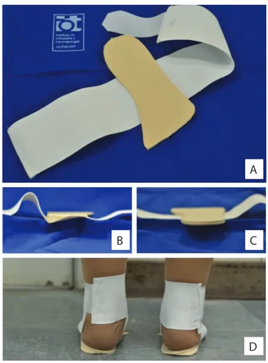
he wedge insoles were made with a full length lateral wedge of 8 mm (equivalent to about eight degrees of inclination) attached to a igure “eight” strap around the ankle (Figure 2). he neutral insoles were exactly the same orthosis, but without a lateral wedge. All the patients were encouraged to use the insoles for 5-10 hours per day.
he VAS, WOMAC and Lequesne questionnaires were again applied during scheduled visits in weeks two, eight and 24. he primary outcomes measured were knee pain and knee
function improvement, which were expressed through the results of the questionnaires applied. he secondary outcomes were the presence of adverse efects (such as ankle pain) and any correla-tion between anthropometric data and clinical outcomes.
he sample size was estimated by calculating n such that it enabled statistical power of 80% and a signiicance level of 5%. For the primary outcome of VAS, we took the standard devia-tion (SD) of 1.9 that had been found in a previous study23 and stipulated that the diference to be detected should be at least 1.5 points. Using a two-tailed hypothesis test, we found that the number of patients per group should be 25. Taking estimates of 20% for dropouts and exclusions, we calculated that 29 patients per group were needed.
he investigator (MUR) who applied the questionnaires was blinded (unaware of the patient’s group). To determine whether the groups difered with regard to the nominal variables, we used absolute and relative frequencies, and checked for associations using chi-square for gender and the likelihood ratio for race. he Mann-Whitney test was used to compare groups regarding
A
B
C
Figure 2. Insoles. (A) Full length ethylene vinyl acetate (EVA) insole attached to an ankle-sprain support; (B) lateral wedge insole (Group W); (C) neutral insole (Group N); (D) patient wearing a lateral wedge insole.
the Kellgren-Lawrence grade. he quantitative characteristics were described in groups through using summary measurements (mean, SD, median, minimum and maximum), and the groups were compared using Student’s t-test. We used a signiicance level of 5% for all analyses.
RESULTS
he patients were recruited between June 2011 and July 2011. Twenty-nine patients were randomly assigned to each group, received the intended treatment and were analyzed for func-tional and pain status using VAS and the WOMAC and Lequesne questionnaires. All the patients were evaluated clini-cally and started using the lateral wedge insoles between August 2011 and September 2011. he trial ended in March 2012, in week 24 of the follow-up. here were no losses.
here were no diferences between the groups in relation to nominal and numerical characteristics (Table 1) or scores
(Table 2) at baseline. Scales were also described as groups and
times through using summary measurements (Table 3). he rela-tionships among the results were analyzed using Pearson’s corre-lations regarding numerical characteristics such as age and body mass index (BMI) and using Spearman’s correlation regarding Kellgren-Lawrence grade (Table 4).
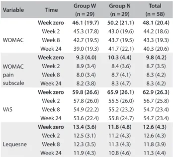
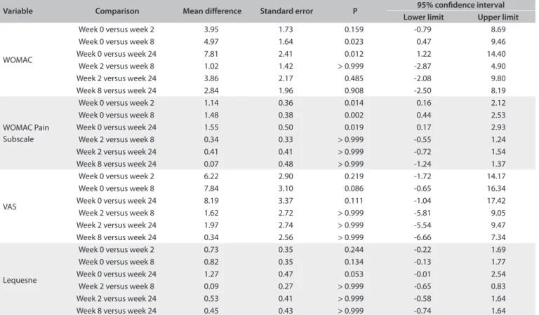
At weeks 8 and 24, both groups showed lower results for WOMAC, with a diference from baseline (P = 0.023 and P = 0.012 respectively). he mean WOMAC pain subscale score showed statistically signiicant reductions at all times. in com-parison with the baseline, in both groups (P < 0.05). here was no diference between the groups at any time regarding any score (Table 3). here was no correlation between anthropomet-ric data and the clinical outcomes (Table 4). Fiteen percent of all the patients reported ankle discomfort (Table 5). One patient in the W group abandoned the treatment due to ankle pain. here were no diferences between the groups regarding adverse efects.
DISCUSSION
Biomechanical and clinical studies have shown that lateral wedge insoles can promote a reduction in the load on the medial knee compartment8-10 and symptomatic beneits for patients with medial knee osteoarthritis.11,24 Likewise, medial wedge insoles have shown beneits for patients with lateral compartment knee osteoarthritis.25 However, despite recommendations in several guidelines,26 neither the present study nor some long-term stud-ies in the literature found any clinical beneit.17,18,23,27 To the best of our knowledge, our study is the irst clinical trial on lateral wedge insoles in a South American population.
Our study has some limitations. First, we did not limit the use of analgesics or any other non-pharmacological treatment. We believe that use of insoles should not exclude any other type of treatment, and therefore, the patients received their
usual care but were asked to keep track of their use of anal-gesics. No diferences were found between the groups in this regard. Second, clinical scores such as WOMAC and Lequesne cannot distinguish one knee from another when the patient has bilateral osteoarthritis. herefore, patients with bilateral disease had both knees treated and only the knee that was
Table 1. Baseline demographics
Variable Study group (n = 29)
Control group (n = 29)
Total (n = 58) P
Gender n (%)
Male 11 (37.9) 10 (34.5) 21 (36.2)
0.785*
Female 18 (62.1) 19 (65.5) 37 (63.8)
Race n (%)
Asian 1 (3.4) 1 (3.4) 2 (3.4)
0.674†
White 20 (69) 23 (79.3) 43 (74.1)
Black 3 (10.3) 3 (10.3) 6 (10.3)
Mixed 5 (17.2) 2 (6.9) 7 (12.1)
Kellgren-Lawrence n (%)
Grade 1 2 (6.9) 2 (6.9) 4 (6.9)
0.481‡
Grade 2 9 (31) 10 (34.5) 19 (32.8)
Grade 3 5 (17.2) 8 (27.6) 13 (22.4)
Grade 4 13 (44.8) 9 (31) 22 (37.9)
Age (years)
Mean (SD) 65.2 (9.6) 63.3 (7.5) 64.3 (8.6) 0.424
Weight (kg)
Mean (SD) 80.9 (17.5) 79.4 (13.6) 80.2 (15.5) 0.710
Height (m)
Mean (SD) 1.6 (0.1) 1.6 (0.1) 1.6 (0.1) 0.915
BMI (kg/m2)
Mean(SD) 30.8 (6.1) 30.3 (5.1) 30.6 (5.6) 0.746
P-values from: Student’s t test; *chi-square test; †likelihood ratio; ‡Mann-Whitney test
Table 2. Scores according to groups and times
Variable Time Group W (n = 29)
Group N (n = 29)
Total (n = 58) Week zero 46.1 (19.7) 50.2 (21.1) 48.1 (20.4)
WOMAC
Week 2 45.3 (17.8) 43.0 (19.6) 44.2 (18.6)
Week 8 42.7 (19.5) 43.7 (19.5) 43.3 (19.3)
Week 24 39.0 (19.3) 41.7 (22.1) 40.3 (20.6)
Week zero 9.3 (4.0) 10.3 (4.4) 9.8 (4.2)
WOMAC pain subscale
Week 2 8.9 (3.4) 8.4 (3.6) 8.7 (3.5)
Week 8 8.0 (3.4) 8.7 (4.1) 8.3 (4.2)
Week 24 8.2 (3.8) 8.3 (4.7) 8.3 (4.2)
Week zero 59.8 (26.6) 65.9 (26.1) 62.9 (26.3)
VAS
Week 2 57.8 (26.0) 55.5 (26.0) 56.7 (25.8)
Week 8 54.9 (22.2) 55.2 (23.2) 54.7 (23.4)
Week 24 53.6 (22.4) 55.8 (24.7) 54.7 (23.4)
Week zero 13.4 (3.6) 11.8 (4.8) 12.6 (4.3)
Lequesne
Week 2 12.5 (3.1) 11.2 (4.3) 12.6 (4.3)
Week 8 12.3 (3.5) 11.3 (4.3) 11.8 (3.9)
Week 24 11.9 (4.3) 10.8 (4.6) 11.3 (4.4)
worse (according to the patient) was taken into consideration and classiied using the Kellgren-Lawrence grade. hird, our patients had some diiculty in responding to the question-naires. Our patients were of low educational level and some were illiterate, which may have had a negative impact on the accuracy of the questionnaire responses. We used the WOMAC and Lequesne questionnaires, which have been validated for the Portuguese language.28,29 However, we believe that studies are
needed to determine whether low educational level could jeop-ardize comprehension of these questionnaires.
Variation in daily usage of wedged insoles may also inluence the clinical outcome. A non-randomized trial26 found that the greatest clinical beneits were obtained from 5-10 hours of daily use, in comparison with less than 5 hours or more than 10 hours. We recommended to our patients that they should use the insoles for 5-10 hours per day, but we are not convinced that this was accomplished, due to extensive use of open footwear such as san-dals and lip-lops in Brazilian culture. Our population had trou-ble using the insoles with open footwear, maybe because of their design (Figure 2).
Toda and Tsukimura24 found that a strapped insole, com-prising a urethane wedge with a 12-mm elevation that was ixed to an ankle-sprain support, provided clinical beneit. Ours was a full-length insole that was also ixed to an ankle-sprain sup-port, and thus it presented some disadvantages as seen in inserted insoles, such as slipping and changing position. It also presented the disadvantage of the necessity for shoes more than one size larger, to accommodate the thickness of the insole. A combined treatment approach, using elastic subtalar strap-ping with lateral wedges, reduces the adduction moment more than wedged insoles alone do, particularly in cases of mild and moderate medial osteoarthritis.10 his may be because strapping
Table 3. Results from Bonferroni’s multiple comparison
Variable Comparison Mean diference Standard error P 95% conidence interval Lower limit Upper limit
WOMAC
Week 0 versus week 2 3.95 1.73 0.159 -0.79 8.69
Week 0 versus week 8 4.97 1.64 0.023 0.47 9.46
Week 0 versus week 24 7.81 2.41 0.012 1.22 14.40
Week 2 versus week 8 1.02 1.42 > 0.999 -2.87 4.90
Week 2 versus week 24 3.86 2.17 0.485 -2.08 9.80
Week 8 versus week 24 2.84 1.96 0.908 -2.50 8.19
WOMAC Pain Subscale
Week 0 versus week 2 1.14 0.36 0.014 0.16 2.12
Week 0 versus week 8 1.48 0.38 0.002 0.44 2.53
Week 0 versus week 24 1.55 0.50 0.019 0.17 2.93
Week 2 versus week 8 0.34 0.33 > 0.999 -0.55 1.24
Week 2 versus week 24 0.41 0.41 > 0.999 -0.72 1.54
Week 8 versus week 24 0.07 0.48 > 0.999 -1.24 1.37
VAS
Week 0 versus week 2 6.22 2.90 0.219 -1.72 14.17
Week 0 versus week 8 7.84 3.10 0.086 -0.65 16.34
Week 0 versus week 24 8.19 3.37 0.111 -1.04 17.42
Week 2 versus week 8 1.62 2.72 > 0.999 -5.81 9.05
Week 2 versus week 24 1.97 2.74 > 0.999 -5.54 9.47
Week 8 versus week 24 0.34 2.56 > 0.999 -6.66 7.34
Lequesne
Week 0 versus week 2 0.73 0.35 0.244 -0.22 1.69
Week 0 versus week 8 0.82 0.35 0.134 -0.13 1.77
Week 0 versus week 24 1.27 0.47 0.053 -0.01 2.54
Week 2 versus week 8 0.09 0.27 > 0.999 -0.65 0.83
Week 2 versus week 24 0.53 0.41 > 0.999 -0.58 1.64
Week 8 versus week 24 0.45 0.43 > 0.999 -0.74 1.64
WOMAC = Western Ontario and McMaster Universities Arthritis Index; VAS = visual analogue scale.
Table 4. Correlation between scores and particular subgroups (P-values)
Correlation Age Body mass index Kellgren-Lawrence*
WOMAC 0.160 0.800 0.836
WOMAC pain subscale
0.369 0.854 0.186
VAS 0.412 0.658 0.897
Lequesne 0.148 0.128 0.121
Pearson’s correlation; *Spearman’s correlation; WOMAC = Western Ontario and McMaster Universities Arthritis Index; VAS = visual analogue scale.
Table 5. Adverse efects
Adverse Efect Study group n (%)
Control group n (%)
Total n (%)
causes valgus angulation of the talus, thereby leading to correc-tion of the femorotibial angle and further reducing the medial joint load.15,24 However, our elderly patients showed diiculty in manipulating the elastic strap, and some were incapable of wear-ing it without help. he degree of change in the femorotibial angle with the insole with subtalar strapping is afected by the tilt of the lateral wedge.16 For constant routine use, wedged insoles with 12-mm elevation and subtalar strapping may be more efec-tive than the 8-mm elevation wedge used in our study.24
he present study did not ind any correlation between anthropometric data and the clinical outcomes. he cohort stud-ied by Baker et al.30 presented high body mass index, with a mean of 33, and a Kellgren-Lawrence score of greater than grade three, with poor results. Conversely, Toda et al.16 predominantly recruited females, with low body mass index (mean = 23.5) and Kellgren-Lawrence scores of grade two or three, with substan-tially better results. Whether these population characteristics are important as confounding variables remains unclear.
At present, there is no evidence to show that lateral wedge insoles are of greater beneit to particular subgroups, such as early or late-stage osteoarthritis or coexisting pathological condi-tions.17 his could be a potential area for future study.
In the present study, both groups showed statistically signii-cant improvement in comparison with the baseline, but with no signiicant diference within the groups. herefore, the clinical beneit of this intervention might only have been due to the pla-cebo efect. We also cannot rule out the possibility of type 2 error, meaning that our sample size might not have been large enough to allow adequate statistical analysis.
We do not believe that the lack of improvement in the study group proves that this particular method was totally inefficient. Furthermore, we are certain that, in fact, our results provide an alert regarding several factors that need to be borne in mind when prescribing insoles, such as insole design, insole material, daily usage, cultural factors and sub-talar strapping, among others.
CONCLUSION
We concluded that use of a lateral wedge insole with subtalar strapping improved patients’ symptoms and function but was not superior to use of placebo insoles.
REFERENCES
1. Lawrence RC, Felson DT, Helmick CG, et al. Estimates of the prevalence
of arthritis and other rheumatic conditions in the United States. Part
II. Arthritis Rheum. 2008;58(1):26-35.
2. Buckwalter JA, Saltzman C, Brown T. The impact of osteoarthritis:
implications for research. Clin Orthop Relat Res. 2004(427
Suppl):S6-15.
3. Zhang W, Nuki G, Moskowitz RW, et al. OARSI recommendations for the
management of hip and knee osteoarthritis: part III: Changes in evidence
following systematic cumulative update of research published through
January 2009. Osteoarthritis Cartilage. 2010;18(4):476-99.
4. Ahlbäck S. Osteoarthrosis of the knee. A radiographic investigation.
Acta Radiol Diagn (Stockh). 1968:Suppl 277:7-72.
5. Ledingham J, Regan M, Jones A, Doherty M. Radiographic patterns
and associations of osteoarthritis of the knee in patients referred to
hospital. Ann Rheum Dis. 1993;52(7):520-6.
6. Hayashi D, Englund M, Roemer FW, et al. Knee malalignment is
associated with an increased risk for incident and enlarging bone
marrow lesions in the more loaded compartments: the MOST study.
Osteoarthritis Cartilage. 2012;20(11):1227-33.
7. Sharma L, Chmiel JS, Almagor O, et al. The role of varus and valgus
alignment in the initial development of knee cartilage damage by
MRI: the MOST study. Ann Rheum Dis. 2013;72(2):235-40.
8. Kakihana W, Akai M, Nakazawa K, et al. Efects of laterally wedged
insoles on knee and subtalar joint moments. Arch Phys Med Rehabil.
2005;86(7):1465-71.
9. Kerrigan DC, Lelas JL, Goggins J, et al. Efectiveness of a lateral-wedge
insole on knee varus torque in patients with knee osteoarthritis. Arch
Phys Med Rehabil. 2002;83(7):889-93.
10. Kuroyanagi Y, Nagura T, Matsumoto H, et al. The lateral wedged insole
with subtalar strapping signiicantly reduces dynamic knee load in
the medial compartment gait analysis on patients with medial knee
osteoarthritis. Osteoarthritis Cartilage. 2007;15(8):932-6.
11. Hinman RS, Payne C, Metcalf BR, Wrigley TV, Bennell KL. Lateral
wedges in knee osteoarthritis: what are their immediate clinical and
biomechanical efects and can these predict a three-month clinical
outcome? Arthritis Rheum. 2008;59(3):408-15.
12. Zhang W, Moskowitz RW, Nuki G, et al. OARSI recommendations
for the management of hip and knee osteoarthritis, Part II: OARSI
evidence-based, expert consensus guidelines. Osteoarthritis
Cartilage. 2008;16(2):137-62.
13. Marks R, Penton L. Are foot orthotics eicacious for treating painful
medial compartment knee osteoarthritis? A review of the literature.
Int J Clin Pract. 2004;58(1):49-57.
14. Toda Y, Tsukimura N, Segal N. An optimal duration of daily wear for
an insole with subtalar strapping in patients with varus deformity
osteoarthritis of the knee. Osteoarthritis Cartilage. 2005;13(4):353-60.
15. Toda Y, Segal N. Usefulness of an insole with subtalar strapping for
analgesia in patients with medial compartment osteoarthritis of the
knee. Arthritis Rheum. 2002;47(5):468-73.
16. Toda Y, Tsukimura N, Kato A. The efects of diferent elevations of laterally
wedged insoles with subtalar strapping on medial compartment
osteoarthritis of the knee. Arch Phys Med Rehabil. 2004;85(4):673-7.
17. Penny P, Geere J, Smith TO. A systematic review investigating the
eicacy of laterally wedged insoles for medial knee osteoarthritis.
18. Malvankar S, Khan WS, Mahapatra A, Down GS. How Efective
are Lateral Wedge Orthotics in Treating Medial Compartment
Osteoarthritis of the Knee? A Systematic Review of the Recent
Literature. Open Orthop J. 2012;6:544-7.
19. Altman R, Asch E, Bloch D, et al. Development of criteria for the
classiication and reporting of osteoarthritis. Classiication of
osteoarthritis of the knee. Diagnostic and Therapeutic Criteria
Committee of the American Rheumatism Association. Arthritis
Rheum. 1986;29(8):1039-49.
20. Bellamy N, Buchanan WW, Goldsmith CH, Campbell J, Stitt LW.
Validation study of WOMAC: a health status instrument for measuring
clinically important patient relevant outcomes to antirheumatic drug
therapy in patients with osteoarthritis of the hip or knee. J Rheumatol.
1988;15(12):1833-40.
21. Lequesne MG. The algofunctional indices for hip and knee
osteoarthritis. J Rheumatol. 1997;24(4):779-81.
22. Kellgren JH, Lawrence JS. Radiological assessment of rheumatoid
arthritis. Ann Rheum Dis. 1957;16(4):485-93.
23. Bennell KL, Bowles KA, Payne C, et al. Lateral wedge insoles for medial
knee osteoarthritis: 12 month randomised controlled trial. BMJ.
2011;342:d2912.
24. Toda Y, Tsukimura N. A six-month followup of a randomized trial
comparing the eicacy of a lateral-wedge insole with subtalar
strapping and an in-shoe lateral-wedge insole in patients with
varus deformity osteoarthritis of the knee. Arthritis Rheum.
2004;50(10):3129-36.
25. Rodrigues PT, Ferreira AF, Pereira RM, et al. Efectiveness of
medial-wedge insole treatment for valgus knee osteoarthritis. Arthritis
Rheum. 2008;59(5):603-8.
26. Zhang W, Moskowitz RW, Nuki G, et al. OARSI recommendations for
the management of hip and knee osteoarthritis, part I: critical appraisal
of existing treatment guidelines and systematic review of current
research evidence. Osteoarthritis Cartilage. 2007;15(9):981-1000.
27. Snyder-Mackler L. Lateral wedge insoles worn for 12 months provided
no symptomatic or structural beneit for people with medial knee
osteoarthritis. J Physiother. 2011;57(3):195.
28. Fernandes MI. Tradução e validação do questionário de qualidade
de vida especíico para osteoartrose WOMAC (Western Ontario
McMaster Universities) para a língua portuguesa [dissertation]. São
Paulo: Universidade Federal de São Paulo; 2003.
29. Marx FC, Oliveira LM, Bellini CG, Ribeiro MCC. Tradução e validação
cultural do questionário algofuncional de Lequesne para osteoartrite
de joelhos e quadris para a língua portuguesa [Translation and
cultural validation of the Lequesne’s algofunctional questionnaire
for osteoarthritis of knee and hip for Portuguese language]. Rev Bras
Reumatol. 2006;46(4):253-60.
30. Baker K, Goggins J, Xie H, et al. A randomized crossover trial of a
wedged insole for treatment of knee osteoarthritis. Arthritis Rheum.
2007;56(4):1198-203.
Sources of funding: None
Conlict of interest: None
Date of irst submission: February 14, 2013
Last received: November 22, 2013
Accepted: December 3, 2013
Address for correspondence: Gustavo Constantino de Campos
Rua Buarque de Macedo, 101 — apto 151 — bl. 2
Jardim Brasil — Campinas (SP) — Brasil
CEP 13073-010
Tel. (+55 19) 8331-8000