Evaluation of liver regeneration by modulation with ischemic
Evaluation of liver regeneration by modulation with ischemic
Evaluation of liver regeneration by modulation with ischemic
Evaluation of liver regeneration by modulation with ischemic
Evaluation of liver regeneration by modulation with ischemic
preconditioning after ischemia and reperfusion and partial
preconditioning after ischemia and reperfusion and partial
preconditioning after ischemia and reperfusion and partial
preconditioning after ischemia and reperfusion and partial
preconditioning after ischemia and reperfusion and partial
hepatectomy
hepatectomy
hepatectomy
hepatectomy
hepatectomy
Avaliação da regeneração hepática com modulação pelo pré-condicionamento
Avaliação da regeneração hepática com modulação pelo pré-condicionamento
Avaliação da regeneração hepática com modulação pelo pré-condicionamento
Avaliação da regeneração hepática com modulação pelo pré-condicionamento
Avaliação da regeneração hepática com modulação pelo pré-condicionamento
isquêmico após isquemia e reperfusão e hepatectomia parcial
isquêmico após isquemia e reperfusão e hepatectomia parcial
isquêmico após isquemia e reperfusão e hepatectomia parcial
isquêmico após isquemia e reperfusão e hepatectomia parcial
isquêmico após isquemia e reperfusão e hepatectomia parcial
LAURA SAMPAIO SALOMÃO1; SILVIA BARBOSA YOUNG,ACBC-RS1; MARIA APARECIDA GALHARDO TCBC-RJ2; LEANDRO ALVES PEREIRA3; ANDRÉA
RODRIGUES CORDOVIL PIRES4; GILSON TELES BOAVENTURA5; ANA MARIA REIS FERREIRA6; JOSÉ MANOEL MARTINHO, TCBC-RJ7
A B S T R A C T A B S T R A C T A B S T R A C T A B S T R A C T A B S T R A C T
Objective Objective Objective Objective
Objective: To evaluate liver regeneration modulated by ischemic preconditioning after ischemia, reperfusion and partial hepatectomy. Methods
Methods Methods Methods
Methods: We used 24 Wistar rats of 12 weeks of age, which were randomly assigned into four groups: control (SHAM), hepatectomy (HEP), ischemia and reperfusion (IRG) and Ischemic Preconditioning (SRG). Analyses were made on liver enzymes ALT and AST, assessment of regeneration through the initial and final weight of the liver and the proliferation of hepatocytes by immunohistochemical analysis with Proliferating Cell Nuclear Antigen (PCNA). ResultsResultsResultsResultsResults: In all groups there was liver regeneration, with no statistically significant difference between them. There were significant differences in ALT and AST between groups HEP and SHAM, PRE and GIR, GIR and SHAM and PRE and SHAM (p <0.05). There were also significant differences in the PCNA labeling of the SHAM group as compared to other groups (p <0.05). ConclusionConclusionConclusionConclusion: The ischemic preconditioning decreased liver injury, but did not influence theConclusion regeneration up to 48 hours.
Key words Key words Key words Key words
Key words: Hepatic regeneration. Ischemic preconditioning. Ischemia. Reperfusion Hepatectomy.
From the Post-Graduation Program in Medical Sciences, Fluminense Federal University (UFF), Laboratory of Experimental Nutrition, Faculty of Nutrition of FFU and the Department of Veterinary Pathology, Faculty of Veterinary UFF, Niterói, Rio de Janeiro State, Brazil.
1. Master´s Degree Graduate, Medical Sciences, Fluminense Federal University,Niterói-RJ-BR; 2. Associate Professor, Souza Marques School of Medicine, Rio de Janeiro-RJ-BR; 3. Statistician, Faculty of Mathematics, Federal University of Uberlândia - Uberlândia-MG-BR; 4. Associate Professor, Department of Pathology, Fluminense Federal University; 5. Assistant Professor III, Department of Nutrition and Dietetics, Faculty of Nutrition, Fluminense Federal University; 6. Professor, Department of Pathology; Coordinator, Post-Graduation Program in Clinical and Animal Reproduction, Faculty of Veterinary Medicine, Fluminense Federal University; 7. Assistant Professor III, Department of General Surgery, Faculty of Medicine, Assistant Professor III.
INTRODUCTION
INTRODUCTION
INTRODUCTION
INTRODUCTION
INTRODUCTION
T
he regeneration capacity of the liver has been widely recognized over time. This process appears in response to surgical resection, trauma, infections or drug intoxication, which results in loss of parenchyma1.In the treatment of liver lesions, resection of segments or lobes is, in most cases, the procedure used. During the surgical procedure bleeding control is of great importance2 by total or partial exclusion of the blood
flow to the liver during resection 1, 2.3. This procedure leads
to deprivation of oxygen supply to the remaining tissue, causing tissue injury, which is called ischemia and reperfusion (IR) 4.5, 6.
In 1908, aiming to control liver bleeding, Pringle 7 created a clamping technique of the portal triad
with induction of hepatic ischemia8. This technique has
been one of the procedures used in operations for hepatic injury and hepatic resections in order reduce blood loss 9. However, the Pringle maneuver induces IR
lesion on the remaining liver, which is associated with increased morbidity and mortality10.
During IR injury, changes occur, such as hepatic microcirculation disorders, elevation of serum aminotransferases and lactic dehydrogenase, mitochondrial dysfunction and lipid peroxidation, related mostly to the duration of ischemia 1 2 1.1.
Ischemic preconditioning (IPC) is one of the procedures used to protect the liver from IR injury. It operates in the period leading to ischemia and consists in inducing a short period of ischemia followed by short period of reperfusion before longer periods of ischemia 2.1 2.13.
In early 2008 a systematic review 14 of a
with clinical trials failed to gain evidence to support or refu-te the use of IPC in liver surgery. Additional studies were recommended to evaluate the role of IPC in hepatectomy involving the period of normothermic ischemia.
The objective of this study was to analyze the influence of IPC on liver regeneration after partial hepatectomy and IR.
METHODS
METHODS
METHODS
METHODS
METHODS
This study was approved by the Ethics Committee on Animal Experiments of UFF (Registration No. 049/08).
Were used 24 rats of the species Rattus norvegicus, Wistar, 12 weeks of age, provided by Center for Animal Breeding Laboratory of UFF. The animals were maintained in the Animal Laboratory of Experimental Nutrition of FFU until completing the ideal age for surgery. They were randomly divided into four surgical groups: Control Group – SHAM (n=6) – animals subjected to laparotomy, surgical simulation and observation under anesthesia for 60 minutes, a period corresponding to the other groups; Group Hepatectomy – HEP (n= 6) – animals that underwent partial hepatectomy after 55 minutes of the opening of the cavity; Group Ischemia and Reperfusion – GIR (n=6) – animals subjected to ischemia for 30 minutes and partial hepatectomy afterwards; Group Preconditioning – PRE (n=6) – animals subjected to IPC 10/10 minutes followed by 30 minutes of ischemia and subsequent partial hepatectomy.
We used surgical microscope in increases of six to 16 times and microsurgical instruments throughout the surgical procedures. All animals underwent laparotomy, starting at the xiphoid process, with 5 cm long. We performed section of falciform and left coronary ligaments and the identification and isolation of the hepatic pedicle, involving the bile duct, hepatic artery and portal vein. In groups HEP, GIR and PRE we performed a 70% partial hepatectomy by removal of the medial and lateral lobes. The resected livers were weighed.
Also in all animals, after review of hemostasis, closure of the abdominal wall consisted 4-0 interrupted nylon suture for the muscular-aponeurosis and 5-0 nylon for the skin.
After 48 hours of observation, they were relaparotomized to obtain blood sampling by puncture of the infrahepatic vena cava and removal of the remaining lobes, and then euthanized.
The remnant liver was removed and weighed. The percentage of regenerated liver mass was calculated using the formula:
% = [C - (A - B)] / A × 100% % = [C - (A - B)] / A × 100% % = [C - (A - B)] / A × 100% % = [C - (A - B)] / A × 100% % = [C - (A - B)] / A × 100%
where:
A = weight estimated at the time of partial hepatectomy; B = weight of the liver resection, (approximately 70% of the total weight of the liver) and C = the weight of the regenerated liver at the time of sacrifice To evaluate the hepatic injury we performed biochemical analyses of ALT and AST.
For immunohistochemical analysis with PCNA, images were captured and analyzed by a pathologist blinded in relationship to the procedure, counting 1000 cells, recording marked and negative nuclei.
All data were expressed in the form of tables and graphs with the mean values ± standard-deviation. To analyze the results, tests were applied according to the nature of the variables. We applied the Mann-Whitney test for analyzes of results and comparison among groups. The adopted significance level was 5% (p <0.05).
RESULTS
RESULTS
RESULTS
RESULTS
RESULTS
The final weights of the remaining liver showed regeneration after hepatectomy around 60% in all groups (Table 1) and there was no statistical difference between them (Table 3).
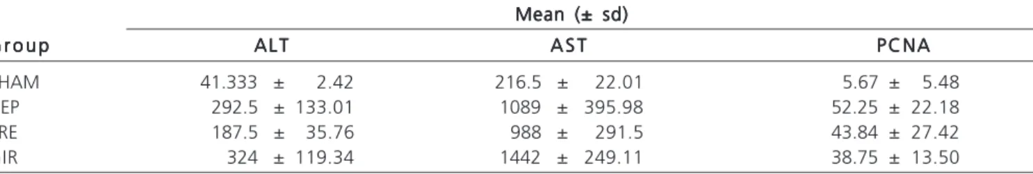
We calculated the mean and standard-deviation of ALT, AST and the percentage of PCNA positivity (Table 2). The GIR group obtained the highest value of ALT and AST (324 and 1442, respectively) and the PRE group had lower ALT and AST (187.5 and 988 respectively) than the GIR. As for transaminases, there were significant differences between the groups HEP and SHAM, GIR and SHAM, PRE and SHAM and PRE and GIR (p <0.05).
In relation to PCNA, the HEP group had the highest percentage (52.25%), whereas the SHAM group displayed the minimum values (5.67%) (Table 2). The SHAM group, when compared to the other groups, showed a significant difference.
DISCUSSION
DISCUSSION
DISCUSSION
DISCUSSION
DISCUSSION
In this study, ischemic preconditioning was not able, within 48 hours, to increase cell proliferation after ischemia and reperfusion followed by partial hepatectomy. However, it decreased hepatic injury after hepatectomy.
One of the procedures used to protect the liver from IR is IPC, based on the premise that tissue acquires resistance to the effects of IR through previous exposure to brief periods of occlusion 12. The short time interval during
IPC ischemia generates oxidative stress, inducing natural defense mechanisms 13.
considered one of the best models for the study of liver regeneration, since it allows the precise definition regenerating the onset of the stimulus. It consists of resection of the left lateral and median lobes, which represent approximately 70% the total liver mass. The right lateral and the caudate lobes, which correspond to about 24% and 6%, respectively, of the total liver mass, are kept intact.
Regeneration of liver hepatocytes is activated by cytokines, particularly to initiate the cell cycle, in which there is activation of genes that cause hepatocytes to go from G0 phase to G1. Several proteins, growth factors and cycle-dependent kinases are involved in this process15.16. The
cells progress through the cell cycle, reaching phase S, with DNA synthesis. Phase G2 (replication) follows, with synthesis of proteins required for cell division, and the end stage of mitosis (M) itself 17. After a phase of intense growth and
reconstruction of the hepatic parenchyma, the regenerative process ceases 16.18. The hepatocytes are the first to
proliferate, constituting about 90% of hepatic mass and 60% of the total number of cells 12, 17. Therefore, these
authors chose to evaluate the cell proliferation of hepatocytes.
For detection of cellular proliferation in various tissues, different methods have been used to provide information about the growth and tissue repair. The technique we used was proliferating cell nuclear antigen (PCNA), which is expressed in the late G1 phase and during phase S15.
Liver injury was assessed through the levels of liver enzymes, ALT and AST, which indicate a higher than normal liver damage 19. In mild hepatocellular injury, the
predominant form in serum is the cytoplasmatic, whereas in severe injury there is release of the mitochondrial enzyme,
Table 1 Table 1 Table 1 Table 1
-Table 1 - Mean values of total liver weight, initial weight, final weight and weight of the remaining lobe of the SHAM, HEP, GIR and PRE groups.
G r u p s G r u p s G r u p s G r u p s
G r u p s Total Weight (A)Total Weight (A)Total Weight (A)Total Weight (A) Initial Weight (B)Total Weight (A)Initial Weight (B)Initial Weight (B)Initial Weight (B)Initial Weight (B) Final Weight (C)Final Weight (C)Final Weight (C)Final Weight (C)Final Weight (C) Weight OfWeight OfWeight OfWeight OfWeight Of Regenerated Part(c-(ab) Regenerated Part(c-(ab)Regenerated Part(c-(ab) Regenerated Part(c-(ab)Regenerated Part(c-(ab) G r a m s
G r a m s G r a m s G r a m s
G r a m s G r a m s G r a m s G r a m s G r a m s G r a m s G r a m sG r a m sG r a m sG r a m sG r a m s PercentagePercentagePercentagePercentagePercentage G r a m sG r a m sG r a m sG r a m sG r a m s PercentagePercentagePercentagePercentagePercentage
SHAM 6.84 6.84
HEP 6.59 4.62 5.95 90.01% 3.97 60.03%
GIR 6.5 4.55 5.85 89.93% 3.9 60.00%
PRE 6.6 4.62 6.14 93.18% 4.16 63.16%
Table 2 Table 2 Table 2 Table 2
-Table 2 - Comparison of ALT, AST, positivity rate of PCNA and regeneration of hepatocytes in groups SHAM, HEP, GIR AND PRE.
M a n n - W h i t n e y M a n n - W h i t n e y M a n n - W h i t n e y M a n n - W h i t n e y
M a n n - W h i t n e y A L TA L TA L TA L TA L T AST AST AST AST AST P C N AP C N AP C N AP C N AP C N A R e g e n e r a t i o nR e g e n e r a t i o nR e g e n e r a t i o nR e g e n e r a t i o nR e g e n e r a t i o n
HEP-GIR 0.69 0.07 0.22 0.81
PRE-HEP 0.14 0.81 0.93 0.47
HEP-SHAM 0.005* 0.005* 0.005*
-PRE-GIR 0.02* 0.02* 0.93 0.23
GIR-SHAM 0.005* 0.005* 0.01*
-PRE-SHAM 0.005* 0.005* 0.005*
-Values with * indicate significant differences (P <0.05)
Table 3 – Table 3 – Table 3 – Table 3 –
Table 3 – Mean and standard-deviation of ALT, AST and percentage of positive PCNA in groups SHAM, HEP, GIR AND PRE.
Mean (± sd) Mean (± sd)Mean (± sd) Mean (± sd)Mean (± sd) G r o u p
G r o u p G r o u p G r o u p
G r o u p A L TA L TA L TA L TA L T A S TA S TA S TA S TA S T P C N AP C N AP C N AP C N AP C N A
SHAM 41.333 ± 2.42 216.5 ± 22.01 5.67 ± 5.48
HEP 292.5 ± 133.01 1089 ± 395.98 52.25 ± 22.18
PRE 187.5 ± 35.76 988 ± 291.5 43.84 ± 27.42
raising AST / ALT ratio. In this case, ALT may have been reduced more rapidly because it has first been released in the bloodstream, the elevated AST remaining longer, resulting in a higher AST / ALT ratio 20.
In this experiment, the animals undergoing hepatectomy obtained an increase in remaining liver weight of about 60%. This percentage is due to the observation time of 48 hours, since the process of restoring the original mass takes from five to seven days 21.
The groups submitted to hepatectomy displayed ALT values above normal (considering the values of 26 and 78 IU/L as physiological to the species) 19. A
significant difference was found between the groups GIR and PRE, demonstrating reduction of liver damage with IPC. Considering the values of 157 IU/l to 246 IU/l as physiological AST for this species 19, only the SHAM group
had values within normal limits. Again, the difference between the GIR and PRE groups reinforces the protection of IPC on the liver, as described by other authors in experi-mental and clinical studies 15,22,23,24.
In respect to the percentage of PCNA positivity, the HEP group had the highest rate, followed by the PRE group, GIR and SHAM, respectively. The SHAM group
differed significantly from the others; however, no significant difference existed when comparing them with one another. This demonstrates that despite the higher cell proliferation of the group with ischemic preconditioning, it did not increase significantly, since it was reduced by IR injury in relation to rats that underwent only the hepatectomy stimulus.
In a prospective study in 2000, Clavien et al.23 showed the effectiveness of IPC in reducing IR lesions,
the first clinical study involving the IPC on the liver in patients undergoing hepatectomy. However, they showed some situations for which IPC was ineffective, as in resections greater than 50%. More recently, Azoulay et al 24 first
reported the use of IPC in clinical transplantation, showing a greater tolerance to IR injury, but they identified the initial worsening of liver function.
This experimental study analyzed only the period of 48 hours after hepatectomy, but it corroborated other clinical studies 14,23,24, which do not definitely advocate
ischemic preconditioning for extended resections.
In conclusion, ischemic preconditioning decreased liver injury after hepatectomy, but did not improve regeneration after ischemia and reperfusion.
R E S U M O R E S U M O R E S U M O R E S U M O R E S U M O
Objetivo: Objetivo: Objetivo: Objetivo:
Objetivo: Avaliar a regeneração hepática com modulação pelo pré-condicionamento isquêmico após isquemia, reperfusão e hepatectomia parcial. Métodos: Métodos: Métodos: Métodos: Foram usadas 24 ratas Wistar, de 12 semanas de idade, distribuídas randomicamente em quatroMétodos: grupos: Grupo Controle (SHAM), Grupo Hepatectomia (HEP), Grupo Isquemia e Reperfusão (GIR) e Grupo Pré-condicionamento Isquêmico (PRE). Foi feita a análise das enzimas hepáticas ALT e AST, avaliação da regeneração através dos pesos inicial e final do fígado e da proliferação dos hepatócitos pela análise imunoistoquímica com o Proliferating Cell Nuclear Antigen (PCNA). Resulta-Resulta-Resulta-Resulta- Resulta-dos:
dos: dos: dos:
dos: Em todos os grupos ocorreu regeneração do fígado, não havendo significância estatística entre eles. Houve diferenças significativas em relação a ALT e AST entre os grupos HEP-SHAM, GIR-PRE, GIR-SHAM E PRE-SHAM (p< 0,05). Também houve diferença significativa em relação à marcação de PCNA do grupo SHAM quando comparado aos demais grupos (p< 0,05). Conclu- Conclu- Conclu- Conclu- Conclu-são
são são são
são: O pré-condicionamento isquêmico diminuiu a lesão hepática, mas não influenciou na regeneração até 48 horas.
Descritores: Descritores: Descritores: Descritores:
Descritores: Regeneração hepática. Precondicionamento isquêmico. Isquemia. Reperfusão. Hepatectomia.
REFERENCES
REFERENCES
REFERENCES
REFERENCES
REFERENCES
1. Fausto N. Liver regeneration. J Hepatol. 2000;32(1 Suppl):19-31. 2. Pacheco EG, Ramalho FS, Ramalho LNZ, Zucoloto S, Castro e Silva Jr O, Oliveira AF. Efeitos do pré-condicionamento hepático em ratos cirróticos, submetidos à isquemia e reperfusão hepática: resultados preliminares. Acta cir bras. 2001;16(Supl 1):41-3. 3. Gurusamy KS, Kumar Y, Sharma D, Davidson BR. Methods of
vascular occlusion for elective liver resections. Cochrane Database Syst Rev. 2007;(4):CD006409. Update in: Cochrane Database Syst Rev. 2009;(1):CD006409.
4. Ishikawa Y, Yamamoto Y, Kume M, Yamagami K, Yamamoto H, Kimoto S, et al. Heat shock preconditioning on mitochondria during warm ischemia in rat livers. J Surg Res. 1999;87(2):178-84. 5. Kohli V, Selzner M, Madden JF, Bentley RC, Clavien PA. Endothelial
cell and heptocyte deaths occur by apoptosis after ischemia-reperfusion injury in the rat liver. Transplantation. 1999;67(8):1099-105.
6. Daemen M, de Vries B, Buurman WA. Apoptosis and inflammation in renal reperfusion injury. Transplantation. 2002;73(11):1693-700. 7. Canalese J, Gove CD, Gimson AE, Wilkinson SP, Wardle EN, Williams R. Reticuloendothelial system and hepatocytic function in fulminant hepatic failure. Gut. 1982;23(4):265-9.
8. Mantovani M, Fontelles MJ, Hirano ES, Morandin RC, Caputo LRG, Schenka AA. Isquemia e reperfusão total associada ao esta-do de choque hemorrágico controlaesta-do: efeitos no sequestro de neutrófilos no pulmão do rato. Acta cir bras. 2002;17(1):46-54. 9. Teixeira ARF, Molan NT, Kubrusly MS, Bellodi-Privato M, Coelho
AM, Leite KR, et al. Pós-condicionamento melhora a peroxidação lipídica na lesão de isquemia-reperfusão hepática em ratos. Acta cir bras. 2009;24(1):52-6.
11. Santos CHM, Pontes JCDV, Miiji LNO, Nakamura DI, Galhardo CAV, Aguena SM. Efeito do pós-condicionamento isquêmico so-bre a isquemia e reperfusão hepática em ratos. Acta cir bras. 2010;25(2):163-8.
12. Lima CX. Efeitos do pré-condicionamento por oxigenoterapia hiperbárica na lesão de isquemia e reperfusão hepática em ratos [dissertação]. Belo Horizonte: Universidade de Minas Gerais, Fa-culdade de Medicina; 2006.
13. Centurion S, Brisotti JL, Oliveira GR, Tolentino E, Pacheco EG, Oliveira AF, et al. Avaliação da função mitocondrial do fígado submetido à isquemia parcial com e sem pré-condicionamento isquêmico. Acta cir bras. 2001;16(Supl 1):61-2.
14. Gurusamy KS, Kumar Y, Sharma D, Davidson BR. Ischaemic preconditioning for liver transplantation. Cochrane Database Syst Rev. 2008;(1):CD006315.
15. Galhardo MA; Júnior CQ, Riboli Navarro PG, Morello RJ, De Jesus Simões M, De Souza Montero EF. Liver and lung late alterations following hepatic reperfusion associated to ischemic preconditioning or N-acetylcysteine. Microsurgery. 2007;27(4):295-9.
16. Tannuri ACA. Modelos de regeneração hepática em animais em crescimento: estudos histológicos, moleculares e avaliação de efei-tos em imunossupressores [tese]. São Paulo: Universidade de São Paulo, Faculdade de Medicina; 2007.
17. Kurir TT, Markoti A, Katalini V, Bozani D, Cikes V, Zemunik T, et al. Effect of hyperbaric oxygenation on the regeneration of the liver after partial hepatectomy in rats. Braz J Med Biol Res. 2004;37(8):1231-7.
18. Jesus RP, Waitzberg DL, Campos FG. Regeneração hepática: pa-pel dos fatores de crescimento e nutrientes. Rev Assoc Med Bras. 2000;46(3):242-54.
19. Jeschke MG, Low JF, Spies M, Vita R, Hawkins HK, Herndon DN, et al. Cell proliferation, apoptosis, NF-kappaB expression, enzyme, protein, and weight changes in liver of burned rats. Am J Physiol Gastrointest Liver Physiol. 2001;280(6):G1314-20.
20. Rüdiger HA, Graf R, Clavien PA. Sub-lethal oxidative stress triggers the protective effects of ischemic preconditioning in the mouse liver. J Hepatol. 2003;39(6):972-7.
21. Fausto N, Laird AD, Webber EM. Liver regeneration. 2. Role of growth factors and cytokines in hepatic regeneration. FASEB J. 1995;9(15):1527-36.
22. Peralta C, Hotter G, Closa D, Prats N, Xaus C, Gelpí E, Roselló-Catafau J. The protective role of adenosine in inducing nitric oxide synthesis in rat liver ischemia preconditioning is mediated by activation of adenosine A2 receptors.
Hepatology. 1999;29(1):126-32.
23. Clavien PA, Yadav S, Sindram D, Bentley RC. Protective effects of ischemic preconditioning for liver resection performed under inflow occlusion in humans. Ann Surg. 2000;232(2):155-162.
24. Azoulay D, Lucidi V, Andreani P, Maggi U, Sebagh M, Ichai P, et al. Ischemic preconditioning for major liver resection under vascular exclusion of the liver preserving the caval flow: a randomized prospective study. J Am Coll Surg. 2006;202(2):203-11.
Received on 20/10/2011
Accepted for publication 19/12/2011 Conflict of interest: none
Source of funding: FAPERJ
How to cite this article: How to cite this article: How to cite this article: How to cite this article: How to cite this article:
Salomão LS, Young SB, Galhardo MA, Pereira LA, Pires ARC, Boaventura GT, Ferreira AMR, Martinho JM. Evaluation of liver regeneration by modulation with ischemic preconditioning after ischemia and reperfusion and partial hepatectomy. Rev Col Bras Cir. [periódico na Internet] 2012; 39(3). Disponível em URL: http://www.scielo.br/rcbc
Address correspondence to: Address correspondence to: Address correspondence to: Address correspondence to: Address correspondence to: José Manuel Martinho