Effects of heparin and hyperbaric oxygenation on necrosis
reduction in an animal model for degloving injuries
Efeitos da heparina e da oxigenação hiperbárica na redução de necrose de
modelo animal para desluvamentos
Douglas Neumar meNoN1; letícia teixeira1; Natalha Bristot Paurosi1; marcio eDuarDo Barros1.
INTRODUCTION
D
eglovings are injuries in which there is detachment of the integument from the underlying tissues1. Themost common cleavage plane is between the fascia and the subcutaneous tissue, but there may be detachment in the muscular, periosteal, subperiosteal planes or even in multiple planes, the latter cases being of high complexity, morbidity and mortality. The most common sites of degloving are the limbs, especially the lower ones, but it may occur in other regions of the body, such as the trunk2 and cephalic region3. Deglovings of the limbs are
usually accompanied by musculoskeletal trauma and not infrequently are also associated with polytrauma of other segments of the body, such as traumatic brain injury, abdominal and chest trauma.
There are several degloving causes reported in the literature, the most frequent being the traffic accidents with mechanisms of grip and rotation4, but
other causes have been described, such as industrial accidents and accidents with agricultural machines5.
Some authors have proposed classification systems for degloving: Hidalgo6 classified these wounds
into three groups, the first group being typical lesions, with a skin continuity solution associated with the detaching wound. The second group consists of the closed detaching lesions, while the third group is cutaneous avulsions, or wounds with loss of substance.
Another classification system was proposed by Arnez7, that considered four patterns of degloving:
pattern 1- limited degloving with abrasion or avulsion. In this pattern, injuries result from abrasion forces, with a slight detachment of the skin. Pattern 2- refers to non-circumferential deglovings, where normally the detached skin is preserved or there is little loss of substance. Usually these lesions have a well-defined cleavage plane, usually between the deep fascia and the subcutaneous tissue. Pattern 3- comprises circumferential deglovings in a single plane, which, just as the type-2 lesions, usually have their cleavage plane between the deep fascia and the subcutaneous tissue, there being open or closed lesions (with or without skin continuity solution). Pattern 4- refers
1 - Federal University of Grande Dourados, Faculty of Health Sciences, Dourados, Mato Grosso do Sul State, Brazil.
A B S T R A C T
Objective: to evaluate the efficacy of the treatment with hyperbaric oxygen therapy or with topical and intralesional heparin in an animal model of degloving lesions. Methods: we conducted an experimental study with adult, male Wistar rats submitted to degloving of the left hind limb and divided into four groups according to the treatment: Group 1 (control) - without treatment; Group 2 (Heparin) - intralesional application at the time of surgery and topically, in the postoperative period, with heparin spray 10,000IU/mL; Group 3 (hyperbaric oxygen-ation) - daily sessions of 30 minutes in a hyperbaric chamber with 100% oxygen and 2 ATA pressure; Group 4 (positive control) - adminis-tration of a single dose of 45 mg/kg of intraperitoneal allopurinol. On the seventh day, we killed the animals, removed the cutaneous flaps and measured the total and necrotic areas, as well as computed the percentage of necrotic area. Results: the mean percentage of necrosis in the control group was 56.03%; in the positive control group it was 51.36% (p<0.45); in the heparin group, 42.10% (p<0.07); and in the hyperbaric oxygen therapy group, 31.58% (p<0.01). Conclusion: both hyperbaric oxygen and heparin therapies were effective in reducing the percentage of necrosis in the model studied, although only the hyperbaric oxygenation showed statistical significance.
between muscle structures or in the musculo-periostal plane. Of course, this latter pattern represents the trauma with the greatest amount of energy applied.
There is a specific category of closed deglovings, in which the skin is detached from the fascia due to liquid collections, such as bruises, seromas, or steatonecrosis material, and receives the eponym of Morel-Lavallée Syndrome or Injury. This usually results from high-energy tangential trauma over richly vascularized tissues, but may also be due to distant lesions with serous and / or blood leakage leading to tissue detachment8.
Various treatment modalities are mentioned in the literature, such as, for example, primary closure, removal of the detached skin and its use as a cutaneous graft, use of dermal matrices9, vacuum dressings10, local
or remote flaps, microsurgical flaps, and even conservative treatment, indicated mainly in cases of closed deglovings. However, the most commonly performed treatment for this type of injury is the immediate removal of the detached skin, followed by removal of the subcutaneous tissue and repositioning of the treated skin as a cutaneous graft. Cryopreservation of the skin may also be performed for use after the trauma, after better stabilization of the patient’s clinical condition11. The choice of the best form
of treatment depends on the characteristics of the lesion and requires an accurate judgment of the surgeon.
Most of the work with deglovings in humans is composed of cross-sectional, descriptive studies or even case reports. The limitation to the emergenge of more elaborate studies on the subject probably resides in the diversity of presentation of this type of injury, causing difficulty in conducts’ standardization12. In view of this,
Milcheski et al.13 developed an experimental model in rats
for studies of this type of injury, in which they promoted a detaching injury of the entire right lower limb of rats, giving rise to a distal flap, susceptible to ischemia and, therefore, useful in the evaluation of aproaches for deglovings. Also in this study the authors tested the protective effect of pentoxifylline and allopurinol on these injuries. The use of pentoxifylline was justified because
perfusion by possibly damaged capillaries, whereas allopurinol was used for antioxidant purposes. After injury induction, rats received a single intraperitoneal dose of these drugs and the necrosis area relative to the total flap area was compared to the control group on the seventh postoperative day. Both agents showed a relative decrease in the necrosis area, demonstrating a protective effect, with better performance for the allopurinol.
Heparin has been used clinically in Plastic Surgery in the treatment of partial thickness burns through topical application. In addition, due to its possible anti-inflammatory effect, it is also used for the treatment of pathological scars, especially those with fibroproliferative disorders such as keloids and hypertrophic scars14. Studies
have also shown an angiogenic effect with the use of topical heparin applied in random flaps in rats15 and
improvement in the healing of chronic venous ulcers16.
The association of these angiogenic, anti-inflammatory and healing properties makes heparin a potentially useful compound in deglovings.
Another therapeutic modality normally applied in the treatment of complicated wounds is hyperbaric oxygenation17, which consists of exposing the patient to
an environment with 100% oxygen concentration under pressure of 2 ATA (absolute atmosphere)18. In clinical practice,
hyperbaric oxygenation is normally used in the management of chronic wounds and/or wounds complicated by infection or necrosis19, although research has studied the application
of this therapeutic modality in other situations where an increase in tissue oxygen concentration is desirable, as in muscle trauma20, fractures21, nerve injuries22, anemia23 and
spinal cord injuries24. The effect of hyperbaric oxygenation
in animal models of degloving (on rats’ tails) was tested by Demirtas et al.25, with positive results. There are descriptions
of hyperbaric chamber models for specific use in research on animals26.
METHODS
The present study was approved by the Ethics Committee with the Use of Animals of the Federal University of Grande Dourados (UFGD) under protocol number 025/2014.
We used adult, male rats from the UFGD vivarium, housed before the procedures in appropriate cages, in groups of six animals, with chow and water ad libitum, kept until the day of surgery. We randomized the animals by draw and divided them into the following groups: Group 1- Control, formed by animals submitted to surgery alone, without additional treatment; Group 2- Heparin, formed by animals treated with spray of topical and intralesional heparin; Group 3- Hyperbaric Oxygen Therapy; and Group 4- Positive Control, submitted to treatment with intraperitoneal injection of allopurinol, in a single dose of 45mg/kg. Group 1 (control) comprised 12 animals, while the other groups were composed of six animals each.
The animals received general anesthesia with ketamine 60mg/kg associated with xylazine10mg/kg, applied intramuscularly to the right animal’sthigh, and the surgical procedure was started after confirmation of the anesthetic plane through the absence of the corneal eyelid reflex. After performing a tricotomy at the base of the left hind limb, we inflicted the wound through a circular incision in the left thigh root and dissection of the skin in the subcutaneous plane. We applied traction at the edge of the flap with Backauss clampsin the craniocaudal
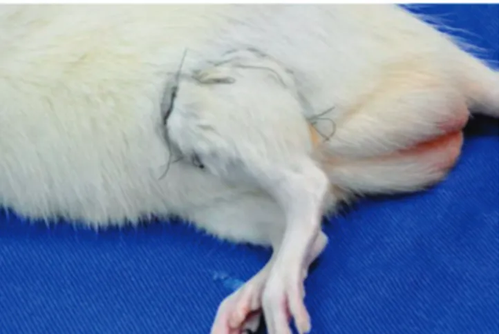
direction, from the root of the thigh to the ankle joint (Figure 1), with later repositioning of the flap and skin suture in the incision area with continuous mononylon 5.0 (Figure 2).
All animals received antibiotic prophylaxis through the intramuscular application of benzathine penicillin at a dose of 40,000IU/kg in the lower limb contralateral to the surgery limb, in addition to analgesia by the addition of dipyrone to the drinking water at a dose of 2mg/ml. After the surgeries, we placed the animals in individual cages in order to avoid aggression or cross autophagy.
The animals of the heparin group received intralesional application of the medication through a specific spraying device. We applied two jets, one anterior and one posterior, before the repositioning and suturing of the wound. This preparation is equivalent to a concentration of 10,000IU/ml. During the first six days
Figure 1. Marking of the flap detachment area. Figure 2. Surgical site on the 7th postoperative day.
of postoperative observation, we performed daily topical applications of the same dose of spray heparin (one anteriorjet and one posterior) on the flap, and euthanized the animals on the seventh postoperative day.
We submitted the animals of the Hyperbaric Oxygen Therapy group to therapy in a hyperbaric chamber specific for animal use of the UFGD Laboratory of Surgical Technique, filled with 100% oxygen under pressure of two atmospheres with daily sessions of 30 minutes, initiated immediately after surgery. During each session, the animals were housed in individual boxes and placed inside the chamber. A flush was performed for five minutes with oxygen in order to remove all the air present inside the chamber, maintaining the oxygen concentration at 100%.
The Positive Control group animals received a single dose of 45mg/kg intraperitoneal allopurinol after the standard surgical procedure.
We monitored the animals daily for changes that could interfere with the study, as well as medication application when applicable. After seven postoperative days, all animals were submitted to euthanasia through intramuscular administration of ketamine 100mg/kg associated with xylazine 10mg/kg, followed by cardiac puncture exsanguination. We performed no application of heparin or hyperbaric oxygenation session on animals on the day of euthanasia.
We then proceeded to the complete removal of the cutaneous flaps by removing the stitches and bluntly dissecting the suture line and the fasciocutaneous
cleavage plane. We sectioned the flaps longitudinally in the craniocaudal direction on the lateral anatomical side to form a flat specimen, allowing the measurement of total and necrosis areas. We duly identified the specimens according to the animal and group (control or intervention) and photographed them. We analyzed the images using the ImageJ® software (Wayne Rasband, National Institutes of Health, USA, 1997). We measured the total area and the necrotic area of the flap, respectively, and calculated the percentage of necrotic area in each specimen and the mean percentage of necrotic area in each group (Figure 3). We performed the statistical analysis through analysis of variance – ANOVA – for comparison between groups, followed by the Student’s T test for comparison of each group with the control one.
RESULTS
We observed cutaneous necrosis in all animals of all groups. In the control group, the percentage of necrotic area ranged from 18.07 to 81.08%, with a mean of 56.04% ± 5.42 and a median of 60.16. There was one death in this group due to anesthetic complications. The mean total flap area was 5.22cm2 ± 0.65, and the mean
necroticarea was 2.77cm2 ± 0.37.
The allopurinol group, used as a positive control, had a mean necrosis area of 51.36% ± 1.99 and a median of 51.57 (p=0.45), ranging from 44.69 to 57.37%, the mean of the total area being 5.50cm2 ± 0.26
and the mean of the necrotic area, 2.85cm2 ± 0.22.
In the group of animals treated with both topical and intralesional heparin, necrosis ranged from 32.41 to 49.95%, with a mean of 42.10% ± 3.56 (p≤0.07) and a mean of 43.02. The mean total area of the flaps was 6.25 cm2 ± 0.12, while the mean necrotic area
was 2.60 cm2 ± 0.24. In this group, two deaths occurred
due to anesthetic complications and was observed hemorrhage in the wounds of all the animals after the application of heparin, although not measured. There was hematoma formation in one of the subjects.
In the Hyperbaric Oxygen Therapy group, there was necrosis ranged from 8.25 to 47.21%, with a mean of 31.58% ± 5.49 (p≤0.01) and a median of 33.49, with a mean total area of 5.15cm2 ± 0.29 and mean necrotic
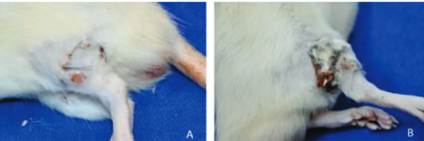
area of 1.88cm2 ± 0.45. We also empirically observed
improved healing in the animals submitted to hyperbaric therapy, with improvement in both scar appearance and flap adhesion to the bed compared with control animals (Figure 4). Table 1 shows the means of the necrotic areas in the four groups.
DISCUSSION
The great doubt in the treatment of patients with deglovings remains whether to remove or preserve the detachedskin. In cases where it is possible to perform a more conservative treatment, with only the primary suture, a more efficient skin coverage is obtained, both from aesthetic and functional points of view. However, keeping an ischemic flap will cause skin necrosis and consequently the loss of an important graft donor area, generating the need for skin capture in other areas, adding morbidity to an already severely compromised patient. On the other hand, an aggressive approach, with early debridement, when ill indicated, imposes unnecessary sequelae to the patient3. In addition, it is very
difficult to precisely define which areas are devascularized and which are viable. Hence, optimal treatment is hardly achieved4,27.
Therapies that improve the blood supply to peripheral tissues may favor more conservative treatments
in degloving cases and may have broad application in Plastic Surgery. In addition to the specific degloving wound cases, their use may be considered in any situation in which an improvement in the vascularization of the involved tissues is desired, as in cases of random skin flaps, skin grafts, chronic wounds, venous or ischemic ulcers and pressure ulcers28.
Regarding the control group, our results were similar to those of Milcheski et al.13, who found
63.34% of necrotic area, while in our study this rate was 56.04%. However, the group chosen as positive control (allopurinol) presented results discordant from these authors’, who observed 34.85% of necrosis, with an important reduction in ischemia, whereas we found 51.36%, showing a much more modest improvement despite the use of the same dosage and route of administration.
We observed a decrease in the percentage of necrotic area in the other two groups tested, with rates of 42.10% for the heparin group and 31.58% for the hyperbaric oxygenation one, although only the hyperbaric oxygenation group showed statistical significance.
In a review article, Berner et al.29 cites several
positive effects of hyperbaric oxygenation that would justify the positive results found in our study, such as neoangiogenesis, increased collagen synthesis, increased stem cell migration and improved local immune response, as well as overall improvement in healing. Another study, produced by Yan et al.30, showed several effects of this
therapy, such as increased blood oxygen concentration and partial pressure, improved blood dispersion of oxygen and its diffusion in tissues, formation of collateral
Table 1 - Comparison between the mean percentages of necrosis in the groups studied.
Group Meanpercentage of necrosis
Control 56.04 ± 5.42
Allopurinol 51.36 ± 1.99 p ≤ 0.45
Heparin 42.10 ± 3.56 p ≤ 0.07
Hyperbaric Oxygenation
cell activation. Evidently, these effects also contributed to the reduction of necrosis in our model.
We also empirically observed that the quality of the scar in the animals of this group, both the suture line and the adherence of the flap to the deep tissues being better, since the flaps presented greater tensile strength in both the cutaneous fascial cleavage plane and in the suture line. Although we did not objectively measure these parameters, they are interesting options for future testing. Zhang et al.31 tested the effects of
hyperbaric therapy in an animal healing model and observed improved healing. The authors evidenced a reduction in healing time and a decrease in the incidence of hypertrophic scars, corroborating our observation.
In the heparin group, the improvement in the necrosis area is due to reduced inflammation and vasodilatation caused by heparin, which are already known and beneficial in the treatment of superficial burns, especially of the face, the usual use of this type of product32. All animals treated with this method showed a
visible increase in bleeding in the immediate postoperative period, although we did not measure it. In one of the heparin treated animals, we observed the formation of a small laminar hematoma after the flap withdrawal, which we believe did not influence the results of this study due to the small volume of the collection. However, these complications (hemorrhage and hematoma) seem sufficient to discourage heparin studies in humans with degloving injuries, since the occurrence of any adverse events in severely compromised patients, as they are, would significantly increase morbidity and mortality.
Cebesoy et al.33 carried out a study with the use
of intraperitoneal heparin in the a rat tail degloving model. They analyzed the cutaneous necrosis depth according to the National Pressure Ulcer Advisory Panel (NPUAP), and demonstrated a reduction in the skin necrosis thickness with the use of both unfractionated and low molecular weight heparin compared with the use of saline solution, as seen in our study. The authors attributed this improvement to the prevention of microthrombus
A similar study analyzed the effects of subcutaneous enoxaparin and rivaroxaban, an oral antithrombotic agent, compared with the subcutaneous application of saline using the same animal model. Both agents showed clinical healing improvement, decrease in the necrotic area and decrease in the histopathological necrosis stage34.
Studies involving human beings present great conduction difficulty, since in large deglovings the treatment should not be delayed, under risk of important sequelae, both of the affected area and of possible tissue donor areas for reconstruction, since even in the best of the hypothesis, large areas of necrosis still occur35. However, the modalities studied in this study are
good options for studies of minor lesions, with a lower possibility of limiting sequelae, such as small degloving wounds or local flaps.
The model used in this study should be understood not only as a model of degloving, but as a model of cutaneous flaps of dubious or deficient vascularity, suggesting the possibility of further studies of these therapeutic modalities in various Plastic Surgery situations, Skin flaps, remote flaps, breast reconstruction and even cosmetic surgery. Because of the distinct mechanisms of action, we believe that it would be interesting to conduct a study with the association of these therapeutic methods to evaluate possible benefits synergism.
The proposed model was successfully reproduced, as the percentages of necrotic areas were similar to those in the literature and our studies showed a decrease in the area of ischemia in the flaps treated with the two methods studied, with advantage for hyperbaric oxygen therapy, although only the latter Presented statistical significance.
complications such as bleeding and bruising. We believe that new studies, capable of measuring inflammatory activity, partial oxygen pressure in tissues, histological
changes, tensile strength in the suture line and in the flap cleavage plane are interesting alternatives to the sequence of this work.
REFERENCES
1. Jadhav NC, Ramdas S, Lingam PP, Sateesh S. Complex traumatic facial degloving injury. J Craniofac Surg. 2016;27(4):1051-2.
2. Zhou YY, Liu W, Yang YJ, Lu GD. Use of hyperbar-ic oxygen on flaps and grafts in China: analysis of studies in the past 20 years. Undersea Hyperb Med. 2014;41(3):209-16.
3. Hakim S, Ahmed K, El-Menyar A, Jabbour G, Peralta R, Nabir S, et al. Patterns and management of deglov-ing injuries: a sdeglov-ingle national level 1 trauma center ex-perience. World J Emerg Surg. 2016;11:35.
4. Milcheski DA, Ferreira MC, Nakamoto HA, Tuma Jr P, Gemperli R. Tratamento cirúrgico de ferimentos desco-lantes nos membros inferiores: proposta de protocolo de atendimento. Rev Col Bras Cir. 2010;37(3):199-203. 5. DeFranzo AJ, Marks MW, Argenta LC, Genecov DG.
Vacuum-assisted closure for the treatment of deglov-ing injuries. Plast Reconstr Surg. 1999;104(7):2145-8. 6. Hidalgo DA. Lower extremity avulsion injuries. Clin
Plast Surg. 1986;13(4):701-10.
7. Arnez ZM, Khan U, Tyler MP. Classification of soft-tis-sue degloving in limb trauma. J Plast Reconstr Aesthet Surg. 2010;63(11):1865-9.
8. Hefny AF, Kaka LN, Salim el NA, Al Khoury NN. Unusual case of life threatening subcutaneous hemorrhage in a blunt trauma patient; Int J Surg Case Rep. 2015;15:119-22.
9. Ozturk CN, Opara P, Ozturk C, Djohan R. Treatment of foot degloving injury with aid of negative pressure wound therapy and dermal regeneration template. J Foot Ankle Surg. 2015;54(6):1132-5.
10. Andres T, von Lübken F, Friemert B, Achatz G. Vac-uum-assisted closure in the management of deglov-ing soft tissue injury: a case report. J Foot Ankle Surg. 2016;55(4):852-6.
11. Dini M, Quercioli F, Mori A, Romano GF, Lee AQ, Agostini T. Vacuum-assisted closure, dermal regen-eration template and degloved cryopreserved skin as useful tools in subtotal degloving of the lower limb. Injury. 2012;43(6):957-9.
12. Krishnamoorthy R, Karthikeyan G. Degloving injuries of the hand. Indian J Plast Surg. 2011;44(2):227-36. 13. Milcheski DA, Nakamoto HA, Tuma P Jr, Nóbrega L, Fer-reira MC. Experimental model of degloving injury in rats: effect of allopurinol and pentoxifylline in improving via-bility of avulsed flaps. Ann Plast Surg. 2013;70(3):366-9. 14. Ng L, Monagle K, Monagle P, Newall F, Ignjatovic V.
Topical use of antithrombotics: review of literature. Thromb Res. 2015;135(4):575-81.
15. Park HJ, Lee S, Kang KH, Heo CY, Kim JH, Yang HS, et al. Enhanced random skin flap survival by sus-tained delivery of fibroblast growth factor 2 in rats. ANZ J Surg. 2013;83(5):354-8.
16. Serra R, Buffone G, Molinari V, Montemurro R, Perri P, Stillitano DM, et al. Low molecular weight heparin
Objetivos: avaliar a eficácia do tratamento com oxigenoterapia hiperbárica ou com heparina tópica e intralesional em modelo animal de desluvamentos. Métodos: estudo experimental, com ratos adultos machos Wistar, submetidos a desluvamento do membro posterior esquerdo e divididos em quatro grupos, de acordo com o tratamento: Grupo 1 (controle) – sem tratamento; Grupo 2 (Heparina) – aplica-ção intralesional no momento da cirurgia e tópica, no pós operatório, com spray de heparina 10.000UI/mL; Grupo 3 (oxigenaaplica-ção hiper-bárica) – sessões diárias de 30 minutos em câmara hiperbárica com 100% de oxigênio e 2 ATA de pressão; Grupo 4 (controle positivo) – administração de dose única de 45mg/kg de alopurionol intraperitoneal. No sétimo dia os animais foram mortos e os retalhos cutâneos foram retirados e realizadas medidas das áreas total e necrótica, bem como cálculo da porcentagem da área de necrose. Resultados: a média da porcentagem de necrose do grupo controle foi 56,03%; no grupo controle positivo, 51,36% (p≤0,45); no grupo da heparina, 42,10% (p≤0,07) e no grupo da oxigenoterapia hiperbárica, 31,58% (p≤0,01). Conclusão: tanto a oxigenoterapia hiperbárica quanto a terapia com heparina mostraram-se eficazes na redução do percentual de necrose no modelo estudado, embora neste trabalho apenas a oxigenação hiperbárica tenha demonstrado significância estatística.
Descritores: Cirurgia Plástica. Ferimentos e Lesões. Traumatologia.
17. Copeland-Halperin LR, Bruce SB, Mesbahi AN. Hy-perbaric oxygen following bilateral skin-sparing mastectomies: a case report. Plast Reconstr Surg Glob Open. 2016;4(4):e680.
18. Dauwe PB, Pulikkottil BJ, Lavery L, Stuzin JM, Rohrich RJ. Does hyperbaric oxygen therapy work in facilitat-ing acute wound healfacilitat-ing: a systematic review. Plast Reconstr Surg. 2014;133(2):208e-15e.
19. Bishop AJ, Mudge E. Diabetic foot ulcers treated with hyperbaric oxygen therapy: a review of the lit-erature. Int Wound J. 2014;11(1):28-34.
20. Cervaens Costa Maia M, Camacho OF, Pinto Marques AF, Barata de Silva Coelho PM. Hyperbaric oxygen therapy treatment for the recovery of muscle injury in-duced in rats. Diving Hyperb Med. 2013;43(4):222-5. 21. Neves PCF, Abib SCV, Neves RF, Pircchio O, Saad
KR, Saad PF, et al. Effect of hyperbaric oxygen ther-apy combined with autologous platelet concentrate applied in rabbit fibula fraction healing. Clinics. 2013;68(9):1239-46.
22. Toros SZ, Karaca ÇT, Günes P, Oysu Ç, Ertugay ÇK, Naiboglu B, et al. Hyperbaric oxygen versus steroid in facial nerve injury: an experimental animal study. Am J Otolaryngol. 2013;34(5):530-6.
23. Van Meter KW. The effect of hyperbaric oxy-gen on severe anemia. Undersea Hyperb Med. 2012;39(5):937-42.
24. Yaman O, Yaman B, Aydin F, Var A, Temiz C. Hyper-baric oxygen treatment in the experimental spinal cord injury model. Spine J. 2014;14(9):2184-94. 25. Demirtas A, Azboy I, Bulut M, Ucar BY, Alabalik U,
Ilgezdi S. Effect of hyperbaric oxygen therapy on healing in an experimental model of degloving injury in tails of nicotine-treated rats. J Hand Surg Eur Vol. 2013;38(4):405-11.
26. Bertoletto PR, Fagundes DJ, Simões Mde J, Oshima CT, Montero EF, Simões RS, et al. Effects of hyper-baric oxygen therapy on the rat intestinal mucosa apoptosis caused by ischemia-reperfusion injury. Mi-crosurgery. 2007;27(4):224-7.
of degloving soft-tissue injuries. J Emerg Trauma Shock. 2014;7(3):228-32.
28. Parente LM, Lino Júnior Rde S, Tresvenzol LM, Vi-naud MC, de Paula JR, Paulo NM. Wound healing and anti-inflammatory effect in animal models of Calendula officinalis L. Growing in Brazil. Evid Based Complement Alternat Med. 2012:2012:375671. 29. Berner JE, Vidal P, Will P, Castillo P. Uso de oxígeno
hiperbárico para el manejo de heridas: bases físicas, biológicas y evidencia disponible. Rev méd Chile. 2014;142(12):1575-83.
30. Yan L, Liang T, Cheng O. Hyperbaric oxygen therapy in China. Med Gas Res. 2015;5:3.
31. Zhang Q, Shao JS, Yue YG, Zhou H, Hua MC, Zhang M. Effect of hyperbaric oxygen on the scar forma-tion at the rabbit ears at an early stage. Zhonghua Zheng Xing Wai Ke Za Zhi. 2013;29(1):55-8.
32. Venkatachalapathy TS. A comparative study of paedi-atric thermal burns treated with topical heparin and without heparin. Indian J Surg. 2014;76(4):282-7. 33. Cebesoy O, Isik M, Erzincan T, Pamukcu U, Bilgin F, Subasi
M. Analysis of the effects of heparin and enoxaparin on degloving injuries. Bratisl Lek Listy. 2014;115(9):550-3. 34. Azboy I, Demirtas A, Bulut M, Alabalik U, Uçar Y,
Alemdar C. Effects of enoxaparin and rivaroxaban on tissue survival in skin degloving injury: an experimental study. Acta Orthop Traumatol Turc. 2014;48(2):212-6. 35. Yan H, Gao W, Li Z, Wang C, Liu S, Zhang F, et al.
The management of degloving injury of lower ex-tremities: technical refinement and classification. J Trauma Acute Care Surg. 2013;74(2):604-10.
Received in: 29/09/2016
Accepted for publication: 03/11/2016 Conflict of interest: none.
Source of funding: none.
Mailing address:
Douglas Neumar Menon