https://doi.org/10.1590/0004-282X20180019 ARTICLE
Propentofylline decreases hypothalamic
astrogliosis induced by hypercaloric diet in the rat
A propentofilina diminui a astrogliose hipotalâmica induzida pela dieta hipercalórica
no rato
Eduardo Fernandes Bondan1,2, Carolina Cardoso Vieira1, Maria de Fátima Monteiro Martins1,2, Thiago Berti
Kirsten1, Maria Martha Bernardi1
1Universidade Paulista, Patologia Ambiental e Experimental, São Paulo SP, Brasil; 2Universidade Cruzeiro do Sul, Medicina Veterinária, São Paulo SP, Brasil.
Correspondence: Eduardo Fernandes Bondan; Rua Caconde, 125 / 51; 01425-011 São Paulo SP, Brasil; E-mail: [email protected]
Conflict of interest: There is no conflict of interest to declare.
Received 27 November 2017; Received in final form 04 January 2018; Accepted 13 January 2018.
ABSTRACT
Obesity is associated with a chronic and low-grade inflammatory response in the hypothalamus, where astrogliosis occurs with the upregulation of the astrocyte structural protein GFAP. As propentofylline (PPF) has inhibitory effects on astrocyte and microglial activation during inflammation, this study aimed to investigate if this xanthine derivative could decrease the astrocyte reaction induced by a hypercaloric diet (HD). Male Wistar rats were divided into four groups: NDS – rats receiving a normocaloric diet (ND) and daily saline solution; NDP – rats receiving ND and daily PPF (12.5 mg/kg/day, intraperitoneal route); HDS – rats receiving HD and saline solution, HDP – rats receiving HD and PPF. On the 21st day, rats were anesthetized, and perfused, and brains were collected for GFAP immunohistochemical study
in the hypothalamus. Results showed that HD induced increased weight gain and hypothalamic astrogliosis. Propentofylline decreased the expression of GFAP in the HDP group, although it did not affect the weight gain induced by this diet.
Keywords: astrocytes; central nervous system; inflammation; obesity; xanthine.
RESUMO
A obesidade está associada com uma resposta inflamatória crônica e de baixo grau no hipotálamo, onde ocorre astrogliose com a super-expressão da proteína astrocitária GFAP. Como a propentofilina (PPF) possui efeitos inibitórios sobre a ativação astrocitária e microglial durante a inflamação, este estudo visou a investigar se esta xantina podia diminuir a reação astrocitária induzida pela dieta hipercalórica (HD). Ratos Wistar machos foram divididos em 4 grupos: NDS- ratos recebendo dieta normocalórica (ND) e solução salina diária; NDP- ratos recebendo ND e PPF diária (12.5 mg/kg/dia, via intraperitoneal); HDS- ratos recebendo HD e solução salina, HDP- ratos recebendo HD e PPF. No 21º dia, os ratos foram perfundidos e os encéfalos, coletados para estudo imuno-histoquímico para a GFAP no hipotálamo. Os resultados mostram que a HD induziu aumento do ganho de peso e astrogliose no hipotálamo. A PPF diminuiu a expressão de GFAP no grupo HD, embora não tenha afetado o ganho de peso induzido por esta dieta.
Palavras-chave: astrócitos; sistema nervoso central; inflamação; obesidade; xantina.
The concept that obesity could affect the central nervous system (CNS), specifically the brain, has recently emerged1
. Cognitive abnormalities, specifically learning and memory def -icits, have been reported in obesity and typical comorbid dis -orders1,2. There are several associations between obesity and
neurological disorders, including sleep apnea, anxiety, manic depressive disorders and increased risk of having a cerebrovas -cular accident3
. It also has been reported that obesity is associ -ated with changes in volume or density of gray and white matter, assessed by magnetic resonance imaging4,5. Obesity-associated
structural abnormalities in the brain5 contribute to a higher
incidence of diseases, such as dementia6 and Alzheimer’s dis
-ease7
. Although the exact cause remains unknown, it has been
assumed that CNS disorders are triggered by multifactorial events including neuroinflammation and oxidative stress8
. Neuroinflammation leads to resident glial cells (astrocytes and microglia) activation, which results in invasion of circulating immune cells and production of proinflammatory cytokines, such as tumor necrosis factor-α (TNF-α), interleukin (IL)-1β, and IL-6, and reactive oxygen species8.
Therefore, obesity is characterized by chronic and low-grade inflammation in several tissues, including the CNS, and particu -larly the hypothalamus, which is known to regulate food intake and energy expenditure9,10. The arcuate and the paraventricular
Astrocytes are known to be the most numerous and diverse neuroglial cells in the CNS12. They are dynamic cells
that respond to changes in the CNS by undergoing morpho -logical and functional alterations that affect neuronal activ -ity9,12. In response to CNS insults, astrocytes develop a hyper -trophic or reactive phenotype termed astrogliosis, which is characterized by the upregulation of specific structural pro -teins, such as glial fibrillary acidic protein (GFAP) and vimen -tin12,13,14,15. Hypothalamic astrogliosis has been widely associ -ated with high-fat diet-induced obesity9,16.
Propentofylline (PPF) [3-methyl-1-(5´-oxohexyl)-7-propylxanthine] is a xanthine derivative that has demon -strated profound neuroprotective, antiproliferative and anti-inflammatory effects in several animal models17
. Clinically, it has shown efficacy in degenerative vascular dementia18
and as a potential adjuvant treatment in Alzheimer’s disease19
, schizo -phrenia20 and multiple sclerosis21. It probably depresses acti -vation of microglial cells and astrocytes, which is associated with neuronal damage during inflammation and hypoxia, and consequently decreases glial production and release of damag -ing proinflammatory factors17
. It has previously been seen that PPF decreased the peripheral astrogliosis observed following gliotoxic injury in the rat brainstem22.
Thus, the objective of the present study was to investigate if PPF could attenuate the hypothalamic astrogliosis induced by a hypercaloric diet (HD) as seen in previous studies.
METHODS
The animal procedures were performed in accordance with the guidelines of the Committee on Care and Use of Laboratory Animal Resources and Brazilian Institutional Ethics Committee guidelines (University Paulista, protocol number 376/15, CEUA/ICS/UNIP). All efforts were made to minimize animal suffering.
Twenty-four adult male Wistar rats, 12 weeks of age and weighing 300-350 g, were divided into four groups (n = 6 in each): NDS group – rats receiving a normocaloric diet (ND) and treated with 0.9% saline solution; NDP group – rats receiving the ND and treated with PPF; HDS group – rats receiving a hypercaloric diet (HD) and treated with 0.9% saline solution, HDP group – rats receiving the HD and treated with PPF.
Upon arrival at our laboratory, rats were housed in micro-isolator cages ( four rats per cage) under controlled temper -atures (22–25oC) and humidity (50–65%), in artificially-lit
rooms on a 12-hour light-dark cycle (lights on 07:00) and food and water ad libitum.
Rats from the HDS and HDP groups received free access to the HD, which was the highly palatable liquid diet sup -plement Ensure®
(Abbott Laboratorios do Brasil, São Paulo, Brazil; total of 1 kcal mL-1
). Each 231-kcal bottle contained 1.7 g polyunsaturated fat, 3.59 g monounsaturated fat, and
2.2 g saturated fat, with no trans fat. It was presented in a graduated cylinder with a stopper, with 600 mL per bottle. The ND (nutritionally balanced food) was irradiated labo -ratory chow (Nuvilab; Sogorb Indústria e Comércio, São Paulo, Brazil, with the following values per 100 g of solid food item: 4.2 kcal g-1
, 56% carbohydrate, 19% protein, 4.5% cellulose, 5% vitamins and 3.5% g total fat). This was given to all experimental groups, including those receiv -ing the HD. Rats from the HD and ND diet groups were housed four per cage and Ensure and laboratory chow were made available to each cage as a whole. Both diets were replenished daily.
The animals were weighed weekly for 21 days and weight gain was defined as the ratio between the final weight and initial weight.
Rats from the NDP and HDP groups were treated daily with a PPF solution (20 mg/ml, Agener União Química, São Paulo - SP) by intraperitoneal route using 12.5 mg/kg/day during the experimental period. Rats from the NDS and HDS groups received 0.9% saline solution intraperitoneal injec -tions daily.
On the 21st day, the rats were anesthetized (thiopental,
60 mg/kg intraperitoneally, Thiopentax®
, Cristália, Itapira, Brazil) and submitted to intracardiac perfusion with 4% glutaraldehyde in 0.1 M Sorensen phosphate buffer (pH 7.4). The brains were collected and fixed in 10% buffered formalin for 72 hours. Coronal sections of each brain were made to reach the periventricular area of the hypothala -mus (using the following coordinates - 5.64 mm from inte -raural line and -3.36 mm from the bregma23). The tissue
was embedded in paraffin for processing for conventional histological procedures. The GFAP immunohistochemis -try was performed using the chain polymer-conjugated staining method (Dako EnVision System). We used poly -clonal rabbit anti-GFAP immunoglobulin (1:50; Z033401, Dako, Glostrup, Denmark) as the primary antibody fol -lowed by the EnVision+ Kit (HRP, Rabbit, DAB+, K4011, Dako, Glostrup, Denmark). Three sections (5 µm thick) per rat were made and, from each individual section and using a 40x objective, ten photomicrographs of the periventricu -lar hypothalamus were taken. The area of GFAP+ cells and their processes, marked in brown, was automatically cal -culated, in pixels, using Image-ProPlus 6.0 software (Media Cybernetics, Silver Spring, USA) calibrated with digital color filters such that only positive cells were included and background staining was excluded from the measurement.
RESULTS
Increased body weight gain by the 21st day was seen in the
HDS and HDP groups relative to the NDS and NDP groups (p < 0.0001) (Figure 1). As for the hypothalamic GFAP expres -sion, differences between diets (p < 0.0001) and treatments (p < 0.0001) were observed, with interaction between factors (p < 0.0001). The Sidak post-test indicated an increased GFAP expression in the HDS group relative to the other experimental groups (NDS, NDP and HDP) (Figure 2). Propentofylline was able to decrease the astrogliosis induced by the HD in the HDP group, although the expression of GFAP in this group was still greater compared to the NDS and NDP groups (p < 0.01).
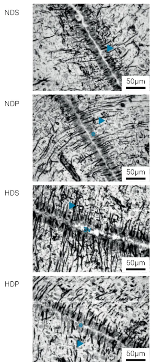
The expression of GFAP in hypothalamic astrocytes from the different groups are seen in Figure 3.
DISCUSSION
Hypothalamic inflammation may exert a paradoxical effect on energy homeostasis depending on the time course, the involved proinflammatory signals, and the degree of inflamma -tion. An intense acute inflammatory response, as in an infection, induces a state of negative energy balance, although chronic and mild inflammation, as seen in obesity, has the opposite effect9
. In our study, an inflammatory response was achieved in the hypothalamus, as suggested by the strong increase in the astrocyte expression of GFAP, indicative of intense astrogliosis in response to the HD. The differences in the weight gain seen in rats fed with the ND or HD from the beginning of the experi -ment until its conclusion 21 days later, confirmed the effect of the HD we used in creating overweight or obese rats.
Weight gain in grams
250
200
150
100
50
0
NDS NDP HDS HDP
Figure 1. Weight gain (in grams) in 21 days. Groups comprise the NDS (normocaloric diet + saline), NDP (normocaloric diet + propentofylline), HDS (hypercaloric diet + saline) and HDP (hypercaloric diet + propentofylline). Data are expressed as means ± standard errors of the means. ****p < 0.0001.
GFAP expression (pixels)
80000
60000
40000
20000
0
NDS NDP HDS HDP
Figure 2. GFAP immunostaining (represented as total count of pixels) in groups comprising the NDS (normocaloric diet + saline), NDP (normocaloric diet + propentofylline), HDS (hypercaloric diet + saline) and HDP (hypercaloric diet + propentofylline). Data are expressed as means ± standard errors of the means. ** p < 0.01; **** p < 0.0001.
Figure 3. Astrocyte GFAP expression in the periventricular zone of the hypothalamus. Groups comprise the NDS (normocaloric diet + saline), NDP (normocaloric diet + propentofylline), HDS (hypercaloric diet + saline) and HDP (hypercaloric diet + propentofylline). 3v - third ventricle; e - ependymal cells; arrowheads - astrocyte processes. GFAP immunohistochemistry.
NDS
NDP
HDS
HDP
50µm
50µm
50µm
50µm
e
3v
Both leptin (an anorexigenic hormone primarily produced by adipose tissue) and ghrelin (the most important orexigenic hormone identified to date) are capable of inducing rapid modi -fications in synaptic inputs to neurons that secrete the anorex -igenic pro-opiomelanocortin and the orex-igenic neuropeptide Y in the arcuate nucleus of the hypothalamus24. However, food
intake and body weight maintenance depend on the proper functioning of several hypothalamic areas, such as the ventro -medial (controlling appetite, body weight and insulin regula -tion), the lateral complex (appetite and body weight control), the arcuate (feeding and control of anterior pituitary, includ -ing GH and TSH secretion), the posterior (thermoregulation), the supraoptic (fluid balance), the suprachiasmatic (biological rhythms), the preoptic and the lateral (lateral anterior ther -moregulation) and the paraventricular (fluid balance, ante -rior pituitary and autonomic control, including regulation of sympathetic induced lipolysis and thermogenesis)9,10,23. Given
the complexity of its circuitry we have chosen to consider the whole hypothalamus as a unique structure of special interest in the observation of astrocytic GFAP expression. It would be simplistic to consider only the arcuate nucleus, the ventrome -dial hypothalamus or the paraventricular nucleus as impor -tant centers for controlling feeding and metabolism.
Hypothalamic astrocytes express various types of leptin receptors, indicating direct effects of the hormones on these cells25. High-fat intake and obesity are associ -ated with activation of inflammatory signaling pathways in the hypothalamus involving astrocytes and microglia and resulting in leptin and insulin resistance and disturbed con -trol of homeostasis10,24. In obesity, increased expression of
leptin receptors in hypothalamic astrocytes may suggest the sequestering of leptin by astrocytes thus leading to decreased leptin signaling to neurons26.
Astrocytes control both glucose and lipid transport and metabolism in the CNS24. Apolipoprotein E, the most abun
-dant lipid transporter in the CNS, is produced mainly in astro -cytes, acting as a satiety factor in the hypothalamus, possibly mediating some of the inhibitory effects of leptin. It is widely known that astrocytes oxidize fatty acids to produce ketone bodies, which constitute an energy source for neurons. On the other hand, ketones mediate leptin and insulin signaling in the hypothalamus thus affecting energy homeostasis24.
It remains to be determined if morphological changes in astrocytes and in the number of synaptic inputs to neurons of the arcuate nucleus are a cause or a consequence of the increased weight gain and its complications24. Exposure to
a high-fat diet was associated with reactive gliosis and this affected the structure of the blood-brain barrier, such that the pro-opiomelanocortin and neuropeptide Y cell bodies and dendrites became less accessible to blood vessels27. It is
unclear whether synaptic inputs are first removed and astro -cytes respond by taking up the vacant space and/or if they actively remove synapses and occupy new spaces27.
The astrogliosis observed in our investigation, due to administration of the HD, is known to be accompanied by increased cytokine expression and attenuation of leptin signaling in the hypothalamic neurons9,16. Activated astro
-cytes may lose their homeostatic functions upon exposure to stressors, decreasing glutamate uptake and increasing the expression of deleterious proinflammatory molecules such as cytokines, nitric oxide, prostaglandins, among others, as an injury response12,15.
Many different types of signaling molecules, including cyto -kines, are able to trigger and/or regulate astrogliosis and can be released by all cell types of the CNS tissue, including invasive inflammatory/immune cells, neurons, microglia, oligodendro -cyte lineage cells, peri-cytes, endothelia and other astro-cytes12,14.
In the CNS, PPF may serve as a glial modulator, with direct actions on microglia, and dose dependently decreases microglial proliferation and expression of inflammatory cyto -kines, such as TNF-α and IL-1β, as seen in response to lipo -polysaccharide stimulation in vitro28,29.
Known mechanisms of PPF include inhibition of cyclic AMP (cAMP) and cyclic GMP phosphodiesterases, and action as a reuptake inhibitor for the purine nucleoside and neu -rotransmitter adenosine by blocking the action of membrane nucleoside transporters. This may lead to increased intracel -lular cAMP levels and greater extracel-lular concentrations of adenosine, which stimulates adenosinergic neurotransmis -sion and adenosine 2 receptor-mediated cAMP synthesis17.
Regulation of cytokine production includes the adenyl -ate cyclase-cAMP-protein kinase pathway29. Propentofylline,
a type III-IV specific phosphodiesterase inhibitor, although it decreases, in a dose-dependent manner, the production of the inflammatory cytokines TNF-α, IL-1 and IL-6 by mouse microglia stimulated by lipopolysaccharide in vitro, increases up to two or three times the production of the inhibitory cyto -kine IL-10, which suppresses cyto-kine release by microglia and macrophages and attenuates astroglial reactivity in vivo29.
A Ca++-dependent and excessive activation of glial cells
is usually found in neuroinflammation and, in this context, increased levels of adenosine induced by PPF administra -tion12 may perform a regulatory role on these Ca++- and
cAMP-dependent molecular signaling pathways that determine many cell-related functions, such as cellular proliferation rate, differentiation state, cytokine production, among others. A strengthening of the cAMP signaling, which can be achieved by adenosine agonists and by PPF, stimulates the production of trophic factors in astrocytes, apparently preventing a delete -rious and secondary astrocytic activation caused by previous microglial upregulation. Although not entirely understood, it has been accepted that drugs that elevate extracellular ade -nosine and/or block the degradation of cyclic nucleotides, like PPF, may be used to counteract glia-related damage in CNS pathological processes22,29,30.
References
1. Bruce-Keller AJ, Keller JN, Morrison CD. Obesity and vulnerability of the CNS. Biochim Biophys Acta. 2009 May;1792(5):395-400. https://doi.org/10.1016/j.bbadis.2008.10.004
2. Mueller K, Sacher J, Arelin K, Holiga S, Kratzsch J, Villringer A et al. Overweight and obesity are associated with neuronal injury in the human cerebellum and hippocampus in young adults: a combined MRI, serum marker and gene expression study. Transl Psychiatry. 2012 Dec;2(12):e200. https://doi.org/10.1038/tp.2012.121
3. Ravanan P, Harry GJ, Awada R, Hoareau L, Tallet F, Roche R et al. Exposure to an organometal compound stimulates adipokine and cytokine expression in white adipose tissue. Cytokine. 2011 Mar;53(3):355-62. https://doi.org/10.1016/j.cyto.2010.11.015
4. Ho AJ, Raji CA, Becker JT, Lopez OL, Kuller LH, Hua X et al. Obesity is linked with lower brain volume in 700 AD and MCI patients. Neurobiol Aging. 2010 Aug;31(8):1326-39. https://doi.org/10.1016/j.neurobiolaging.2010.04.006
5. Kullmann S, Schweizer F, Veit R, Fritsche A, Preissl H. Compromised white matter integrity in obesity. Obes Rev. 2015 Apr;16(4):273-81. https://doi.org/10.1111/obr.12248
6. García-Ptacek S, Faxén-Irving G, Cermáková P, Eriksdotter M, Religa D. Body mass index in dementia. Eur J Clin Nutr. 2014 Nov;68(11):1204-9. https://doi.org/10.1038/ejcn.2014.199
7. Praticò D, Trojanowski JQ. Inflammatory hypotheses: novel mechanisms of Alzheimer’s neurodegeneration and new therapeutic targets? Neurobiol Aging. 2000 May-Jun;21(3):441-5. https://doi.org/10.1016/S0197-4580(00)00141-X
8. Golden TR, Hinerfeld DA, Melov S. Oxidative stress and aging: beyond correlation. Aging Cell. 2002 Dec;1(2):117-23. https://doi.org/10.1046/j.1474-9728.2002.00015.x
9. García-Cáceres C, Yi CX, Tschöp MH. Hypothalamic astrocytes in obesity. Endocrinol Metab Clin North Am. 2013 Mar;42(1):57-66. https://doi.org/10.1016/j.ecl.2012.11.003
10. Thaler JP, Yi CX, Schur EA, Guyenet SJ, Hwang BH, Dietrich MO et al. Obesity is associated with hypothalamic injury in rodents and humans. J Clin Invest. 2012 Jan;122(1):153-62. https://doi.org/10.1172/JCI59660
11. Becskei C, Riediger T, Hernádfalvy N, Arsenijevic D, Lutz TA, Langhans W. Inhibitory effects of lipopolysaccharide on hypothalamic nuclei implicated in the control of food intake. Brain Behav Immun. 2008 Jan;22(1):56-64. https://doi.org/10.1016/j.bbi.2007.06.002
12. Sofroniew MV, Vinters HV. Astrocytes: biology and pathology. Acta Neuropathol. 2010; 119:7-35. https://doi.org/10.1007/s00401-009-0619-8
13. Pekny M, Pekna M. Astrocyte intermediate filaments in CNS pathologies and regeneration. J Pathol. 2004 Nov;204(4):428-37. https://doi.org/10.1002/path.1645
14. Sofroniew MV. Molecular dissection of reactive astrogliosis and glial scar formation. Trends Neurosci. 2009 Dec;32(12):638-47. https://doi.org/10.1016/j.tins.2009.08.002
15. Cregg JM, DePaul MA, Filous AR, Lang BT, Tran A, Silver J. Functional regeneration beyond the glial scar. Exp Neurol. 2014 Mar;253:197-207. https://doi.org/10.1016/j.expneurol.2013.12.024
16. Buckman LB, Thompson MM, Moreno HN, Ellacott KL. Regional astrogliosis in the mouse hypothalamus in response to obesity. J Comp Neurol. 2013 Apr;521(6):1322-33. https://doi.org/10.1002/cne.23233
17. Sweitzer S, De Leo J. Propentofylline: glial modulation, neuroprotection, and alleviation of chronic pain. Handb Exp Pharmacol.
2011;200(200):235-50. https://doi.org/10.1007/978-3-642-13443-2_8
18. Kittner B, Rössner M, Rother M. Clinical trials in dementia with propentofylline. Ann N Y Acad Sci. 1997 Sep;826 Cerebrovascul:307-16. https://doi.org/10.1111/j.1749-6632.1997.tb48481.x
19. Bachynsky J, McCracken P, Lier D, Alloul K, Jacobs P. Propentofylline treatment for Alzheimer disease and vascular dementia: an economic evaluation based on functional abilities. Alzheimer Dis Assoc Disord. 2000 Apr-Jun;14(2):102-11. https://doi.org/10.1097/00002093-200004000-00008
20. Salimi S, Fotouhi A, Ghoreishi A, Derakhshan MK, Khodaie-Ardakani MR, Mohammadi MR et al. A placebo controlled study of the propentofylline added to risperidone in chronic schizophrenia. Prog Neuropsychopharmacol Biol Psychiatry. 2008 Apr;32(3):726-32. https://doi.org/10.1016/j.pnpbp.2007.11.021
21. Suzumura A, Nakamuro T, Tamaru T, Takayanagi T. Drop in relapse rate of MS by combination therapy of three different phosphodiesterase inhibitors. Mult Scler. 2000 Feb;6(1):56-8. https://doi.org/10.1177/135245850000600111
22. Bondan EF, Martins MF, Dossa PD, Viebig LB, Cardoso CV, Martins JL et al. Propentofylline reduces glial scar development following gliotoxic damage in the rat brainstem. Arq Neuropsiquiatr. 2016 Sep;74(9):730-6. https://doi.org/10.1590/0004-282X20160108
23. Paxinos & Watson. The rat brain in stereotaxic coordinates. 5th ed. San Diego: Elsevier; 2005.
24. Chowen JA, Argente J, Horvath TL. Uncovering novel roles of nonneuronal cells in body weight homeostasis and obesity. Endocrinology. 2013 Sep;154(9):3001-7. https://doi.org/10.1210/en.2013-1303
25. Hsuchou H, He Y, Kastin AJ, Tu H, Markadakis EN, Rogers RC et al. Obesity induces functional astrocytic leptin receptors in hypothalamus. Brain. 2009 Apr;132(Pt 4):889-902. https://doi.org/10.1093/brain/awp029
26. Pan W, Hsuchou H, Xu C, Wu X, Bouret SG, Kastin AJ. Astrocytes modulate distribution and neuronal signaling of leptin in the hypothalamus of obese A vy mice. J Mol Neurosci. 2011 Mar;43(3):478-84. https://doi.org/10.1007/s12031-010-9470-6
27. Horvath TL, Sarman B, García-Cáceres C, Enriori PJ, Sotonyi P, Shanabrough M et al. Synaptic input organization of the melanocortin system predicts diet-induced hypothalamic reactive gliosis and obesity. Proc Natl Acad Sci USA. 2010 Aug;107(33):14875-80. https://doi.org/10.1073/pnas.1004282107
28. Si Q, Nakamura Y, Ogata T, Kataoka K, Schubert P. Differential regulation of microglial activation by propentofylline via cAMP signaling. Brain Res. 1998 Nov;812(1-2):97-104. https://doi.org/10.1016/S0006-8993(98)00954-8
29. Yoshikawa M, Suzumura A, Tamaru T, Takayanagi T, Sawada M. Effects of phosphodiesterase inhibitors on cytokine production by microglia. Mult Scler. 1999 Apr;5(2):126-33. https://doi.org/10.1177/135245859900500210
30. Schubert P, Morino T, Miyazaki H, Ogata T, Nakamura Y, Marchini C et al. Cascading glia reactions: a common pathomechanism and its differentiated control by cyclic nucleotide signaling. Ann N Y Acad Sci. 2000 Apr;903 1 VASCULAR FACT:24-33. https://doi.org/10.1111/j.1749-6632.2000.tb06346.x
decreased astrocytic activation 21 days after the onset of administration of the HD, probably by simultaneously sup -pressing the release of proinflammatory molecules, such as the above-mentioned TNF-α, IL-1β and IL-6, which may trig -ger and promote astrogliosis during inflammation, and by