Specimen processing during laparoscopic renal
sur-gery: a review of techniques and technologies
Saleh Binsaleh
King Saud University, Faculty of Medicine, Department of Surgery, Division of Urology, Riyadh, Saudi Arabia.
Laparoscopic surgery has well-defined benefits for patients and has become accepted over time as a standard access strategy for the management of benign and malignant urologic diseases. Unlike in open surgery, the surgeon is often faced with the additional challenges of specimen retrieval and extraction at the end of laparoscopic extirpative procedures. This final step often requires significant laparoscopic skill to entrap and safely extract the laparoscopic specimens. Failure to apply safe exit steps at the end of a laparoscopic procedure may lead to significant morbidity. The aim of this review is to explore the different techniques and technologies available for laparoscopic kidney retrieval, entrapment and safe extraction.
KEYWORDS: Extraction Site; Intact Specimen; Morcellation; Specimen Retrieval; Laparoscopy; Nephrectomy.
Binsaleh S. Specimen processing during laparoscopic renal surgery: a review of techniques and technologies. Clinics. 2014;69(12):862-866. Received for publication onAugust 24, 2014;First review completed onOctober 1, 2014;Accepted for publication onOctober 1, 2014 E-mail: [email protected]
Tel.: 00966 11 4671575
& INTRODUCTION
Laparoscopic surgery first became widely accepted with the advent of laparoscopic cholecystectomy (1). The benefits of small trocar site incisions versus large muscle-cutting sub-costal incisions were immediately apparent. Subsequently, laparoscopic techniques have been used for advanced renal surgeries, such as simple nephrectomy, radical nephrectomy, radical nephroureterectomy and donor nephrectomy. However, because of the large specimen sizes, simple extraction cannot be performed in a manner similar to that used during a cholecystectomy. Thus, various steps in laparoscopic renal surgery remain technically demanding, including specimen retrieval and extraction.
Laparoscopic specimen entrapment and extraction occur at what is falsely considered the ‘‘end of the procedure’’. In comparison to open extirpative surgery, where the speci-men is simply lifted out with minimal effort through a larger incision that has been created to accomplish the surgical objectives, significant laparoscopic skill is required to entrap and safely extract laparoscopic specimens. Disregarding this important step of the procedure may lead to significant morbidity. Therefore, it is imperative for laparoscopic procedures that the ‘‘end of the procedure’’ be strictly defined as the completion of skin closure and dressing placement.
In the following review, the available technologies and techniques for renal specimen entrapment and extraction will be explored; these techniques can minimize morbidity while maintaining the inherent advantages of a minimally invasive surgical approach.
Specimen entrapment and retrieval devices
The initial steps of laparoscopic nephrectomy include establishing laparoscopic access, exposing the kidney, vascular control and dissection. The next step is entrapping the specimen in a retrieval device for safe extraction while minimizing possible tumor spillage (in the case of cancer) or intra-abdominal contamination (from infected tissues).
A variety of different retrieval devices are commercially available (Table 1), each of which has its own particular characteristics. Their common features include the follow-ing: sac permeability, resistance, stability within the abdominal cavity and easy handling from trocar insertion to opening, closure and removal. Some of these retrieval devices are simple sacs made of polyurethane and are available in a variety of sizes, such as the LapSac (Cook Urological, USA) and the Endobag (Covidien, USA). Others are sacs with opening and closing mechanisms, such as the EndoCatch bag (Covidien, USA) or the Endopouch bag (Ethicon, USA). The use of specifically designed retrieval bags and impermeable sacs, such as the LapSac (Cook Urological, USA), for both intact and morcellated specimen removal decreases the risk of port site recurrence, which is a rare but serious complication that has been reported after organ retrieval without protection (2). Tumor spillage during extraction of a cancerous specimen is an obvious risk, especially for high-grade or advanced-stage tumors. Ankem et al. examined laparoscopic retrieval bag washings for malignant cells after hand-assisted laparoscopic radical Copyrightß2014CLINICS– This is an Open Access article distributed under
the terms of the Creative Commons Attribution Non-Commercial License (http:// creativecommons.org/licenses/by-nc/3.0/) which permits unrestricted non-commercial use, distribution, and reproduction in any medium, provided the original work is properly cited.
No potential conflict of interest was reported.
nephrectomy and intact specimen removal and found that, with minimal manipulation, low-stage, low-grade tumors did not exfoliate cells into the retrieval sac; however, higher grade or stage tumors may have different characteristics in terms of cell exfoliation and should be properly addressed (3).
Most commercially available retrieval bags are available in only one size and are expensive. It is also sometimes difficult to manipulate the retrieval bags within the body because of the limited space available in the surgical field. For these reasons, several types of improvised retrieval bags, including surgical gloves, condoms and re-closable zipper bags, have been used, although they are mainly used for small-sized specimens; furthermore, these bags do not always include a purse-string closure mechanism around the bag’s opening (4-8). An example of a cheap and easily produced retrieval bag is the Nadiad device, which is manually constructed using a 5F ureteral catheter, a nylon thread and a polyethylene bag. This bag is sealed at one end with an auto-seal device and a tunnel is created around the open end of the bag to thread the ureteral catheter with a nylon thread. For specimen entrapment, the device is inserted through the 10-mm working port using a non-traumatic grasper; after the specimen is placed in the bag, the ureteral catheter is removed and the nylon thread is used to tie the bag tightly. This cheap device is easy to make and deploy and is effective at removing surgical specimens. However, attention should be paid to its lack of perme-ability tests and stperme-ability tests and this bag should not be used for morcellation (9).
Generally, these alternative entrapment devices are not currently designed for use during laparoscopic specimen retrieval; however, they may open new industry channels for customized, inexpensive surgical and medical applica-tions.
Intact specimen extraction or morcellation
When possible, intracorporeal morcellation in an entrap-ment device (for non-donor nephrectomy cases) has been employed at many centers to minimize the size of the extraction incision (10). Morcellators use a sharp cylindrical blade over the specimen that divides the tissue into small strips. Morcellators that use diathermy instead of blades are also available (11). To achieve safe morcellation, the entrapment sac must be leak-proof and strong enough to prevent perforation. Additionally, the tissues should be kept under direct or laparoscopic view, with careful attention to protecting the tissue, trocar and port site through which the fragmented tissue will be retrieved. Safe morcellation without tissue spillage or bag perforation has been achieved with the use of the LapSac or EndoCatch II sac (Table 1). These sacs are made of impermeable materials that prevent tissue and cell dissemination (12,13). Proponents of frag-mented specimen removal have suggested that this techni-que is associated with improved cosmesis, a reduced risk of incisional hernia and a minimal risk of peritoneal contam-ination appropriate technical safeguards are used (14). However, questions have arisen regarding the adequacy of surgical staging and the risk of tumor implantation when cancer surgery is performed after specimen destruction (15). Additionally, serious bowel injury can occur following an unexpected rupture of the entrapment sac, leading to the morcellation of an adjacent bowel loop.
Ono et al. reviewed their 5-year experience with laparo-scopic radical nephrectomy in 57 patients. Although intact specimen extraction was initially used, the investigators removed the specimen in a fractionated fashion in the final 34 patients. There were no significant differences between the intact and the fractionated removal techniques in terms of postoperative analgesia use (29 mg of pentazocineversus
29 mg) or convalescent time (22.7 daysversus23.3 days) (16).
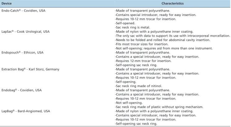
Table 1 -Laparoscopic specimen entrapment and retrieval devices.
Device Characteristics
Endo-CatchH- Covidien, USA -Made of transparent polyurethane.
-Contains special introducer, ready for easy insertion. -Requires 10-12 mm trocar for insertion.
-Self-opened. -Sac neck ring is metal.
LapSacH- Cook Urological, USA -Made of nylon with a polyurethane inner coating.
-The only sac with data to support its use with intracorporeal morcellation. -Needs to be folded and rolled for abdominal cavity insertion.
-Fits most trocar sizes for insertion.
-Not self-opening; requires aid from more than one instrument.
EndopouchH- Ethicon, USA -Made of transparent polyurethane.
-Contains a special introducer, ready for easy insertion. -Requires 12-mm trocar for insertion.
-Self-opening sac neck ring. Extraction BagH- Karl Storz, Germany -Made of transparent polyurethane.
-Contains a special introducer, ready for easy insertion. -Requires 10-12 mm trocar for insertion.
-Self-opening.
-Sac neck ring made of nitinol.
EndobagH- Covidien, USA -Made of transparent polyurethane.
-Contains a special introducer, ready for easy insertion. -Requires 10-12 mm trocar for insertion.
-Not self-opening.
-Sac neck ring made of plastic without spring mechanism.
LapBagH- Bard-Angiomed, USA -Made of nylon with a polyurethane inner coating.
-Contains special introducer, ready for easy insertion. -Requires 10-12 mm trocar for insertion.
Similarly, Gettman et al. reported that there was no subjective or objective advantage associated with kidney fragmentation (compared with intact specimen extraction through an infraumbilical incision) in a prospective series of 12 patients who underwent transperitoneal laparoscopic radical nephrectomy (17). Another series that analyzed the potential benefits of the smaller incision associated with morcellated specimen extraction dealt primarily with cytoreductive nephrectomies in patients with metastatic disease. Of the 11 patients, extractions were performed intact in five patients and after morcellation in six. The morcellation group had lower analgesic requirements than the intact extraction group (243¡72 mg of morphine sulfate
equivalentversus332¡75 mg;p= 0.049), as well as a shorter interval to the commencement of interleukin-2 therapy (40¡4.3 days versus58¡3.7 days;p= 0.006). However, the study is somewhat biased by the inclusion of three patients who required open conversion in the intact specimen extraction group. This bias is illustrated well by the greater blood loss in the intact extraction group compared with the morcellation group (3620¡1160 mL versus 1333¡552 mL;
p= 0.017) (18).
With respect to oncological safety, there are limited numbers of case reports of tumor seeding after kidney morcellation for malignancies (14,19). Possible contributing factors to these recurrences are the use of sacs that are not specifically designed for morcellation and the failure to recognize micro-perforations of these sacks. Long-term studies evaluating the oncologic results of morcellation have shown that this technique did not significantly impact the ability to detect pT3 disease and that there were no significant differences in recurrence-free, cancer-specific or overall survival compared to intact specimen retrievals (12,20,21).
The pathological validity of morcellated cancerous kidney specimens, including cases with perinephric fat invasion, was evaluated in vitro. The study included 11 renal cell carcinomas (mean tumor size 4.2 cm, range 2–7 cm), of which seven were formalin-fixed specimens and four were fresh specimens. There were no differences in the observed histology, grade, or stage when intact specimens were compared to a second analysis of the same specimens after morcellation. Important pathologic characteristics for prog-nosis, such as micro-vascular invasion, can also be evalu-ated using morcellevalu-ated fragments; however, no information can be provided on the pathological tumor size or the status of the specimen’s surgical margins (22).
The clinical pathologic staging after morcellation may be improved by removing larger fragments through a small extension in the skin incision (23). In this ‘‘modified morcellation’’ technique, a 2.5 cm incision is used, and the specimen is morcellated ex vivounder direct vision. With this approach, entrapment can be expedited and facilitated by the use of a less durable entrapment sac and reasonable histopathologic results can still be achieved due to the larger fragment size that can be achieved with this technique.
The final decision to morcellate should be made in conjunction with the patient, who must understand the risks and benefits of specimen morcellation.
Extraction site choice: where and how?
For morcellated specimens, the extraction is straightfor-ward and is usually performed inside a retrieval bag through one of the 12-mm ports. Similarly, for hand-assisted
laparoscopic renal procedures, such as donor nephrectomy, extraction is achieved through the incision site of the hand-assist device.
Intact specimen extraction after laparoscopic radical nephrectomy has generally been performed as an extension of a port site incision, a connection of two port sites, an incision into prior abdominal scars, or the creation of a new incision site.
Intact specimen extraction through the transverse lower flank muscle incision (either by port site extension or the connection of two ports) may be associated with a higher risk of incisional hernia, especially in patients with other risk factors (24).
Creating a new incision depends on multiple factors that are related to the patient, the type of proposed surgery and the surgeon’s experience. Traditionally, vertical median subumbilical incisions have been used for most open abdominal surgeries. For these procedures, the skin is incised at the midline between the umbilicus and the pubic symphysis. The rectus sheath and peritoneum are incised at the midline, which is the least vascular area. This type of incision has the presumed advantage of speedy abdominal entry and less bleeding, and the incision can be extended upwards if more space is required for access. The disadvantages of a vertical midline incision include a greater risk of postoperative wound dehiscence and the development of an incisional hernia. The resulting scar is also cosmetically less pleasing (25).
In a paramedian incision, the skin incision is made to one side of the midline (usually right). The anterior rectus sheath is opened under the skin incision. The belly of the underlying rectus abdominis muscle is then retracted laterally and the posterior rectus sheath and peritoneum are opened. Because of a shutter-like effect, there is presumably less stress on the scar. Paramedian incisions reportedly heal more strongly than midline incisions but have no cosmetic advantage (25). Bird et al. analyzed a cohort of patients undergoing laparoscopic radical nephrectomy with intact specimen extraction through three different sites (lower quadrant, umbilical and paramedian sites). The risk of incisional hernia was significantly associated with a higher body mass index (BMI) and a paramedian extraction site (26).
muscles are adherent, sharp dissection is necessary to separate them. The peritoneum is then opened sharply at the midline. The initial entry is then widened sharply with fine scissors to expose the intraperitoneal contents. Matin and Gill described a simple modification of the standard Pfannenstiel incision for performing intact specimen extrac-tion during retroperitoneal laparoscopic radical nephrect-omy. In this technique, after completing the retroperitoneal laparoscopic kidney dissection, the specimen is entrapped in a specimen retrieval bag. Then, a 5- to 7-cm Pfannenstiel skin incision is made over the symphysis pubis, lateralized slightly toward the side of the surgery. A vertical incision is then made in the anterior rectus fascia near the lateral aspect of the ipsilateral rectus muscle from the level of the pubic bone, extending cephalad for approximately 5 to 7 cm. The transversalis fascia is then perforated near the level of the pubis to enter the pelvic extraperitoneal space and is developed to the upper retroperitoneum. The drawstring of the closed bag is grasped, allowing the delivery of the entrapped intact specimen through the modified Pfannenstiel incision. This group did not conduct a formal analysis but believed that this approach provided increased patient comfort and cosmesis over the use of an expanded lateral port site (29).
The advantages of the Pfannenstiel incision compared to vertical incisions include decreased pain, improved cosm-esis and better healing (27,30,31). The resulting scar can be hidden under most types of clothing, including a bathing suit. Additionally, the Pfannenstiel incision may be asso-ciated with a decreased risk of incisional hernia (27,32,33). However, this type of incision may not be completely benign. The incidence of ilioinguinal or iliohypogastric neuropathy is as high as 3.7% after such incisions and such injuries have multiple potential causes, including: incor-poration of the nerve into the sutures during the facial closure, direct nerve trauma with or without neuroma formation, laparoscopic trocar injury, closure of laparo-scopic sites, or constriction of the nerve with scar or wound healing. Symptoms may occur immediately or can be delayed; typically, patients have burning pain in the lower abdomen, upper medial thigh and pelvic region, with altered skin sensitivity in the inguinal area (34). Simforoosh et al. reported a series of fifty patients who underwent a mini-laparoscopic live donor nephrectomy with kidney extraction through a 6- to 8-cm Pfannenstiel incision. Two 3.5-mm trocars were inserted above and lateral to the umbilicus for grasping and scissoring. One 5-mm trocar with a camera was inserted in the umbilicus and an 11-mm trocar for bipolar coagulation and vascular clip applier was inserted through the fascia in the Pfannenstiel incision. The mean warm ischemia time was 4.41 (range, 2.35-9) minutes and the mean hospital stay was 2.2 (range, 2-5) days. Clavien grades I and II complications were noted in three and two donors, respectively and no major periopera-tive or immediate postoperaperiopera-tive complications were reported (35). Tisdale et al. retrospectively compared the intact specimen extraction sites formed by expanded port incisions to a Pfannenstiel incision in 150 patients undergoing laparos-copic radical nephrectomy, nephroureterectomy and donor nephrectomy. The Pfannenstiel group had a shorter hospital stay (2.84versus3.37 days,p,0.05) and used significantly less morphine (23.7versus47.3 mg,p,0.006). An incisional hernia developed in three patients (2.9%) who underwent extrac-tions through the port site extension, whereas no hernias
were observed in the Pfannenstiel group (27). Gupta et al. compared different incision locations (modified iliac fossa and Pfannenstiel incisions) for kidney retrieval during laparoscopic transperitoneal donor nephrectomy in 343 patients. In their study, the warm ischemia time (3 versus
3.5 min), mean hospital stay (3.35 versus 3.8 days) and analgesic requirements were comparable. In the Pfannenstiel group, two patients sustained a bladder injury and one patient sustained a bowel injury. Furthermore, the mean length of the Pfannenstiel incision was longer than the iliac fossa incision (7.3 cm versus 5.8 cm). However, the Pfannenstiel incision was found to be superior in terms of cosmesis (36). Wound-related complications have also been reported in other abdominal surgeries and are dependent on the extraction site. In a multivariate analysis of risk factors for surgical site infection and incisional hernia after laparoscopic colorectal surgery, the use of Pfannenstiel extraction sites was associated with lower infection rates, although the difference was not statistically significant (37). Samia et al. reported a 7% incidence of incisional hernia after laparoscopic colorectal surgery. Midline incisional hernias accounted for 84% of all hernias, occurring in 8.9% of midline extractions (p,0.05
versusnon-midline extractions). The hernia rates for muscle-splitting, Pfannenstiel and ostomy site extractions were 2.3%, 3.8% and 4.8%, respectively (32). Similarly, Orcutt et al. retrospectively analyzed 171 patients who had undergone laparoscopic colorectal cancer surgery requiring a specimen extraction, either through a Pfannenstiel or a midline incision. Patients in the Pfannenstiel group had significantly lower rates of wound disruption (0 versus 13%, p= 0.02), superficial surgical site infection (7versus22%,p= 0.03) and overall wound complications (13versus30%,p= 0.04) (33).
Another possible extraction technique in selected patients is transvaginal specimen retrieval. The main driving force behind this technique is the reduction of abdominal scar development in women. This approach has been proposed as a reproducible technique for donor or radical nephrect-omy, with excellent patient acceptance, high satisfaction and low morbidity. However, this technique should not be performed in young nulliparous women, in patients with atrophic vaginitis, in cases involving extremely large speci-mens, in patients with vaginal infection or in patients with vaginal prolapse (38-41).
& ACKNOWLEDGMENTS
This study was supported by a grant from the College of Medicine Research Center, Deanship of Scientific Research, King Saud University, Riyadh, Saudi Arabia.
& REFERENCES
1. Perissat J. Laparoscopic cholecystectomy: The European experience. Am J Surg. 1993;165(4):444-9.
2. Iwamura M, Tsumura H, Matsuda D, Kurosaka S, Yoshida K, Baba S. Port site recurrence of renal cell carcinoma following retroperitoneo-scopic radical nephrectomy with manual extraction without using entrapment sac or wound protector. J Urol. 2004;171(3):1234-5. 3. Ankem MK, Hedican SP, Pareek G, Waterman BJ, Moon TD, Selvaggi
SM, et al. Examination of laparoscopic retrieval bag washings for malignant cells after hand-assisted laparoscopic radical nephrectomy and intact specimen removal. Urology. 2006;68(1):50-2, http://dx.doi. org/10.1016/j.urology.2006.01.032.
4. Chung SC, Li MK, Li AK. Lost stone during laparoscopic cholecystect-omy: retrieval using a condom. HPB Surg. 1993;7(1):67-8, http://dx.doi. org/10.1155/1993/10187.
Endosc. 1998;8(6):457–9, http://dx.doi.org/10.1097/00019509-199812000-00012.
6. Leung WS, Ng PS, Yuen PM. Morcellation inside a zipper bag pouch: An efficient and safe way to retrieve a large, solid ovarian mass laparoscopically. J Gynecol Surg. 2007;23(1):31–6, http://dx.doi.org/10. 1089/gyn.2007.B-02252-1.
7. Yano H, Okada K, Kinuta M, Iwazawa T, Kanoh T, Monden T. Use of non-powder surgical glove for extraction of gallbladder in laparoscopic cholecystectomy. Digest Endosc. 2003;15:315–9, http://dx.doi.org/10. 1046/j.1443-1661.2003.00263.x.
8. Yuen PM, Rogers MS. Laparoscopic removal of ovarian cysts using a zipper storage bag. Acta Obstet Gyncol Scand. 1994;73(10):829–31, http://dx.doi.org/10.3109/00016349409072514.
9. Ganpule AP, Gotov E, Mishra S, Muthu V, Sabnis R, Desai M. Novel cost-effective specimen retrieval bag in laparoscopy: Nadiad bag. Urology. 2010;75(5):1213-6, http://dx.doi.org/10.1016/j.urology.2008.09.057. 10. Cadeddu JA, Ono Y, Clayman RV, Barrett PH, Janetschek G, Fentie DD,
et al. Laparoscopic nephrectomy for renal cell cancer: Evaluation of efficacy and safety: A multicenter experience. Urology. 1998;52(5):773–7, http://dx.doi.org/10.1016/S0090-4295(98)00391-4.
11. Erian J, Hassan M, Pachydakis A, Chandakas S, Wissa I, Hill N. Efficacy of laparoscopic subtotal hysterectomy in the management of menor-rhagia: 400 consecutive cases. BJOG. 2008;115(6):742-8, http://dx.doi. org/10.1111/j.1471-0528.2008.01698.x.
12. Wu SD, Lesani OA, Zhao LC, Johnston WK, Wolf JS Jr, Clayman RV, et al. A multi-institutional study on the safety and efficacy of specimen morcellation after laparoscopic radical nephrectomy for clinical stage T1 or T2 renal cell carcinoma. J Endourol. 2009;23(9):1513-8, http://dx.doi. org/10.1089/end.2009.0387.
13. Varkarakis I, Rha K, Hernandez F, Kavoussi LR, Jarrett TW. Laparoscopic specimen extraction: morcellation. BJU Int. 2005;95 Suppl 2:27-31. 14. Barrett PH, Fentie DD, Taranger LA. Laparoscopic radical nephrectomy
with morcellation for renal cell carcinoma: The Saskatoon experience. Urology. 1998;52(1):23-8, http://dx.doi.org/10.1016/S0090-4295(98)00159-9. 15. Kaouk JH, Gill IS. Laparoscopic radical nephrectomy: morcellated or
leave intact? Leave intact. Rev Urol. 2002;4(1):38-42.
16. Ono Y, Kinukawa T, Hattori R, Yamada S, Nishiyama N, MizutaniK, et al. Laparoscopic radical nephrectomy for renal cell carcinoma: A five-year experience. Urology. 1999;53(2):280-6, http://dx.doi.org/10.1016/ S0090-4295(98)00505-6.
17. Gettman MT, Napper C, Corwin TS, Cadeddu JA. Laparoscopic radical nephrectomy: prospective assessment of impact of intact versus fragmen-ted specimen removal on postoperative quality of life. J Endourol. 2002; 16(1):23-6, http://dx.doi.org/10.1089/089277902753483673.
18. Walther MM, Lyne JC, Libutti SK, Linehan WM. Laparoscopic cytoreductive nephrectomy as preparation for administration of systemic interleukin-2 in the treatment of metastatic renal cell carcinoma: A pilot study. Urology. 1999;53(3):496-501, http://dx.doi.org/10.1016/S0090-4295 (98)00562-7.
19. Castillo OA, Vitagliano G. Port site metastasis and tumor seeding in oncologic laparoscopic urology. Urology. 2008;71(3):372-8, http://dx.doi. org/10.1016/j.urology.2007.10.064.
20. Gabr AH, Gdor Y, Strope SA, Roberts WW, Wolf JS Jr. Approach and specimen handling do not influence oncological perioperative and long-term outcomes after laparoscopic radical nephrectomy. J Urol. 2009;182 (3):874-80.
21. Lesani OA, Zhao LC, Han J, Okotie O, Desireddi NV, Johnston WK, et al. Safety and efficacy of laparoscopic radical nephrectomy with manual specimen morcellation for stage cT1 renal-cell carcinoma. J Endourol. 2008;22(6):1257-9, http://dx.doi.org/10.1089/end.2008.0171.
22. Landman J, Lento P, Hassen W, Unger P, Waterhouse R. Feasibility of pathological evaluation of morcellated kidneys after radical nephrect-omy. J Urol. 2000;164(6):2086-9.
23. Landman J, Venkatesh R, Kibel A, Vanlangendonck R. Modified renal morcellation for renal cell carcinoma: laboratory experience and early clinical application. Urology. 2003;62(4):632-4, http://dx.doi.org/10.1016/ S0090-4295(03)00680-0.
24. Elashry OM, Giusti G, Nadler RB, McDougall EM, Clayman RV. Incisional Hernia after Laparoscopic Nephrectomy with Intact Specimen Removal: Caveat Emptor. J Urol. 1997;158(2):363-9.
25. Kendall SW, Brennan TG, Guillou PJ. Suture length to wound length ratio and the integrity of midline and lateral paramedian incisions. Br J Surg. 1991;78(6):705-7.
26. Bird VG, Au JK, Sandman Y, De Los Santos R, Ayyathurai R, Shields JM. Comparison of different extraction sites used during laparoscopic radical nephrectomy. J Urol. 2009;181(4):1565-70.
27. Tisdale BE, Kapoor A, Hussain A, Piercey K, Whelan JP. Intact specimen extraction in laparoscopic nephrectomy procedures: Pfannenstiel versus expanded port site incisions. Urology. 2007;69(2):241-4, http://dx.doi. org/10.1016/j.urology.2006.09.061.
28. Pfannenstiel HJ. Uber die Vortheile des suprasymphysaren Fascienquerschnitts fur die gynakologischen Koliotomien, zugleig ein Beitrag zu der Indikationsstellung der Operationswege. In: Sammlung Klinischer Vortrage N.F. no. 268, Gynakologie (Leipzig). 1900;97:1735-56. 29. Matin SF, Gill IS. Modified Pfannenstiel Incision for Intact Specimen Extraction after Retroperitoneoscopic Renal Surgery. Urology. 2003; 61(4):830-2, http://dx.doi.org/10.1016/S0090-4295(02)02579-7. 30. Grantcharov TP, Rosenberg J. Vertical compared with transverse
incisions in abdominal surgery. Eur J Surg. 2001;167(4):260-7.
31. Brown SR, Goodfellow PB. Transverse verses midline incisions for abdominal surgery. Cochrane Database Syst Rev. 2005;19:CD005199. 32. Samia H, Lawrence J, Nobel T, Stein S, Champagne BJ, Delaney CP.
Extraction site location and incisional hernias after laparoscopic color-ectal surgery: should we be avoiding the midline? Am J Surg. 2013;205(3):264-7.
33. Orcutt ST, Balentine CJ, Marshall CL, Robinson CN, Anaya DA, Artinyan A, et al. Use of a Pfannenstiel incision in minimally invasive colorectal cancer surgery is associated with a lower risk of wound complications. Tech Coloproctol. 2012;16(2):127-32, http://dx.doi.org/10.1007/s10151-012-0808-7.
34. Whiteside JL, Barber MD, Walters MD, Falcone T. Anatomy of ilioinguinal and iliohypogastric nerves in relation to trocar placement and low transverse incisions. Am J Obstet Gynecol. 2003;189(6):1574-8, http://dx.doi.org/10.1016/S0002-9378(03)00934-7.
35. Simforoosh N, Soltani MH, HosseiniSharifi SH, Ahanian A, Lashay A, Arab D, et al. Mini-laparoscopic live donor nephrectomy: initial series. Urol J. 2014;10(4):1054-8.
36. Gupta M, Singh P, Dubey D, Srivastava A, Kapoor R, Kumar A. A comparison of kidney retrieval incisions in laparoscopic transperitoneal donor nephrectomy. Urol Int. 2008;81(3):296-300, http://dx.doi.org/10. 1159/000151407.
37. Drosdeck J, Harzman A, Suzo A, Arnold M, Abdel-Rasoul M, Husain S. Multivariate analysis of risk factors for surgical site infection after laparoscopic colorectal surgery. Surg Endosc. 2013;27(12):4574-80, http://dx.doi.org/10.1007/s00464-013-3126-x.
38. Gill IS, Cherullo EE, Meraney AM, Borsuk F, Murphy DP, Falcone T. Vaginal extraction of the intact specimen following laparoscopic radical nephrectomy. J Urol. 2002;167(1):238-41.
39. Ghezzi F, Raio L, Mueller MD, Gyr T, Buttarelli M, Franchi M. Vaginal extraction of pelvic masses following operative laparoscopy. Surg Endosc. 2002;16(12):1691-6, http://dx.doi.org/10.1007/s00464-002-9043-z. 40. Pietrabissa A, Abelli M, Spinillo A, Alessiani M, Zonta S, Ticozzelli E, et al. Robotic-assisted laparoscopic donor nephrectomy with transva-ginal extraction of the kidney. Am J Transplant. 2010;10(12):2708-11. 41. Serrano-Ysern A, Lopez A, Mendez F, Perez L, Acosta J. Laparoscopic