Laparoendoscopic Pfannenstiel Nephrectomy using Conventional
Laparoscopic Instruments - Preliminary Experience
Anibal W. Branco, William Kondo, Luciano C. Stunitz, Jarbas Valente, Alcides J. Branco
Filho
Cruz Vermelha Hospital and Vita Batel Hospital, Curitiba, Parana, Brazil
ABSTRACT
Purpose: To conirm the feasibility of the laparoendoscopic Pfannenstiel nephrectomy using conventional laparoscopic
instruments.
Materials and Methods: Since March 2009, laparoscopic nephrectomy through a Pfannenstiel incision has been performed
in selected patients in our service. The Veress needle was placed through the umbilicus which allowed carbon dioxide inlow. One 5 mm (or 10 mm) trocar was placed at the umbilicus for the laparoscope, to guide the placement of three trocars over the Pfannenstiel incision. Additional trocars were placed as follows: a 10 mm in the midline, a 10 mm ipsilateral to the kidney to be removed (2 cm away from the middle one), and a 5 mm contralateral to the kidney to be removed (2 cm away from the middle one). The entire procedure was performed using conventional laparoscopic instruments. At the end of the surgery, trocars were removed and all three incisions were united into a single Pfannenstiel incision for specimen retrieval.
Results: Five nephrectomies were performed following this technique: one atrophic kidney, one kidney donation, two
renal cancers and one bilateral renal atrophy. Median operative time was 100 minutes and median intraoperative blood loss was 100 cc. No intraoperative complications occurred and no patients required blood transfusion. Median length of hospital stay was 1 day (range 1 to 2 days).
Conclusions: The use of the Pfannenstiel incision for laparoscopic nephrectomy seems to be feasible even when using conven
-tional laparoscopic instruments, and can be considered a potential alternative for tradi-tional laparoscopic nephrectomy.
Key words: laparoscopy; nephrectomy; Pfannenstiel; transumbilical surgery; minimally invasive surgery Int Braz J Urol. 2010; 36: 719-23
INTRODUCTION
Recently, many authors, including ourselves, have successfully described the role of the laparoendo -scopic single-site surgery (LESS) by umbilical approach for urological pathologies (1-4). The potential beneits are related to the minimization of skin morbidity (temporary incision pain and muscle spasms, avoidance of epigastric vessel injury) and the improved cosmesis. The use of an alternative location of the incision (Pfannenstiel) for a LESS nephrectomy was recently reported in animal
doi: 10.1590/�1677-55382010000600010
model and humans by Steinway et al. (6) and Ponsky et al. (7), respectively. The aim of this study was to conirm the feasibility of this approach for different indications of laparoscopic nephrectomy using only conventional laparoscopic instruments.
MATERIALS AND METHODS
selected patients in our service. All patients signed the informed consent for the surgery. Data were collected prospectively for subsequent analysis. Patient selec -tion was determined by any situa-tion, pathological or not (i.e. kidney donation), for which laparoscopic nephrectomy was deemed appropriate as the standard of care in our practice. The previous history of mul -tiple cesarean sections was not a contraindication for the procedure.
All cases are summarized in Table-1.
SURGICAL TECHNIQUE
Under general anesthesia, the patient was placed in a 60-degree lateral decubitus position, contralateral to the kidney to be operated. The axilla was protected with a small pillow and the arm was maintained on an armrest in a functional position. The Veress needle was placed through the umbilicus allowing insuflation of the abdominal cavity with car -bon dioxide. The pneumoperitoneum was maintained between 10 and 12 mmHg. One 5 mm trocar (or a 10 mm trocar) was placed in the umbilicus for the lapa -roscope, to guide the placement of three trocars over the Pfannenstiel incision, avoiding damage to adjacent structures. Trocars were placed as follows: a 10 mm at the midline, a 10 mm ipsilateral to the kidney to be removed (two centimeters away from the middle one), and a 5 mm contralateral to the kidney to be
removed (two centimeters far from the middle one). The laparoscope was introduced into the abdominal cavity through the middle port over the Pfannenstiel incision. The surgeon always worked with the trocars placed over the Pfannenstiel incision. There was no need to change the position of the camera during the procedure.
The surgical steps were:
• Mobilization of the colon;
• Identiication of the psoas muscle and the ure -ter;
• Dissection and ligature of the renal vessels;
• Releasing the kidney from its attachments, start -ing medially, followed by the inferior renal pole and the lateral aspect of the kidney;
• Ligature of the ureter;
• The ureter was held by the assistant close to the diaphragm allowing the posterior and the superior dissection;
• The kidney was placed inside an endoscopic bag;
• All skin incisions were united by a single 6 cm Pfannenstiel incision and the kidney was retrieved from the abdominal cavity.
The assistant retracted the ureter towards the diaphragm exposing the posterior aspect of the kidney. After releasing the posterior attachments, the upper renal pole could be reached and released.
In the case of live donor nephrectomy, the kidney was irst released and then the renal vessels Table 1 – Patients’ demographics and indications for the procedure.
Age (yrs) Gender Indication Previous Surgery
Case 1 42 Female Non-functional right kidney 2 Cesarean sections (Pfannenstiel incision scar) Case 2 48 Female Live-donor nephrectomy
(mother to son) (Pfannenstiel incision scar)2 Cesarean sections Case 3 60 Male 5-cm mass in the middle third of
the left kidney No Case 4 62 Male 4.8-cm mass in the middle third
of the left kidney No Case 5 32 Female End-stage renal failure with
recurrent episodes of acute pyelonephritis
and the ureter were ligated, as usually performed in cases for renal transplantation.
RESULTS
All ive procedures were successfully per -formed using the described technique. We did not use any special instruments or single-port devices in our patients.
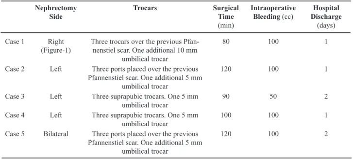
Median operative time was 100 minutes (range 80 to 120 minutes), median intraoperative blood loss was 100 cc (range 50 to 100 cc), and me -dian length of hospital stay was 1 day (range from 1 to 2 days) (Table-2).
In the second case (live donor nephrectomy), the warm ischemia time was 3.5 minutes. The graft presented immediate diuresis. The postoperative course of the recipient was uneventful and the patient was discharged seven days after the transplantation with a creatinine level of 0.9 mg/dL.
The pathological examination of the specimen demonstrated renal atrophy in the irst patient, a 5.5 cm renal cell carcinoma (pT1b) in the third patient, a 4.8 cm renal cell carcinoma (pT1b) in the fourth pa -tient, and bilateral renal atrophy in the ifth patient.
No intraoperative or postoperative complica -tions occurred in this initial series.
COMMENTS
Nephrectomy has been performed by minimally invasive surgery using transumbilical laparoendoscopic single-site and hybrid transvagi -nal approaches (1-5,8). Recently, Ponsky et al. (7) described the irst series of LESS nephrectomy and nephroureterectomy performed through a Pfannenstiel incision with no additional ports for renal cancer. In this article, we present our initial experience with this approach using no special devices for different indications of laparoscopic nephrectomy.
In 2008, Gupta et al. (9) compared differ -ent locations of incisions (modiied iliac fossa and Pfannenstiel incisions) to retrieve the kidney during laparoscopic transperitoneal donor nephrectomy and they found that there was less morbidity associated with the iliac fossa incision. Two patients had bladder injury and 1 patient had bowel injury while making the Pfannenstiel incision. Also, the mean length of the Pfannenstiel incision was longer than the iliac fossa incision (7.3 cm vs. 5.8 cm). However, Pfannenstiel
Table 2 – Intraoperative and early postoperative outcomes.
Nephrectomy Side
Trocars Surgical
Time
(min)
Intraoperative Bleeding (cc)
Hospital Discharge
(days)
Case 1 Right
(Figure-1) Three trocars over the previous Pfannenstiel scar. One additional 10 mm -umbilical trocar
80 100 1
Case 2 Left Three ports placed over the previous Pfannenstiel scar. One additional 5 mm
umbilical trocar
120 100 1
Case 3 Left Three suprapubic trocars. One 5 mm
umbilical trocar 90 50 2 Case 4 Left Three suprapubic trocars. One 5 mm
umbilical trocar 100 100 1 Case 5 Bilateral Three ports placed over the previous
Pfannenstiel scar. One additional 5 mm umbilical trocar
incision was found to be superior in terms of cosme -sis.
Considering these risks of bladder and bowel injuries during the performance of the Pfannenstiel incision, we decided to start all our procedures using a transumbilical trocar to guide the placement of the trocars over the Pfannenstiel incision. We did not feel comfortable in placing the suprapubic trocars without the laparoscopic guidance achieved by the umbilical puncture.
Advantages of the Pfannenstiel incision compared to vertical incisions include decreased pain, improved cosmesis (10-12), and better healing. The transverse suprapubic scar can be hidden with most types of clothing, including a bathing suit. In addition, the Pfannenstiel incision may be associ
-ated with a decreased rate of incisional hernia (12). When Bird et al. (13) analyzed a cohort of patients undergoing laparoscopic radical nephrectomy with intact specimen extraction through 3 different sites (lower quadrant site, umbilical site, and paramedian site), they found that the occurrence of incisional hernia was signiicantly associated with the parame -dian extraction site. Also, cesarean section delivery rates in Brazil are the highest in the world. In 1992, Moraes and Goldenberg (14) found an estimated in -cidence of cesarean sections of 80.5% in a Brazilian city. Cesarean rates increased according to income and were higher among women using private health care. Therefore, most Brazilian women already have a Pfannenstiel scar and it is interesting to beneit from an existing incision whenever possible.
Figure 1 – A) Position of all trocars. B) Cutting the left renal artery. C) Ligature of the left renal vein using Hem-o-lok® clips. D) Final aspect of the scar.
A
B
As previously observed by Ponsky et al. (7), the distance from the Pfannenstiel incision to the kidney was not a major challenge during surgery because longer laparoscopic instruments (those used for bariatric surgeries, with 45 cm working length) were available for the procedure. In their series, Ponsky et al. (7) used extra-long laparoscopic stapling devices to divide the superior renal pole at -tachments and hilar vessels; however, we conirmed the feasibility of the procedure without using any special devices, which is very interesting to reduce costs of the procedure.
The surgeon must be familiar with the visu -alization of the structures in a more medial perspec -tive than the standard transperitoneal approach when using the Pfannenstiel LESS (7). This dificulty can be overcome using a 30-degree laparoscope that provides a wider range of viewing angles. Also, the use of the Pfannenstiel incision allows the surgeon to place one trocar 2 cm away from the other, and this facilitates the movements inside the abdominal cavity when compared to transumbilical LESS, reducing the external clash of the instruments. The umbilical trocar was important during the procedure to facilitate the dissection of the posterior aspect of the kidney and the superior renal pole.
CONCLUSIONS
In this series, we report our initial experi -ence using the Pfannenstiel incision for laparoscopic nephrectomy with success. Prospective studies are necessary to conirm the real beneits and indications of this alternative surgical approach.
CONFLICT OF INTEREST
None declared.
REFERENCES
1. Branco AW, Kondo W, Stunitz LC, Filho AJ, de George MA: Transumbilical laparoscopic urological surgery:
are special devices strictly necessary? BJU Int. 2009; 104: 1136-42.
2. Desai MM, Stein R, Rao P, Canes D, Aron M, Rao PP, et al.: Embryonic natural orifice transumbili
-cal endoscopic surgery (E-NOTES) for advanced reconstruction: initial experience. Urology. 2009; 73: 182-7.
3. Gill IS, Canes D, Aron M, Haber GP, Goldfarb DA, Flechner S, et al.: Single port transumbilical (E-NOTES) donor nephrectomy. J Urol. 2008; 180: 637-41; discussion 641.
4. Desai MM, Rao PP, Aron M, Pascal-Haber G, Desai MR, Mishra S, et al.: Scarless single port transumbili
-cal nephrectomy and pyeloplasty: irst clini-cal report. BJU Int. 2008; 101: 83-8.
5. Sanchez-Salas RE, Barret E, Watson J, Stakhovskyi O, Cathelineau X, Rozet F, Galiano M, Rane A, De
-sai MM, Sotelo R, Vallancien G: Current status of natural orifice trans-endoscopic surgery (NOTES) and laparoendoscopic single site surgery (LESS) in urologic surgery. Int Braz J Urol. 2010; 36: 385-400.
6. Steinway ML, Lengu IJ, Cherullo EE, Ponsky LE: Laparoendoscopic single-site (LESS) nephrectomy through a Pfannenstiel incision: porcine model. J Endourol. 2009; 23: 1293-6.
7. Ponsky LE, Steinway ML, Lengu IJ, Hartke DM, Vourganti S, Cherullo EE: A Pfannenstiel single-site nephrectomy and nephroureterectomy: a practical application of laparoendoscopic single-site surgery. Urology. 2009; 74: 482-5.
8. Branco AW, Branco Filho AJ, Kondo W, Noda RW, Kawahara N, Camargo AA, et al.: Hybrid transvaginal nephrectomy. Eur Urol. 2008; 53: 1290-4.
9. Gupta M, Singh P, Dubey D, Srivastava A, Kapoor R, Kumar A: A comparison of kidney retrieval incisions in laparoscopic transperitoneal donor nephrectomy. Urol Int. 2008; 81: 296-300.
10. Grantcharov TP, Rosenberg J: Vertical compared with transverse incisions in abdominal surgery. Eur J Surg. 2001; 167: 260-7.
11. Brown SR, Goodfellow PB: Transverse verses midline incisions for abdominal surgery. Cochrane Database Syst Rev. 2005; 19: CD005199.
12. Tisdale BE, Kapoor A, Hussain A, Piercey K, Whelan JP: Intact specimen extraction in lapa
-roscopic nephrectomy procedures: Pfannenstiel versus expanded port site incisions. Urology. 2007; 69: 241-4.
extraction sites used during laparoscopic radical nephrectomy. J Urol. 2009; 181: 1565-70.
EDITORIAL COMMENT
Nowadays laparoscopists are very interested in reducing skin scars. In this article, at least two or three portal scars could be avoided. In my opinion, the method proposed may be useful when the surgeon intends to remove the kidney without morcellation and in such cases the Pfannenstiel incision is a very good cosmetic option.
This is a hybrid approach that combines an umbilical access for the camera, at the beginning of the procedure, and the “LESS” at the transverse suprapubic skin incision used at the end for removal of the kidney. The initial umbilical port followed the introduction of trocars in the lower abdomen and permitted the introduction of instrument to facilitate the dissection of upper pole and posterior surface of the kidney.
This article has some aspects that should be pointed out as 1. Introduction of trocars at the Pfan
-nenstiel incision under endoscopic orientation; 2. No need of any special device other than regular trocars; 3. The trocars can be a little bit more distant than in the LESS umbilical procedure which can facilitate the procedure.
Before the laparoendoscopic Pfannenstiel nephrectomy could be considered more than feasible, its safety, results and complications should be com -pared to the traditional laparoscopic nephrectomies, exactly as laparoscopic and open nephrectomies was compared.
Dr. Anuar Ibrahim Mitre
Associate Professor of Urology University Sao Paulo, USP Sao Paulo, SP, Brazil E-mail: [email protected]
14. de Moraes MS, Goldenberg P: Cesarean sections: an epidemic proile. Cad Saude Publica. 2001; 17: 509-19.
Accepted after revision: June 21, 2010
Correspondence address:
Dr. William Kondo