79
CLINICS 2006;61(1):79-82
Department of Ophthalmology, Hospital das Clínicas, São Paulo University Medical School - São Paulo/SP, Brazil.
Email: deborahsalerno@yahoo.com.br
LETTER TO THE EDITORS
ATYPICAL CHOROIDAL MELANOMA: REPORT OF 3
CASES
André Gustavo Bombana Nicoletti, Deborah Salerno Costa, Ramon Coral Ghanem, Pedro Carlos Carricondo, Ruth Miyuki Santo, and Suzana Matayoshi
Choroidal melanoma is the most common primary ocu-lar malignant neoplasm among adults.1 However,
innumer-able benign and malignant lesions may mimic its ophthalmoscopic features.2 Moreover, atypical presentations
of the tumor may also occur, in some cases making diag-nostic elucidation even more difficult and thus increasing the importance of supplementary tests, especially ultrasonographic methods. Three cases of choroidal melanoma with uncommon clinical features are presented in which the supplementary tests and the clinical follow-up were essential for the correct diagnosis of the lesions.
CASE 1
A female patient, 44 years old, leukodermatous, pre-sented with a 2-week history of sudden decrease in visual acuity of the right eye, preceded by photopsia for approxi-mately 2 weeks. On examination of visual acuity by fin-ger counting at 20 cm, an afferent pupillary defect was ob-served; intraocular pressure (IOP) was 12 mm Hg. On bi-omicroscopy, retro-lens retinal detachment was observed, which was better evidenced on retinal mapping, where an inferior exudative detachment with involvement of the pos-terior pole was found. No abnormalities were detected on examination of the left eye. Echography of the right eye revealed the presence of an elevated vitreous lesion with a base diameter of approximately 16 mm, anteriorly limited by the retina and/or choroid, and with high internal reflec-tivity. In view of these findings, diagnostic hypotheses such as malignant choroidal melanoma and suprachoroidal
hemorrhage were considered. The option was echography every 2 weeks, upon which the presence of low internal reflectivity, forming an angle kappa highly suggestive of melanoma of the choroid3 was evidenced. The presence of
arterial vascularization in the interior of the lesion on Dop-pler ultrasound reinforced this hypothesis. Clinical staging tests did not show systemic dissemination of the tumor.
The proposed treatment was enucleation of the of the right eyeball, which was initially rejected by the patient; however, within 30 days, she progressed with signs of pain, total retinal detachment, light perception visual acuity, 58 mm Hg IOP, rubeosis iridis on biomicroscopy, and angle neovascularization on gonioscopy, characterizing neovascular glaucoma. Enucleation of the right eyeball was performed, and histopathologic examination revealed a neo-plasm of choroidal origin predominantly occupying the in-ferior quadrants with an approximate thickness of 15 mm at the greater axis associated with total retinal detachment. The tumor was extensively necrotic (over 90%) with hemorrhagic areas and lymphocyte infiltration. The search for melanin in the whole tumor, including necrotic cells, was positive. The immunohistochemical tests were posi-tive for S-100, vimentin, and HMB-45, confirming the di-agnosis of choroidal melanoma.
The patient is without evidence of local or systemic re-lapse after 18 months of follow-up.
CASE 2
80
CLINICS 2006;61(1):79-82 Atypical choroidal melanoma: report of 3 cases
Nicoletti AGB et al.
of light perception visual acuity, fixed medial mydriasis, glaukomflecken, congested iris vessels, and a shallow an-terior chamber were observed. On funduscopy, the retina appeared fibrotic and completely detached. Intraocular pressure measured by applanation tonometry was 58 mm Hg. On gonioscopy, a 360o angle closure without presence
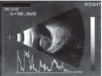
of new blood vessels on indentation was observed. The left eye presented visual acuity of 20/20 with –1.00 D sph, a deep anterior chamber, and an IOP 14 of mm Hg. Echography the right eye was then performed showing closed funnel retinal detachment and the presence of a solid lesion in the temporal sector with a base diameter of ap-proximately 8 mm (in the B mode), with low to moderate internal reflectivity forming the angle kappa observed in the A mode (Figure 1). After clinical evaluation, enuclea-tion of the right eyeball was indicated. The histopathologic examination confirmed the established hypothesis of ma-lignant choroidal melanoma.
The patient is being followed up as an outpatient and is without evidence of systemic dissemination 1 year after the diagnosis.
CASE 3
A female patient, 50 years old, leukodermatous, pre-sented with complaints of pain of moderate intensity and reduction in visual acuity for approximately 2 months. On examination, the visual acuity measured in the left eye was 20/80. The direct pupillary light reflex was decreased on the left, with a relative afferent defect. On biomicroscopy a pigmented nodule was detected in the inferior temporal sector of the iris of the left eye, associated with mild
con-junctival hyperemia and anterior chamber reaction (Figure 2). The mean IOP was 14 mm Hg. On funduscopy, the pres-ence of an inferior exudative retinal detachment was ob-served with involvement of the posterior pole. Examina-tion of the right eye did not reveal alteraExamina-tions. On echography, an elevated, solid, and homogeneous lesion with medium to low echogenicity was detected, delineat-ing the angle kappa in the region anterior to the inferior temporal equator, associated with inferior retinal detach-ment involving the posterior pole. The lengh of the lesion basement was 13.7 mm transversally, and the height was 6 mm. Ultrasound biomicroscopy (UBM) showed rectifi-cation and anterior detachment of the inferior temporal sec-tor of the iris, with disorganization of the ciliary processes between 3 and 5 hours. In addition, an increase in the thick-ness of the ciliary body in a solid form and with variable echogenicity with impairment of the root of the iris was observed (Figure 3).
Figure 1 - Case 2 echography: Closed funnel retinal detachment and the presence of a solid lesion in the temporal sector with a diameter of approximately 8 mm, with low to moderate internal reflectivity forming the angle kappa
Figure 2 - Case 3 biomicroscopy: a pigmented nodule was detected in the inferior temporal sector of the iris
81
CLINICS 2006;61(1):79-82 Atypical choroidal melanoma: report of 3 cases
Nicoletti AGB et al.
In view of the described features, the established diag-nosis was choroidal melanoma with extension to the iris through the ciliary body. After systemic examination, the patient underwent enucleation of the left eyeball. His-topathologic examination confirmed uveal involvement by primary choroidal melanoma.
After approximately 1 year, the patient is without any evidence of local or systemic relapse.
DISCUSSION
Innumerable benign and malignant lesions may simu-late the ophthalmoscopic features of choroidal melanoma.2
Historically, 20% of enucleated eyes with a clinical diag-nosis of malignant choroidal melanoma presented benign lesions on histological examination. More recent studies, based on improved supplementary tests, have reported a much better diagnostic precision, with 95% to 100%
cor-rect diagnoses.2-5 The Collaborative Ocular Melanoma
Study Group (COMS) found only 2 false positive cases (0.48%) in 413 eyes with a clinical diagnosis of malignant choroidal melanoma.2
Among the lesions that might simulate ophthalmoscopic features of malignant melanoma, limited hemorrhages of the choroid should be noted, which may result from ab-normal conditions such as hypotonia, inflammation, trauma, vascular diseases, or which might even occur spontaneaously.6-9 Recent choroidal hemorrhages present a
reddish color, but after their organization, hemosiderin con-tent and associated proliferation of the retinal pigment epi-thelium result in an ophthalmoscopic aspect similar to melanoma of the choroid.10 In addition, this lesion may be
presented as a typical hemorrhagic choroidal detachment.
Reese et al11 reported a case that seemed to be a
hemorrhagic process and was later correctly diagnosed as melanoma. This fact was attributed to the initial presence of an associated choroidal hemorrhage that masked the presence of the tumor. Indeed, the patient of case 1 had choroidal hemorrhage concomitant with the tumoral lesion, which made the initial diagnosis difficult, both by impair-ing adequate identification of the lesion and by producimpair-ing a high internal reflectivity, a characteristic contrary to that of melanoma. The extensive necrosis observed in case 1 of over 90% is an atypical histologic presentation and could also be responsible for the detected high internal reflec-tivity, as has been proposed by Bujara et al.12 However,
choroidal hemorrhages may also present with low internal reflectivity, making the differential diagnosis even more difficult.
Using color Doppler, the presence of arterial vasculari-zation was observed in the interior of the tumor, providing
further support for the elucidation of the case. Lieb et al,13
studying 44 intraocular masses, were able to show blood in the interior of 39 of them. In addition, they were not able to identify a vascular supply to benign lesions.
Some authors have reported cases of patients with a clinical diagnosis of malignant choroidal melanoma who actually presented limited hemorrhages that were observed only during the follow-up of these patients after detection of reduction in the diameter of these lesions.6,8 Augsburger
et al observed that such hemorrhages completely disap-peared within periods of less than 2 months.7
Association between uveal melanomas and secondary glaucomas has been established by several authors.14-17 The
occurrence of glaucomas secondary to intraocular tumors has been known for many years. In 1896, Marshall et al18
observed that 57% of eyes enucleated because of uveal
melanomas presented intraocular hypertension. Yanoff17
studied 96 eyes with a diagnosis of uveal melanoma and detected ocular hypertension in 19 cases (20%). These au-thors also observed that tumors of the anterior segment or those of the posterior segment presenting a large volume, mainly those accompanied by total retinal detachment, were more frequently associated with secondary glaucomas. Indeed, the patient of case 2 presented a choroidal lesion of large volume and associated with total retinal detach-ment, although she had first been diagnosed 10 years after a blunt ocular trauma.
Crises of angular closure typically occur in anatomi-cally predisposed eyes, which, among other particularities, present a decrease in the axial length of the eye.19 In
gen-eral, such characteristics are observed bilaterally. The pa-tient of case 2 presented an anterior chamber with normal depth in the noninvolved eye, which led to the suspicion of a secondary cause of angular closure and increase in IOP, although angular closure without neovascularization asso-ciated with choroidal melanoma is an atypical clinical pres-entation. Because of the presence of total retinal detach-ment, fundoscopic examination of the posterior segment was not possible, and echography was required. In the B mode, a roundish choroidal lesion was observed, with a base diameter of approximately 8 mm, localized at the posterior pole. In the A mode, low internal reflectivity was observed determining the angle kappa; such characteristics
were compatible with malignant choroidal melanoma,3
which was confirmed afterwards by histopathologic exami-nation. According to Yanoff,17 the presence of glaucoma
82
CLINICS 2006;61(1):79-82 Atypical choroidal melanoma: report of 3 cases
Nicoletti AGB et al.
patients of the latter group presenting opacification of the optical media.
Shield et al20 evaluated 200 patients (208 eyes) referred
to them because of lesions of the iris suspected to be ma-lignant melanoma. The clinical diagnosis of melanoma of the iris was confirmed in only 40 eyes (24%). The other lesions were classified as pseudomelanomas, the most fre-quent findings being primary cysts, nevus, essential atro-phy, and foreign bodies.
Secondary involvement of the iris by a choroidal melanoma is a rare condition and must also be considered in the differential diagnosis of pigmented nodules of the
iris, as was observed in the patient of case 3. In these situ-ations, UBM is of great value because it is able to identify the tumor’s extent by contiguity of the choroidal melanoma to the anterior uvea.
The 3 described cases illustrate different clinical presen-tations of uveal melanoma, which, together with the different benign lesions simulating their clinical aspects, contributed to the difficult diagnosis of these and certain other cases. Ultrasonographic methods (echography and UBM) and out-patient follow-up make a correct diagnosis and treatment pos-sible and practical for these patients with a clinical suspicion - even if it is a remote possibility - of a malignant tumor.
REFERENCES
1. Scotto J, Fraumeni FJ, Lee JH. Melanomas of the eye and other noncutaneous sites: epidemiologic aspects. J Natl Cancer Inst. 1976;56:489-91.
2. Accuracy of diagnosis of choroidal melanomas in the Collaborative Ocular Melanoma Study. COMS report no. 1. Arch Ophthalmol. 1990;108:1268-73.
3. Fuller DG, Snyder WB, Hutton WL, Vaiser A. Ultrasonographic features of choroidal malignant melanomas. Arch Ophthalmol. 1979;97:1465-72.
4. Char DH, Stone RD, Irvine AR, Crawford JB, Hilton GF, Lonn LI, et al. Diagnostic modalities in choroidal melanoma: sensitivity, specificity, and reproducibility. Am J Ophthalmol. 1980;89:223-30.
5. Ferry AP. Lesions mistaken for malignant melanoma of the posterior uvea. A clinicopathologic analysis of 100 cases with ophthalmoscopically visible lesions. Arch Ophthalmol. 1964;72:463-9.
6. Morgan CM, Gragoudas ES. Limited choroidal hemorrhage mistaken for a choroidal melanoma. Ophthalmology. 1987;94:41-46. 7. Augsburger JJ, Coats TD, Lauritzen K. Localized suprachoroidal
hematomas - ophthalmoscopic features, fluorescein angiography, and clinical course. Arch Ophthalmol. 1990;108:968-72.
8. Williams DF, Mieler WF, Lewandowski M. Resolution of an apparent choroidal melanoma. Retina. 1989;9:131-35.
9. Brubaker RF. Intra-ocular surgery and choroidal hemorrhage. Arch Ophthalmol. 1984;102: 1753-4.
10. Reese AB. Tumors of the Eye. 3rd edition, Hagerstown, MD. Harper and Row. 1976, p 174-262.
11. Reese AB. The differential diagnosis of malignant melanoma of the choroid. Arch Ophthalmol. 1957;58:477-82.
12. Bujara K, Domarus VD, Guthoff R. Necrotic malignant melanoma of the choroid with unusual clinical course. A clinical echographical and histological case study. Ophthalmologica. 1980;180:222-7.
13. Lieb WE, Shields JA, Cohen SM. Color Doppler imaging in the management of intraocular tumors. Ophthalmology. 1990;97:1660-4. 14. Jensen OA. Malignant melanomas of the uvea in Denmark 1943-1952. A clinical, histopathological, and prognostic study. Acta Ophtalmol. 75 (Suppl), 1963;75:1-220.
15. Shields MB, Klintworth GK. Anterior uveal melanomas and intraocular pressure.Ophthalmology. 1980;87:503-17.
16. Shields MB, Proia AD. Neovascular glaucoma associated with an iris melanoma. Arch Ophthalmol. 1987;105:672-74.
17. Yanoff M. Glaucoma mechanisms in ocular malgnant melanomas. Am J Ophtalmol. 1970;70:898-904.
18. Marshall CD. On tension in cases of intraocular tumors. Tr Ophtalmol Soc. 1896;16:155.
19. George R, Paul PG, Baskaran M, Ve Ramesh S, Raju P, Arvind H, et al. Ocular biometry in occludable angles and angle closure glaucoma: a population based survey. Br J Ophthalmol 2003;87:399-402. 20. Shields JA, Sanborn ES, Augsburger JJ. The differential diagnosis of