*Correspondência: Rua Luciana de Abreu, 323/206 - Moinhos de Vento
Porto Alegre – RS, Brazil CEP: 90570-060
AbstrACt
Celiac disease (CD) is found in genetically predisposed individuals, and characterized by intolerance to gluten ingestion, contained in cereals such as barley, rye, wheat and malt. Clinical manifestations vary from asymptomatic patients to severe forms of malabsorption syndromes, which may involve multiple systems and increase the risk of some neoplasias. Diagnosis of CD often requires a high degree of suspicion. There is not a single test for the diagnosis, which is reached after a combination of clinical
and laboratory data. The irst step may be a serum test, such as the antibodies anti- tissue transglu -taminase, or antiendomisio. If serum result is positive, duodenal biopsy is necessary for diagnostic
conirmation. IgA deiciency, which occurs in 3% of patients with DC, may lead to false-negatives
because serology is based on IgA antibodies. Another cause of false-negative tests is diet restriction of gluten; therefore diagnostic investigation must be carried out during a diet containing gluten. The screening for CD in asymptomatic individuals is not indicated.
KeywordS: Celiac disease. Diagnosis. Adult
diagnosis
of
celiac
disease
in
adults
tatiana sudbrack da gama e silva1, tania Weber furlanetto2
Study conducted at Universidade Federal do Rio Grande do Sul, Porto Alegre, RS, Brazil
1. Médica – Mestranda pela Universidade Federal do Rio Grande do Sul, Porto Alegre, RS
2. Pós-doutorado - Chefe do Serviço de Medicina Interna do Hospital de Clínicas de Porto Alegre, Porto Alegre, RS
i
ntroductionCeliac disease (CD) is characterized by intolerance to gluten ingestion, which is contained in cereals such as barley, rye, hay, and malt, happening in genetically predisposed individuals and
presents an inlammatory process that involves the small intes -tine mucosa, leading to atrophy of intestinal villi, malabsorption and a variety of clinical manifestations. Gluten proteins are relatively resistant to digestive enzymes, resulting in peptide deri-vatives that may lead to immunogenic response in CD patients. CD’s clinical manifestations may involve the gastrointestinal tract, as well as skin, liver, nervous system, reproductive system, bones, and endocrine system.1,2 Herpetiform dermatitis occurs in
10 to 20% of the patients and is a pathognomonic manifestation.3
Until recently, CD diagnosis was recognized only in patients who presented typical clinical manifestations or high degree of suspicion. Diagnosis is generally performed in children with malabsorption syndrome. After the appearance of high accuracy serum tests and bigger attention by the physicians to atypical manifestations, CD’s prevalence increased, as well as its
diag-nosis outside the ield of pediatrics. Estimated prevalence in
general population is around 1:100.4
CD’s clinical manifestations may vary, as described in Table 1.5,6
There is an important genetic predisposition in patients with CD, characterized by HLA-DQ2 and HLA-DQ8 surface markers.
Gluten interacts with HLA markers, provoking an abnormal immune response of the mucosa and tissue injury.
A review has showed that the relation of patients with diag-nosed and not diagdiag-nosed CD may be 1:7.7
A study indicates that over 36% CD patients had had a
previous diagnosis of IBS (irritable bowel syndrome).8
If not treated, celiac disease has a high morbimortality. Anemia, infertility, osteoporosis, and cancer, mainly intestinal lymphoma, are among the risks of complication in patients not treated.
CD diagnostic investigation must be performed before the introduction of treatment, which is gluten-free diet, for it may negatively alter the serum tests’ results and improve histology.9
CD diagnosis is not always easy to be performed. In around
10% of cases, there is dificulty in diagnosis due to conlicting serology, histology, and clinical indings. CD’s diagnosis must be
considered in every patient presenting chronic diarrhea,
abdo-minal distention, latulence, iron-deiciency anemia, early onset osteoporosis, elevated transaminases, irst and second degree
deiciency, Turner syndrome, Down syndrome and peripheral
neuropathy.4,6,10-15
There is no justiication in literature, at the moment, for
population screening for CD diagnosis.
s
erologyMarkers used are the antibodies antiendomisio (EMA) and anti-tissue transglutaminase (anti-tTG), for being sensitive and
speciic for early CD’s diagnosis.4 Several studies have evidenced
a high correlation among their results, thus it is not necessary to research both of them.
The research of antigliadin antibody (AGA) performance is not comparable to test mentioned above and is disused.
Serum tests are responsible for recognizing that CD is not rare.9
Positive serum test suggests CD diagnosis, but duodenal biopsy is still gold standard.9
Positive serology may become negative after 6-12 months from the gluten-free diet introduction.
Serum markers sensitivity is related to the extent of histo-logical damage in CD, both at diagnosis and in the follow up to gluten-free diet adherence. Serum tests sensitivity will be high when there is the presence of total villous atrophy and its
progressive decrease, as histological indings are less altered.
Thus, negative serology does not exclude CD diagnosis. Serum tests may be used to evaluate the patient’s adherence to gluten-free diet. Antibodies become negative after 3-12 months of diet.16
Anti-tissue transglutaminase (anti-ttG IgA)
The antigen against which antiendomisio antibodies are directed is the transglutaminase enzyme. Anti-tTG IgA is the anti-body against tissue transglutaminase (the enzyme responsible for deamination of gliadin the lamina propria).
This test is performed by the ELISA method and uses as
subtract the guinea pig protein – irst generation (90% sensitivity and 95.3% speciicity), cells derived from human erythrocytes (95.1 sensitivity and 98.3 speciicity) or human recombinant –
second generation.9 Some diseases may interfere in the results,
leading to false-positives, such as chronic hepatic disease, heart
failure, arthritis, diabetes mellitus, and intestinal inlammatory
disease. This interference has been decreased with newer tests.9
Separately, it is the most eficient serum test for detecting
CD. It may be performed with a small blood sample collected
from the inger.
It was recently demonstrated that tTG-Abs RIA may be detected in human saliva, which can prevent the need of collec-ting blood, making the CD diagnosis easier, especially in children. The research on anti-tTG IgA is highly sensitive to CD diagnosis and for the follow up of patients under gluten-free diet.19
Antiendomisio IgA (EMA)
EMA IgA antibodies bind endomisio, the conjunctive tissue enveloping the smooth muscle, producing a characteristic
pattern. It is detected by indirect immunoluorescence. It is a
method that requires more time if compared to the ELISA method, besides being operator-dependent.9 For its performance, monkey
esophagus (EMA IgA 97.4% sensitivity and 99.6% speciicity) or human umbilical cord (EMA IgA 90.2 sensitivity and 99.6% speciicity) as subtracts for performing the test.9
It is recognized that the presence of EMA is a predictor of progression in direction of villous atrophy.17,18
Antigliadin antibodies (AGA IgA)
This is the oldest marker and is determined by the ELISA method. Reference values are not constant among laboratories.
Its eficacy is dificult to deine, for available data in literature are heterogeneous and do not permit comparison. Its speciicity is approximately 90%, and the sensitivity is around 85%-90%,
presenting low positive predictive value.9 There are other tests
with higher diagnostic performance.
IgA selective deiciency
IgA deiciency is the most common human immunodeiciency
and it is 10-15 times more common in CD patients. However, IgA dosage must be performed only if there is high suspicion of its
deiciency. Approximately 3% of CD patients have this deiciency,
table 1 – Celiac disease’s clinical manifestations
Classic form: Symptomatic intestinal malabsorption. Chronic diarrhea, abdominal pain, abdominal distention, weight loss,
and latulence may occur.
Atypical form: Absence or few gastrointestinal symptoms,
pres-ence of atypical symptoms, such as anemia due to iron deiciency,
osteoporosis or osteopenia, infertility, low stature. It is the most common presentation.
Silent form: Occasional diagnosis, histological or serological, in asymptomatic individuals.
Latent form: There are 2 forms: 1 – Patients with previous CD diagnosis who responded to gluten-free diet and presented a normal histology or only intraepithelial lymphocytes increase. 2 – Individuals with normal intestinal mucosa, under diet including gluten, who will subsequently develop CD.
which may produce false-negative in serum tests EMA, anti-tTG IgA and AGA IgA, all of them based on IgA.20
In patients with IgA selective deiciency serology can be
performed with IgG, both EMA IgG and tTGA IgG have
excel-lent sensitivity (close to 100%) and speciicity. Nevertheless, IgG-based tests have lower sensitivity and speciicity in relation
to those based on IgA, those with normal IgA levels.4 Thus, if
serology (EMA IgA or tTGA IgA) is negative in patients with high CD suspicion, seric IgA must be dosed.4,9
If CD suspicion is high, with persistently negative tests, individuals must perform typing for HLA and, if positive, they must perform duodenal biopsy or alternatively perform biopsy directly.4,5
HLA typing
It is the irst step for investigating relatives of CD patients.
HLA typing excludes one third of 1st degree relatives and identiies
individuals for evaluation with biopsy. It is also the clinical exam indicated if the individual presents negative serology and refuses
to undergo biopsy. HLA allele DQ2 is identiied in 90%-95% of
celiac patients, and HLA DQ8, in most of the others. Therefore,
their absence has a negative predictive value next to 100%.4,9
HLA typing is useful also to exclude the disease in patients who, unwittingly, are already undergoing gluten-free diet or individuals in which diagnosis is not clear.
Duodenal biopsy
CD diagnosis and lifelong gluten-free diet introduction must
not be irmed without compatible histological indings, regardless
of the results of serological tests. However, it is also not advised
to afirm a diagnosis based only on the histological diagnosis,
because the disease does not compromise uniformly intestine, and alterations are not observed exclusively in CD. In spite of these problems, intestinal biopsy is considered ‘gold standard’ diagnosis.4
Patients who present persistently positive serology and nega-tive biopsy probably have latent CD.
The proper number of biopsy fragments from the duodenal second portion or the more distal part is between 4 and 6.10,20
A recent study demonstrated that four biopsies may be suficient for CD diagnosis in 100% of cases.21,22 Mucous alterations have
an irregular pattern, well demonstrated in magniication,23,24
mainly associated with chromoendoscopy;24 Brunner glands
and peptic alterations may hinder histological exam, if biopsies are too proximal.
The pathologist must be familiarized with the spectrum of alte-rations compatible with CD, evaluate and describe lymphocytic
iniltration, pattern of crypts, and villous atrophy. Classiication is done using modiied Marsh’s criteria25 and Oberhuber et al.26
Classiication proposed by Marsh in 1992 is the most widely used
until today. The patient’s symptom frequently correlates with the
degree of tissue injury, according to what is described below:9
• Marsh I: iniltrative lesion, normal villous architecture
and mucosa, IEL increase (>30-40 lymphocytes/enterocytes counted).
• Marsh II: hyperplasic lesion; similar to Marsh I, but it also presents crypt hyperplasia.
• Marsh III: destructive lesion, subdivided in IIIa – partial villous atrophy; IIIb – subtotal villous atrophy, and IIIc – total villous atrophy.
Intraepithelial lymphocytes (IEL) increase, with normal mucosa architecture may be observed in autoimmune diseases, such as systemic lupus erythematosus, rheumatoid arthritis, and Hashimoto’s thyroiditis, in patients using non-hormonal
anti-inlammatories, in CD’s initial presentation and latent CD.4,27 An
increase in LIE may also relect a state of T cells activation trig -gered by gluten, immune disturbs, drugs, and infectious agents. Patients with CD who present only IEL increase, with no alte-rations in the architecture of the mucosa, may be symptomatic and be under increased osteoporosis risk.
Studies suggesting that bulb biopsies seem to be adequate and this may be the only one to show villous atrophy.28-31
In patients that have already initiated GFD, even before the
conirmation biopsy, with high CD suspicion and negative sero -logy, a test with a diet containing gluten may be performed, in this case for at least four weeks and, afterwards, biopsy. However, some patients are late responders and may take years to have their histology altered.4
It must be very clear that GFD must be established only after
a conirmed CD diagnosis.
Diagnosis may be dificult, because the serology may be nega -tive, the disease may have an irregular histological behavior or the number or place of biopsies may not be adequate. Biopsies may have an adequate size, be well oriented with villi turned upside,
on ilter paper, enabling for crossing and non-tangent sections,
for tangential cuts may lead to misinterpretations. The type of
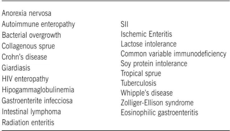
table 2 – Celiac disease differential diagnosis
Anorexia nervosa Autoimmune enteropathy Bacterial overgrowth Collagenous sprue Crohn’s disease Giardiasis HIV enteropathy Hipogammaglobulinemia Gastroenterite infecciosa Intestinal lymphoma Radiation enteritis
SII
Ischemic Enteritis Lactose intolerance
Common variable immunodeiciency
clamp does not seem to be relevant.21,32 Mucosa inlammation
and architecture alterations may be concealed using corticoids and immunosuppressive agents.
Duodenal mucosa inspection, during upper gastrointestinal
endoscopy, is important and may reveal signiicant indings; the endoscopist must be attentive to indings related to villous
atrophy, despite of low sensitivity of the tests. During
endos-copy the following indings, suggesting CD, may be identiied: thickened mucous folds, mosaic pattern, lat folds, smaller size and disappearance of maximum insuflated folds. Patients who
undergo upper gastrointestinal endoscopy for weight reduction, anemia, diarrhea, and those with high CD risk (irritable bowel
syndrome, inlammatory bowel disease, chronic hepatic disease,
Down syndrome, several autoimmune diseases, mainly diabetes mellitus type 1) must take intestinal biopsy.
In Table 2 some diseases that are part of differential diagnosis are presented.5,33,34
Other exams – Endoscopic capsule
Abnormalities in CD patients’ mucosa without previous diagnosis may be observed through the endoscopic capsule
exam, for investigating anemia and iron deiciency.34 In these
cases serology and duodenal biopsies could probably eliminate the need of endoscopic capsule exam. Duodenum, evaluated by upper gastrointestinal endoscopy (UGE), may appear entirely
normal, while in proximal and distal intestines classic indings
of CD are discovered with endoscopic capsule.34
Other exams – Magniication endoscopy
Recently an article was published demonstrating that high
resolution UGE with magniication (with OBI – optimal band
imaging) permits a clear visualization of duodenal villi pattern
(sensitivity, speciicity, positive and negative predictive value are 100%). OBI system may play a role in optimizing UGE accuracy
in CD.33
Discussion/Conclusion
CD diagnosis is complex, especially in asymptomatic patients or those with atypical manifestations. Intestinal biopsy is needed for this diagnosis, even in face of a positive serology. However,
histological indings are not speciic, then the diagnosis can be
established only after clinical correlation. Still today most patients with CD do not have this diagnosis, although, in the last years, prevalence has grown due to higher suspicion degree and higher accuracy of serum tests. The meaning of the great number of patients not diagnosed is not well established, as well as that of those patients who present only extraintestinal or non-classic symptoms.
No conlicts of interest declared concerning the publication of this article.
r
eferences1. Rewers M, Liu E, Simmons J, Redondo MJ, Hoffenberg EJ. Celiac disease
associated with type 1 diabetes mellitus. Endocrinol Metab Clin North Am.
2004;33(1):197-214.
2. Rewers M. Epidemiology of celiac disease: what are the prevalence, incidence, and progression of celiac disease? Gastroenterology. 2005;128(4 Suppl 1):S47-S51.
3. Alaedini A, Green PH. Narrative review: celiac disease: understanding a complex
autoimmune disorder. Ann Intern Med. 2005;142(4):289-98.
4. AGA Institute Medical Position Statement on the Diagnosis and Management of Celiac Disease. Gastroenterology. 2006;131(6):1977-80.
5. Presutti RJ, Cangemi JR, Cassidy HD, Hill DA. Celiac disease. Am Fam Physi-cian. 2007;76(12):1795-802.
6. Torres MI, Lopez Casado MA, Rios A. New aspects in celiac disease. World J
Gastroenterol. 2007;13(8):1156-61.
7. Sdepanian VL, Morais MBD, Fagundes-Neto U. Doença celíaca: a evolução dos conhecimentos desde sua centenária descrição original até os dias atuais.
Arq Gastroenterol. 1999; 36(4):244-57.
8. Green PH. The many faces of celiac disease: clinical presentation of celiac disease in the adult population. Gastroenterology. 2005;128(4 Suppl 1):S74-S8.
9. Rostom A, Murray JA, Kagnoff MF. American Gastroenterological Association (AGA) Institute technical review on the diagnosis and management of celiac disease. Gastroenterology. 2006;131(6):1981-2002.
10. Green PH, Cellier C. Celiac disease. N Engl J Med. 2007;357(17):1731-43.
11. Mackey J, Treem WR, Worley G, Boney A, Hart P, Kishnani PS. Frequency of celiac disease in individuals with Down syndrome in the United States. Clin Pediatr (Phila). 2001;40(5):249-52.
12. Murray JA. Celiac disease in patients with an affected member, type 1 diabetes,
iron-deiciency, or osteoporosis? Gastroenterology. 2005; 128(4 Suppl
1):S52-S6.
13. Rubio-Tapia A, Murray JA. The liver in celiac disease. Hepatology. 2007; 46(5):1650-8.
14. Sanders DS, Carter MJ, Hurlstone DP, Pearce A, Ward AM, McAlindon ME, et al. Association of adult coeliac disease with irritable bowel syndrome: a
case-control study in patients fulilling ROME II criteria referred to secondary
care. Lancet. 2001;358(9292):1504-1508.
15. Yang A, Chen Y, Scherl E, Neugut AI, Bhagat G, Green PH. Inlamma
-tory bowel disease in patients with celiac disease. Inlamm Bowel Dis.
2005;11(6):528-32.
16. Collin P, Kaukinen K, Valimaki M, Salmi J. Endocrinological disorders and celiac disease. Endocr Rev. 2002; 23(4):464-83.
17. Breyer H, Maguilnik I. Doença celíaca - “Procura e encontratás”. Rev AMRIGS. 2008;52:138-43.
18. Ladinser B, Rossipal E, Pittschieler K. Endomysium antibodies in coeliac disease: an improved method. Gut. 1994; 35(6):776-8.
19. Bonamico M, Nenna R, Luparia RP, Perricone C, Montuori M, Lucantoni F,
et al. Radioimmunological detection of anti-transglutaminase autoantibodies in human saliva: a useful test to monitor celiac disease follow-up. Aliment Pharmacol Ther. 200828(3):364-70.
20. Cataldo F, Marino V, Ventura A, Bottaro G, Corazza GR. Prevalence and clinical
features of selective immunoglobulin A deiciency in coeliac disease: an
Italian multicentre study. Italian Society of Paediatric Gastroenterology and Hepatology (SIGEP) and “Club del Tenue” Working Groups on Coeliac Disease. Gut. 1998;42(3):362-65.
21. Mee AS, Burke M, Vallon AG, Newman J, Cotton PB. Small bowel biopsy
for malabsorption: comparison of the diagnostic adequacy of endo-scopic forceps and capsule biopsy specimens. Br Med J (Clin Res Ed). 1985;291(6498):769-72.
22. Pais WP, Duerksen DR, Pettigrew NM, Bernstein CN. How many duodenal
biopsy specimens are required to make a diagnosis of celiac disease? Gastro-intest Endosc. 2008;67(7):1082-7.
23. Lo A, Guelrud M, Essenfeld H, Bonis P. Classiication of villous atrophy with enhanced magniication endoscopy in patients with celiac disease and tropical
sprue. Gastrointest Endosc. 2007;66(2):377-82.
24. Siegel LM, Stevens PD, Lightdale CJ, Green PH, Goodman S,
Garcia-Carras-quilho RJ, et al. Combined magniication endoscopy with chromoendoscopy in
the evaluation of patients with suspected malabsorption. Gastrointest Endosc. 1997;46(3):226-30.
25. Marsh MN. Gluten, major histocompatibility complex, and the small intestine. A
molecular and immunobiologic approach to the spectrum of gluten sensitivity (celiac sprue). Gastroenterology. 1992;102(1):330-54.
26. Oberhuber G, Granditsch G, Vogelsang H. The histopathology of coeliac disease: time for a standardized report scheme for pathologists. Eur J Gastroenterol Hepatol. 1999;11(10):1185-94.
Artigo recebido: 24/02/09
Aceito para publicação: 08/09/09
28. Bonamico M, Mariani P, Thanasi E, Ferri M, Nenna R, Tibeti C, et al. Patchy
villous atrophy of the duodenum in childhood celiac disease. J Pediatr
Gastro-enterol Nutr. 2004;38(2):204-7.
29. Ravelli A, Bolognini S, Gambarotti M, Villanacci V. Variability of histologic lesions in relation to biopsy site in gluten-sensitive enteropathy. Am J Gastroenterol. 2005;100(1):177-85.
30. Vogelsang H, Hanel S, Steiner B, Oberhuber G. Diagnostic duodenal bulb biopsy in celiac disease. Endoscopy. 2001;33(4):336-40.
31. Bonamico M, Thanasi E, Mariani P et al. Duodenal bulb biopsies in celiac disease:
a multicenter study. J Pediatr Gastroenterol Nutr. 2008;47(5):618-622.
32. Dandalides SM, Carey WD, Petras R, Achkar E. Endoscopic small bowel mucosal biopsy: a controlled trial evaluating forceps size and biopsy location in the diagnosis of normal and abnormal mucosal architecture. Gastrointest Endosc. 1989;35(3):197-200.
33. Cammarota G, Cesaro P, Cazzato A, Sparano L, Vecchio FM, Larocca LM, et al. Optimal band imaging system: a new tool for enhancing the duodenal villous pattern in celiac disease. Gastrointest Endosc. 2008;68(2):352-7.
34. Muhammad A, Pitchumoni CS. Newly Detected Celiac Disease by Wireless Capsule Endoscopy in Older Adults With Iron Deiciency Anemia. J Clin