475
IMAGES IN NEUROLOGY
Panayiotopoulos syndrome and
continuous spike-wave during slow sleep
Síndrome de Panayiotopoulos y punta-onda continua durante el sueño lento
Antonio Díaz-Negrillo
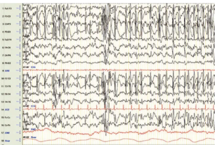
A 6-year-old girl suddenly developed a loss of concious-ness episode with head deviation to the left, generalized hypertonia, and clonic movements when waking up. Then, she presented vomit and bladder sphincter incontinence. Therefore, the Panayiotopoulos syndrome was suspected. Brain magnetic resonance imaging (MRI) was normal. The electroencephalography (EEG) performed after 48 hours of the episode revealed spike-wave paroxysms in parieto-oc-cipital regions of the left hemisphere (Fig 1). During non-rapid eye movement (NREM) sleep, the EEG showed epi-leptiform activity of continuous spike-wave greater than 85% (Fig 2).
The Panayiotopoulos syndrome can occur in very rare cases with atypical clinical and EEG findings, being the lat-ter central in delat-termining the patient’s prognosis1.
Department of Clinical Neurophysiology, Infanta Elena Hospital, Madrid, Spain.
Correspondence: Antonio Díaz Negrillo; Unidad de Neurofisiología Clínica; Avenida Reyes Católicos 21; 28340 Valdemoro - Madrid; E-mail: [email protected]
Conflict of interest: There is no conflict of interest to declare. Received 13 December 2011; Accepted 23 December 2011
Sens: 7 μV/mm. Time constant: 0.3 seconds. High-frequency filter: 30 Hz. Fig 1. Waking EEG manifests the presence of some paroxysms of spike-wave in parieto-occipital areas of the left hemisphere.
Sens: 7 μV/mm. Time constant: 0.3 second. High-frequency filter: 30 Hz. Fig 2. NREM sleep EEG. Epileptiform activity in the left hemisphere: continuous spike-wave greater than 85%.
1. Caraballo R, Astorino F, Cersósimo R, Soprano AM, Fejerman N. Atypical evolution in childhood epilepsy with occipital paroxysms. Epileptic Disord2001;3:57-62.