Original Article
3 3 Arq Bras Oftalmol. 2016;79(1):33-6 http://dx.doi.org/10.5935/0004-2749.20160010
INTRODUCTION
Glaucoma is a multifactorial disease characterized by progressive optic neuropathy with loss of visual field. Historically, elevated intrao-cular pressure (IOP) is regarded as the primary cause of glaucomatous optic nerve damage; however, there is now evidence that other fac-tors are involved in the pathogenesis of glaucoma, such as changes in blood flow, perfusion, and oxygen delivery(1). There is compelling
accumulated evidence supporting the theory that decreased ocular blood flow contributes to glaucomatous optic neuropathy(1,2). In a
model of optic nerve injury, it was demonstrated that alterations of perfusion and oxygen delivery to the retinal ganglion cells may signi-ficantly contribute to vision loss(3). Therefore, detailed hemodynamic
evaluation of the orbital circulation is needed to better understand this complex disorder.
The hemodynamics of orbital blood vessels can be quantified by color Doppler imaging (CDI), which allows estimations of velo-city and resistance to blood flow in unanesthetized subjects. This method has been widely used to study these blood flow parameters in retrobulbar blood vessels in humans and animals(4-8). The Doppler
waveform represents changes in the velocity of the blood flow during the cardiac cycle, and deflections in the late systolic [peak systolic velocity (PSV)] or early diastolic flow [end diastolic velocity (EDV)] are characteristic of high resistance arterial blood flow waveforms(6,9).
The resistive index (RI), also known as the Pourcelot ratio, is calculated
Effects of prostaglandin analogs on blood flow velocity and resistance in the
ophthalmic artery of rabbits
Efeitos dos análogos da prostaglandina na velocidade do luxo sanguíneo e resistência na
artéria oftálmica de coelhos
AmáliA Turner GiAnnico1, leAndro limA1, GilliAn c. ShAw2, heloiSA h. A. ruSS3, Tilde rodriGueS FroeS1, FAbiAno monTiAni-FerreirA1
Submitted for publication: May 28, 2015 Accepted for publication: October 20, 2015
1 Universidade Federal do Paraná, Curitiba, PR, Brazil.
2 Department of Molecular and Comparative Pathobiology, School of Medicine, Johns Hopkins Uni
versity, Baltimore, MD, USA.
3 Instituto Graefe de Oftalmologia, Curitiba, PR, Brazil.
Funding: No specific financial support was available for this study.
Disclosure of potential conflicts of interest: None of the authors have any potential conflict of interest to disclose.
Corresponding author: Amália T. Giannico. Rua dos Funcionários, 1.540 Curitiba, PR 80035050 Brazil Email: [email protected]
Approved by the following research ethics committee: Universidade Federal do Paraná, protocol #011/2011.
ABSTRACT
Purpose: The aim of this study was to investigate the effects of prostaglandin analogs on blood flow in the ophthalmic artery of clinically healthy rabbits. Methods: Fifty-five clinically healthy New Zealand white rabbits were divided into six groups, and the left eyes were treated for four weeks with the preservative benzalkonium chloride (BAK) only or a topical formulation of different prosta-glandin analogs (bimatoprost BAK, tafluprost BAK-free, travoprost BAK, travoprost POLYQUAD, and latanoprost BAK). Color Doppler imaging was performed before and after the treatments. The mean values of the peak systolic velocity (PSV) and end diastolic velocity and the resistive index (RI) were calculated. Statistical analysis was performed to compare the differences pre- and post-treatment for each drug and post-treatment among the drugs.
Results: The prostaglandin analogs did not affect PSV. Bimatoprost BAK, travoprost POLYQUAD, and latanoprost BAK did not change RI. Tafluprost BAK-free and tra-voprost BAK therapy resulted in similar reductions in RI. No significant differences pre- and post-treatment were found when BAK was administered alone. Conclusion: The prostaglandin analogs tafluprost BAK-free and travoprost BAK improved blood flow in the ophthalmic artery in healthy New Zealand white rabbits, which suggests that these drugs enhance the prevention of the progres-sion the progresprogres-sion of glaucoma.
Keywords: Color Doppler imaging; Orbital hemodynamics; Glaucoma; Orycto-lagus cuniculus
RESUMO
Objetivo:O objetivo deste estudo foi investigar os efeitos dos análogos da prosta-glandina (PGAs) no fluxo sanguíneo da artéria oftálmica em coelhos.
Métodos: Cinquenta e cinco coelhos da raça Nova Zelândia clinicamente saudáveis foram divididos em seis grupos para tratamento com formulação tópica de diferentes APGs (bimatoprosta BAK, tafluprosta BAK-free, travoprosta BAK, travoprosta POLYQUAD e latanoprosta BAK) e formulações contendo apenas o conservante cloreto de benzal-cônio (BAK). Foi realizada ultrassonografia com Doppler antes e após os tratamentos. Os valores do pico da velocidade sistólica (PSV) e da velocidade diastólica final foram obtidos e o índice de resistência (RI) foi então calculado. A análise estatística foi rea-lizada para comparar as diferenças entre cada droga no pré e pós-tratamento, além das diferenças no pós-tratamento entre as drogas.
Resultados: Estes colírios PGAs não afetaram o PSV. A bimatoprosta com o conservante BAK, travoprosta com o conservante POLYQUAD e latanoprosta com o conservante BAK não alteraram o RI. Já o tratamento com tafluprosta sem conservante (BAK-free) e travoprosta com o conservante BAK promoveram redução similar dos valores do RI. Não houve diferença significativa na comparação entre valores pré e pós-tratamento quando BAK foi administrado isoladamente.
Conclusão: Os PGAs tafluprosta BAK-free e travoprosta BAK melhoraram o fluxo san-guíneo na artéria oftálmica em coelhos da raça Nova Zelândia sugerindo que estes medicamentos possam contribuir na prevenção da progressão do glaucoma.
Effects of prostaglandin analogs on blood flow velocity and resistance in the ophthalmic artery of rabbits
3 4 Arq Bras Oftalmol. 2016;79(1):33-6
from the blood flow velocities. This index, expressed by the formula [(PSV - EDV) / PSV], indicates the downstream resistance in arteries (ranging from 0 to 1, where 0 is no resistance and 1 is the maximum resistance)(9). CDI research has shown that through increased RI,
retrobulbar blood flow is reduced in patients with glaucoma and that there may be a predictive value for the progression of the disease(10,11).
A high RI value correlates with an increase in vascular resistance, leading to decreased perfusion, which may in turn contribute to glaucomatous optic neuropathy(2,5,7,9,12,13).
Prostaglandin analogs (PGAs) are IOP-lowering agents commonly prescribed in glaucoma treatment. PGAs act primarily by enhancing uveoscleral outflow of aqueous humor; however, PGAs also appear to act on the trabecular meshwork to facilitate aqueous humor outflow through the conventional outflow pathway(14).
The majority of topical treatments for elevated IOP contain a preservative, the most common of which is benzalkonium chloride (BAK), a quaternary ammonium salt. Chronic exposure to BAK has been associated with symptoms of ocular discomfort attributed to BAK-induced instability of the tear film, reduced density of superfi-cial epithelial cells, disruption of corneal epithelial barrier function, and conjunctival inflammation(15,16). These undesirable effects may
be reversible in glaucoma patients who are switched to BAK-free medications(15). BAK is absorbed and accumulates in ocular structures
involved in glaucoma pathogenesis, but the potential effects of pre-servatives in the vasculature of the eye are unknown(17).
Considering the growing evidence supporting a vascular patho-genesis for glaucoma, a decrease in blood flow could potentially accelerate disease development. Therefore, in addition to their IOP-lo wering capabilities, these medications may be detrimental or beneficial to ocular hemodynamics. The purpose of this investigation was to examine the potential effects of topical solutions of PGAs with and without preservatives on the blood flow of the ophthalmic artery (OA) in clinically healthy rabbits.
METHODS
A
NIMALSThe investigation was carried out using 55 clinically healthy, six- month-old New Zealand white rabbits (Oryctolagus cuniculus), with 32 females and 23 males weighing 2.2 kg to 3.0 kg. The animals were selected randomly from a commercial breeder. All procedures using live rabbits were conducted in accordance with the Federal Universi-ty of Parana’s Animal Use Committee (Curitiba ciUniversi-ty, Paraná state, Brazil) and with the ARVO Statement for the Use of Animals in Ophthalmic and Vision Research.
The rabbits were housed under a 12:12 hour light: dark cycle for one week prior to and for the duration of the study. Food and water were given ad libitum, and the humidity (70%) and temperature (22.5°C) were controlled.
Physical examinations were performed before the ocular exami-nations to exclude animals with any indications of systemic disease. Rabbits with evidence of ocular or systemic diseases were excluded from this research. To avoid inter-investigator discrepancies, the same masked investigator performed the CDI, and another investigator, who was not masked, instilled the eye drops.
T
REATMENTSThe rabbits received a number from one to 55 and were then divided into six groups using the randomized function in Microsoft Office Excel (Microsoft Office 2007 for Windows). The left eyes were treated daily for four weeks with one drop of topical PGA eye drops applied to the conjunctival fornix. The ophthalmic drugs and num-bers of females and males are listed in table 1.
C
OLORD
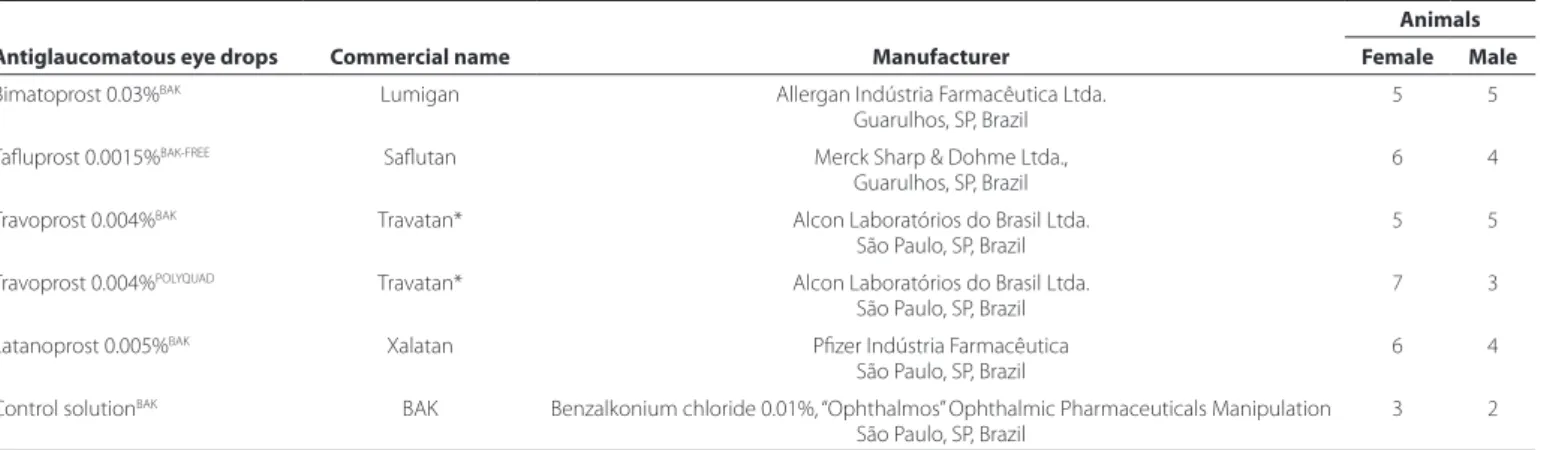
OPPLERIMAGINGCDI was performed before and after the treatment with eye drops using an ultrasound system (MyLab 30; Esaote, Genova, Italy) equip-ped with a 12-MHz linear ultrasound transducer. The animals were not anesthetized, and the eye and orbit were imaged with the animal in sternal recumbency. All CDI examinations were performed by the same two people, one of whom restrained the rabbit, while the other performed the imaging. Ultrasound gel was applied to the dorsal region and to the zygomatic arch, and the transducer was gently positioned with minimal pressure in the horizontal plane after instillation of one drop of topical anesthesia (tetracaine ophthalmic drops, Anestalcon®, Alcon). The long axis of the transducer was held horizontally between the lateral and medial canthus with the marker pointing nasally (Figure 1A).
We used the muscular cone and power Doppler mode to de-termine the relative position of the OA. After the detection of the blood flow by the power Doppler, the spectral Doppler sampling volume was placed in the center of the imaged vessel. The OA is readily identifiable by its characteristic dicrotic notch in the Doppler waveform associated with the closure of the aortic valve(5). The
ultra-sound beam and the OA were parallel, and the sample volume was set at 1 mm inside the vessel (Figure 1B). The mean values of PSV and EDV from three pulse waveforms were calculated (Figure 1B), and the RI of the blood flow was calculated using the formula [(PSV - EDV) / PSV].
Table 1. Prostaglandin analog eye drops, commercial names, manufacturers, and numbers of female and male New Zealand white rabbits
Antiglaucomatous eye drops Commercial name Manufacturer
Animals
Female Male
Bimatoprost 0.03%BAK Lumigan Allergan Indústria Farmacêutica Ltda.
Guarulhos, SP, Brazil
5 5
Tafluprost 0.0015%BAK-FREE Saflutan Merck Sharp & Dohme Ltda.,
Guarulhos, SP, Brazil
6 4
Travoprost 0.004%BAK Travatan* Alcon Laboratórios do Brasil Ltda.
São Paulo, SP, Brazil
5 5
Travoprost 0.004%POLYQUAD Travatan* Alcon Laboratórios do Brasil Ltda.
São Paulo, SP, Brazil
7 3
Latanoprost 0.005%BAK Xalatan Pfizer Indústria Farmacêutica
São Paulo, SP, Brazil
6 4
Control solutionBAK BAK Benzalkonium chloride 0.01%, “Ophthalmos” Ophthalmic Pharmaceuticals Manipulation
São Paulo, SP, Brazil
3 2
*= there is no difference in the commercial name between the eye drops TravatanBAK and TravatanPOLYQUAD.
Giannico AT, et al.
3 5 Arq Bras Oftalmol. 2016;79(1):33-6
S
TATISTICALANALYSISThe Shapiro-Wilk normality test demonstrated that the data errors were normally distributed. The data were statistically analyzed using the computer software StatView (SAS Institute, Cary, NC, USA). ANOVA was used to compare the pre-treatment results to show that the groups were homogeneous. Paired t-tests were used to compare the potential differences between pre- and post-treatment results for each drug, and the Tukey-Kramer post hoc test was used to compare potential post-treatment differences among the drugs. P-values of <0.05 were considered to be statistically significant.
RESULTS
The means and standard deviations of the pre- and post-treatment values for RI, PSV, and EDV are presented in table 2. PSV and EDV are expressed in centimeters per second (cm/s), whereas RI has no units.
No significant differences were found in the pre-treatment PSV, EDV, and RI of the left eye of all the rabbits (P>0.05), showing that these parameters were homogeneous amongst the groups before treatment. No significant differences were found between pre- and post-treat ment PSV or EDV in all the animal groups nor for the pre- and post-treatment RI in the rabbits that received bimatoprost BAK, tra-voprost POLYQUAD, or latanoprost BAK (P>0.05) (Table 2). However,
eyes that received tafluprost BAK-free and travoprost BAK exhibited a significant RI decrease after treatment (P=0.003 and P=0.005, res-pectively) (Table 2). This decrease in RI was similar in magnitude for the two drugs because the post-treatment values in the eyes that received tafluprost BAK-free and travoprost BAK were not significan-tly different (P=0.6264).
DISCUSSION
In general, prostaglandins play an important role in the local re-gulation of blood flow, and both endogenous prostaglandins as well as PGAs can have potent vasodilator effects(18). Previous experiments,
both in human and animal models, have reported how PGAs may affect ocular blood flow. Depending on the drug or formulation used, PGAs have been reported to increase, decrease, or not affect ocular blood flow. However, the reports are conflicting, with opposing re-sults in some cases(19,20).
In the work presented here, both tafluprost BAK-free and travo-prost BAK significantly reduced the RI of the OA in rabbits. This de-crease in RI can be interpreted as a beneficial effect on ocular blood flow. In primates, chronic optic nerve ischemia has been shown to induce retinal ganglion cell loss independently of high IOP(3).
Con-sequently, this reduction of the RI of the OA could also be helpful in glaucoma treatment, by potentially preventing retinal ganglion cell
Table 2. Results of retrobulbar blood low velocity (cm/s) and resistive index pre- and post-treatment with prostaglandin analog eye drops as mea-sured with color Doppler imaging in the ophthalmic artery of New Zealand white rabbits
Antiglaucomatous eye drops
PSV EDV RI
Pre Post P-value Pre Post P-value Pre Post P-value
BimatoprostBAK 31.19 ± 4.88 34.89 ± 07.17 0.194 08.21 ± 2.89 09.94 ± 3.38 0.234 0.74 ± 0.08 0.74 ± 0.10 0.941
TafluprostBAK-FREE 36.64 ± 5.50 30.03 ± 08.92 0.061 09.80 ± 2.81 11.22 ± 4.71 0.424 0.73 ± 0.07 0.63 ± 0.07 0.003*
TravoprostBAK 29.21 ± 5.79 31.91 ± 11.14 0.548 08.49 ± 3.45 13.62 ± 6.55 0.066 0.71 ± 0.09 0.59 ± 0.07 0.005*
TravoprostPOLYQUAD 34.81 ± 9.34 33.01 ± 10.44 0.689 09.68 ± 3.46 10.48 ± 5.29 0.694 0.72 ± 0.05 0.68 ± 0.09 0.244
LatanoprostBAK 34.57 ± 6.23 30.07 ± 08.57 0.196 10.94 ± 3.51 09.20 ± 4.73 0.362 0.69 ± 0.06 0.70 ± 0.06 0.551
Control solutionBAK 33.88 ± 7.37 34.26 ± 11.95 0.953 08.10 ± 1.67 09.10 ± 2.84 0.517 0.75 ± 0.07 0.73 ± 0.04 0.552
Values are the mean ± standard deviation.
PSV= peak systolic velocity; EDV= end diastolic velocity; RI= resistive index; BAK= benzalkonium chloride. *= P<0.05, paired t-test between pre- and post-treatment results.
PSV= peak systolic velocity; EDV= end diastolic velocity.
Figure 1. (A) Image of a New Zealand white rabbit during the ultrasound exam. The transducer was positioned in the horizontal plane, with the long axis of the transducer held parallel with a line connecting the medial and lateral canthus, and the marker pointing nasally. (B) The top panel shows an image of retrobulbar color Doppler imaging and pulse waveform with dicrotic notches showing blood low velocities in a rabbit’s ophthalmic artery. Blood low toward the ultrasound transducer was encoded in red by the power Doppler mode. The ophthalmic artery was detected and the spectral Doppler sampling volume (gate of 1 mm) was placed in the center and parallel to the imaged vessel (arrow). The bottom panel is a pulse waveform showing relative blood low velocities.
Effects of prostaglandin analogs on blood flow velocity and resistance in the ophthalmic artery of rabbits
36 Arq Bras Oftalmol. 2016;79(1):33-6
death. Although these two formulations decreased the RI to similar extents, other factors need to be considered when choosing which one to prescribe, such as which one is more effective for decreasing IOP and, in this specific case, whether the presence of a preserva tive is detrimental to each patient in question. The presence of pre servatives in topical antiglaucoma drug formulations is currently under scru-tiny(21). Chronic exposure to the preservative BAK has been associated
with ocular discomfort, causing changes in the tear film, cornea (in-cluding the corneal epithelium), and conjunctiva(15,16).
No effects on CDI parameters were found for topical bimatoprost BAK, travoprost POLYQUAD, or latanoprost BAK. Some research has also revealed no changes in the blood flow in the OA in humans and rabbits in response to PGAs, although some PGAs show good results in other vascular beds, such as on the optic nerve head and in the central retinal artery(20,22-24). Nevertheless, our research showed that
the results with travoprost are controversial. Interestingly, formula-tion of this drug with two different preservatives showed disparate results, and more studies are needed to confirm the effect of this drug on RI. Other research groups have shown that travoprost is effective for improving blood flow, as found in the present study with travoprost BAK(25-27). Further, in the present research, travoprost with
BAK as a preservative significantly decreased RI, while travoprost with POLYQUAD did not; therefore, the preservative BAK may have some effect in this instance. Little is known about the long-term effects of different preservatives concerning their penetration and distribution in the eye, but it is recognized that BAK is itself absorbed and accu-mulates in ocular surface structures as well as in deeper structures involved in glaucoma(17). Interestingly, our findings suggest that the
combination of PGA with BAK decreases RI, ultimately improving ocu-lar blood flow. Despite this RI alteration observed in the travoprost with BAK group, no significant decreases in RI were found in the other groups receiving drugs preserved with BAK or groups receiving only BAK. It is possible that BAK specifically and synergistically enhances the effect of travoprost by an as-yet-unknown mechanism. Neverthe-less, some investigators have argued that eye drops formulated with preservatives other than BAK or with no preservatives demonstrate little or no ocular toxicity, and may always be preferred(28).
Few studies have evaluated the effect of tafluprost on ocular cir-culation. In the present study, a significant decrease was found in the RI of the OA of rabbits, and similar results have also been found in the optic nerve and retinal circulation in rabbits, cats, and humans (27,29,30).
In the present study, both eyes were analyzed, but the eye drops were instilled in only one of the eyes. The main advantage of using the fellow eye as a control is that the experimental eye and the control eye are both in the same animal, therefore allowing direct comparison, balancing out any variations of the treatment and con-trol groups.
A limitation of our study is that no glaucomatous eyes were eva-luated. Healthy eyes, without increased IOP, may facilitate changes in Doppler parameters. Thus, we suggest that further studies be carried out in rabbits with induced glaucoma to assess whether the changes would be similar to those found in the present study.
Our study reveals the effect of different PGAs used to treat glauco-ma on OA blood flow in healthy New Zealand white rabbits. Further studies on human patients and patients with glaucoma (humans and animals) are necessary to establish the effects of each treatment and to assess whether improvement of retrobulbar blood flow enhance the prevention of the progression of this disease.
REFERENCES
1. Drance S, Anderson DR, Schulzer M. Risk factors for progression of visual field abnor-malities in normaltension glaucoma. Am J Ophthalmol. 2001;131(6):699-708. 2. Carter CJ, Brooks DE, Doyle DL, Drance SM. Investigations into a vascular etiology for
low-tension glaucoma. Ophthalmology. 1990;97(1):49-55.
3. Cioffi GA. Ischemic model of optic nerve injury. Trans Am Ophthalmol Soc. 2005; 103:592-613.
4. Galassi F, Nuzzaci G, Sodi A, Casi P, Cappelli S, Vielmo A. Possible correlations of ocular blood flow parameters with intraocular pressure and visual-field alterations in glaucoma: a study by means of color Doppler imaging. Ophthalmologica. 1994; 208(6):304-8.
5. Williamson TH, Harris A. Color doppler ultrasound imaging of the eye and orbit. Surv Ophthalmol. 1996;40(4):255-67.
6. Gelatt-Nicholson KJ, Gelatt KN, MacKay E, Brooks DE, Newell SM. Doppler imaging of the ophthalmic vasculature of the normal dog: blood velocity measurements and reproducibility. Vet Ophthalmol. 1999;2(2):87-96.
7. Liu JH, Li R, Nelson TR, Weinreb RN. Resistance to blood flow in the rabbit ophthalmic artery after topical treatment with timolol. J Ocul Pharmacol Ther. 2007;23(2):103-9. 8. Yang Q, Shen J, Guo W, Wen J, Wang Z, Yu D. Effect of acute intraocular pressure
elevation on blood flow velocity and resistance in the rabbit ophthalmic artery. Vet Ophthalmol. 2011;14(6):353-7.
9. Pourcelot L. Velocimetrie ultrasonore doppler. Séminaire INSERM. Paris, France: Editions INSERV; 1974. p.213-40.
10. Martinez A, Sanchez M. Predictive value of colour Doppler imaging in a prospective study of visual field progression in primary open-angle glaucoma. Acta Ophthalmol Scand. 2005;83(6):716-22.
11. Zeitz O, Galambos P, Wagenfeld L, Wiermann A, Wlodarsch P, Praga R, et al. Glaucoma progression is associated with decreased blood flow velocities in the short posterior ciliary artery. Br J Ophthalmol. 2006;90(10):1245-8.
12. Hayreh SS, Revie IH, Edwards J. Vasogenic origin of visual field defects and optic nerve change in glaucoma. Br J Ophthalmol. 1970;54(7):461-72.
13. Pozniak MA, Kelcz F, Stratta RJ, Oberley TD. Extraneous factors affecting resistive index. Invest Radiol. 1988;23(12):899-904.
14. Schachtschabel U, Lindsey JD, Weinreb RN. The mechanism of action of prostaglan-dins on uveoscleral outflow. Curr Opin Ophthalmol. 2000;11:112-5.
15. Pisella PJ, Pouliquen P, Baudouin C. Prevalence of ocular symptoms and signs with pre-served and preservative free glaucoma medication. Br J Ophthalmol. 2002;86:418-23. 16. Ishibashi T, Yokoi N, Kinoshita S. Comparison of the short-term effects on the human
corneal surface of topical timolol maleate with and without benzalkonium chloride. J Glaucoma 2003;12:486-90.
17. Champeau EJ, Edelhauser HF. The effect of ophthalmic preservatives on the ocular surface: conjunctival and corneal uptake and distribution of Benzalkonium chloride and chlorhexidine digluconate. In: Holly FJ, editor. The Preocular Tear Film in Health, Disease and Contact Lens Wear. Lubbock, TX: Dry eye Institute, Inc.; 1986. 18. Kimura T, Yoshida Y, Toda, N. Mechanisms of relaxation induced by prostaglandins in
isolated canine uterine arteries. Am J Obstet Gynecol. 1992;67:1409-16.
19. Ishikawa H, Yoshitomi T, Mashimo K, et al. Pharmacological effects of latanoprost, prostaglandin E2, and F2alpha on isolated rabbit ciliary artery. Graefes Arch Clin Exp Ophthalmol. 2002;240:120-5.
20. Akaishi T, Kurashima H, Odani-Kawabata N, et al. Effects of repeated administrations of tafluprost, latanoprost, and travoprost on optic nerve head blood flow in conscious normal rabbits. J Ocul Pharmacol Ther. 2010;26:181-6.
21. Stalmans I, Sunaric Mégevand G, Cordeiro MF, et al. Preservative-free treatment in glaucoma: who, when, and why. Eur J Ophthalmol. 2013;23:518-25.
22. Alagoz G, Gürel K, Bayer A, Serin D, Celebi S, Kukner S. A comparative study of bima-toprost and travoprost: effect on intraocular pressure and ocular circulation in newly diagnosed glaucoma patients. Ophthalmologica. 2008;222:88-95.
23. Harris A, Garzozi HJ, McCranor L, Rechtman E, Yung CW, Siesky B. The effect of lata-noprost on ocular blood flow. Int Ophthalmol. 2009;29:19-26.
24 García-Pérez JL, Puerto-Hernández B, Rebolleda Fernández G, Muñoz-Negrete FJ, González-Gordaliza C. Evaluation of the effect of bimatoprost/timolol fixed combina-tion on ocular blood flow in patients with ocular hypertension using colour Doppler imaging. Preliminary study. Arch Soc Esp Oftalmol. 2010;85:131-7.
25. Koz OG, Ozsoy A, Yarangumeli A, Kose SK, Kural G. Comparison of the effects of tra-voprost, latanoprost and bimatoprost on ocular circulation: a 6-month clinical trial. Acta Ophthalmol Scand. 2007;85:838-43.
26. Ohashi M, Mayama C, Ishii K, Araie M. Effects of topical travoprost and unoprostone on optic nerve head circulation in normal rabbits. Curr Eye Res. 2007;32:743-9. 27. Kurashima H, Watabe H, Sato N, Abe S, Ishida N, Yoshitomi T. Effects of prostaglandin
F(2α) analogues on endothelin-1-induced impairment of rabbit ocular blood flow: comparison among tafluprost, travoprost, and latanoprost. Exp Eye Res. 2010;91:853-9. 28. Ammar DA, Noecker RJ, Kahook MY. Effects of benzalkonium chloride-preserved, polyquad- preserved, and sofZia-preserved topical glaucoma medications on human ocular epithelial cells. Adv Ther. 2010;27:837-45.
29. Izumi N, Nagaoka T, Sato E, Mori F, Takahashi A, Sogawa K, Yoshida A. Short-term effects of topical tafluprost on retinal blood flow in cats. J Ocul Pharmacol Ther. 2008;24:521-6.