Session editor: Alfredo José Mansur
Associate editors: Desiderio Favarato; Vera Demarchi Aiello
Mailing address: Alfredo José Mansur Incor Av. Dr. Enéas C. Aguiar, 44 -05403-000 - São Paulo, SP, Brazil
English version by Stela Maris C. Gandour
Case 5/ 2000 – A 73-year-old woman with retrosternal pain but no obstructive coronary artery lesion on coronary angiography (Instituto do Coração of Hospital das Clínicas - FMUSP - São Paulo)
Clinicopathologic Session
A 73-year-old woman sought medical assistance at another hospital complaining that she had experienced re-trosternal pain that irradiated to the mandible on the day before. She also reported sweating, nausea, vomiting, and dizziness. Acute ischemic heart syndrome was then diagnosed. The laboratory assessment showed the follo-wing results: hemoglobin, 12.3g/dL; hematocrit, 36.3%; leu-kocytes, 11,800/mm3 (neutrophils, 83%; eosinophils, 4%;
lymphocytes, 11%; monocytes, 2%); platelets, 179,000/ mm3; and the following serum values: creatinine, 0.9mg/dL; urea, 68mg/dL; sodium, 140mEq/L; potassium, 4.4mEq/L; total CK, 132 U/L (reference range values: 25 to 170 U/L); MB fraction of creatine kinase, 13 U/L; aspartate amino-transferase (AST), 16 U/L; lactate dehydrogenase 355 U/L (reference range values: 180 to 460 U/L). As part of the medi-cal treatment, a coronary angiography was indicated.
On the following day, she had the same symptoms again, and the pain lasted approximately 3 hours. She then sought medical treatment at InCor. She denied other symp-toms, arterial hypertension, diabetes mellitus, or dyslipi-demia.
On physical examination the patient was in regular condition, eupneic, and pale. Her pulse frequency was 76bpm, her blood pressure was 130/60mmHg, and the pul-ses were symmetrical with normal amplitudes in the limbs. The lungs, heart, and abdomen showed no abnormalities on physical examination.
The electrocardiogram showed atrioventricular disso-ciation with a variation in the shape of the QRS complex, a heart rate of 72bpm, QRS duration of 80 msec, and QRS axis + 30º backwards. The ST segment and the T waves showed plus-minus T waves from V4 to V6 (fig. 1). The chest X-ray (June 7) raised the suspicion of mediastinal enlargement.
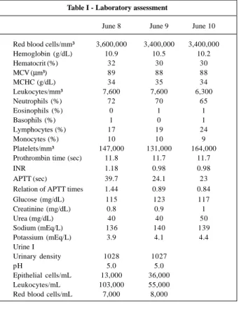
Serum levels of the MB fraction of creatine kinase 3, 11, 16, and 24 hours after the pain started were, respectively, 11, 7, 4, and 11 U/L. The remaining results of the laboratory as-sessment are shown in table I.
The transthoracic echocardiogram showed a moderate concentric hypertrophy of the left ventricle and normal sys-tolic function (diassys-tolic diameter, 46mm; syssys-tolic diameter,
30 mm; and shortening fraction, 34%). The filling pattern showed an E<A indicative of relaxation disorder. The dia-meters of the aortic root and of the left atrium were 40mm. The valves and the pericardium were considered within the normal range.
The hemodynamic study revealed the pressures 140/0/ 10mmHg in the left ventricle and 140/80/100mmHg in the aorta. Coronary angiography showed coronary arteries with no obstructive lesions and a normal left ventricle. An electrode of a prophylactic endocardial pacemaker was im-planted in the right ventricle because of persistent atrioven-tricular dissociation.
The long-lasting electrocardiogram obtained with Holter monitoring revealed a predominantly sinus rhythm. The minimum and maximum heart rates were 76bpm and 130bpm, respectively. It disclosed 1055 ventricular extra-systoles (mean of 52/hour), 861 isolated extraextra-systoles, 72 pairs, 14 ventricular tachycardias, the longest with 7 beats and a rate of 130bpm, the fastest with 4 beats and a rate of 169bpm, and the slowest with 3 beats and a rate of 99bpm. There were also 305 isolated atrial extrasystoles, 179 pairs, and 55 atrial tachycardias (the longest with 21 beats and a rate of 207bpm, the fastest with 3 beats and a rate of 250bpm, and the slowest with 3 beats and a rate of 162bpm). The patient did not report palpitations or pain during recording. The patient did not have any more pain and was at first medicated with intravenous nitroglycerin and 120mg of pro-pranolol, which was later replaced by 120mg of verapamil daily.
The chest X-ray performed at bedside showed an en-larged cardiac silhouette (++/4) and an elongated and dila-ted aorta. On the same day, the patient had intense chest pain and dyspnea followed by cardiopulmonary arrest, whi-ch did not respond to resuscitation maneuvers. The elec-trocardiogram at the beginning of the final event showed car-diac electrical activity with a mean rate of 76bpm and QRS wi-th 80 msec in wi-the absence of a pulse or cardiac noises (fig. 2).
Discussion
Clinical findings - The patient is a 73-year-old woman
sug-gest an ischemic cardiac etiology. The alterations in ventri-cular repolarization more evident in the anteroseptal and in-ferior walls also strengthen this diagnostic hypothesis,
even though they are not specific findings of ischemic heart disease. The presence of junctional rhythm, most of the time, indicates an underlying heart disease, even though it may occur in individuals with a normal heart. The junctional rhythm may be found in ischemia of the inferior wall because of impairment of the atrioventricular node. The presence of angiographically normal coronary arteries does not exclude myocardial ischemia; mechanisms such as vasospasm, microvascular dysfunction, and a disproportional increase in myocardial oxygen consumption in relation to coronary artery flow – the so-called nonobstructive coronary heart di-sease – have been implicated. A large number of these pa-tients, however, have other causes for chest pain, such as esophageal and psychiatric disorders. The prognosis is known to be more favorable in the absence of obstruction of the epicardial coronary arteries, which is not compatible with the adverse and fulminating evolution of this patient.
The occurrence of chest pain with a rapidly fatal evo-lution suggests the diagnosis of pulmonary thromboembo-lism, which is an underdiagnosed condition with 70% of its fatal cases not diagnosed in vivo. Deaths resulting from pulmonary thromboembolism occur early; according to so-me studies, 80% of the deaths occur within the first 2.5 hours of the event. Dyspnea is the most frequent symptom (approximately 70% of cases), but chest pain is also found in 60% of cases and usually has pleuritic characteristics, resulting from pleural irritation due to underlying pulmona-ry infarction. Pulmonapulmona-ry thromboembolism with a signifi-cant right ventricular overload may cause ischemia of this chamber, mainly if an association with coronary obstruction exists, triggering anginal findings. Massive pulmonary thromboembolism may also commonly determine electric activity without a pulse, which happened as the final event in the present case. Our patient, however, had no risk fac-tors for pulmonary thromboembolism except age. The chest X-ray had no signs suggestive of pulmonary thromboem-bolism, such as oligemia, laminar atelectasis, or wedge-sha-ped images, dilation of the trunk of the pulmonary artery, or elevation of the diaphragm. These typical alterations, ho-wever, are infrequent. On echocardiography, no signs of overload or right ventricular dysfunction were evident, whi-ch does not support the occurrence of massive pulmonary thromboembolism 1,2.
Finally, the presence of intense chest pain, aortic dila-tion, the rapidly fatal outcome with cardiac arrest in electri-cal activity without a pulse, as well as the rapid drop in he-moglobin levels (Hb 12.3g/dL in 6/6 and 10.2g/dL in 6/10) point to the diagnosis of aneurysm rupture or acute dissec-tion of the thoracic aorta.
Aneurysm of the thoracic aorta has various causes, ac-cording to the aortic segment involved. The ascending aortic aneurysm is associated with cystic degeneration of the tunica media of a vessel, which is a condition associated with arterial hypertension and with structural deficiencies of the connective tissue (as observed in Marfan’s and Ehlers-Danlos syndromes). On the other hand, the descending aortic aneurysm is more commonly of atherosclerotic origin. Fig. 1 - Electrocardiogram on hospital admission.
Fig. 2 - Electrocardiogram on the last day of hospitalization.
Table I - Laboratory assessment
June 8 June 9 June 10 Red blood cells/mm³ 3,600,000 3,400,000 3,400,000 Hemoglobin (g/dL) 10.9 10.5 10.2
Hematocrit (%) 32 30 30
MCV (µm³) 89 88 88
MCHC (g/dL) 34 35 34
Leukocytes/mm³ 7,600 7,600 6,300
Neutrophils (%) 72 70 65
Eosinophils (%) 0 1 1
Basophils (%) 1 0 1
Lymphocytes (%) 17 19 24
Monocytes (%) 10 10 9
Platelets/mm³ 147,000 131,000 164,000 Prothrombin time (sec) 11.8 11.7 11.7
INR 1.18 0.98 0.98
APTT (sec) 39.7 24.1 23
Relation of APTT times 1.44 0.89 0.84
Glucose (mg/dL) 115 123 117
Creatinine (mg/dL) 0.8 0.9 1
Urea (mg/dL) 40 40 50
Sodium (mEq/L) 136 140 139
Potassium (mEq/L) 3.9 4.1 4.4
Urine I
Urinary density 1028 1027
pH 5.0 5.0
considered. This method is also subjected to artifacts resulting from obesity and thoracic conformation.
Aortography was previously the examination of choice for diagnosing aortic dissection. In the present case, we do not know whether the hemodynamic study included aortography. Currently, reports in the literature attribute a low sensitivity to aortography (around 75%); false-negative results probably are due to thrombosis and consequent absence of blood flow through the false lumen, preventing it from being visualized. Another disadvantage of aortogra-phy is the use of nephrotoxic contrast medium and risk of catheterization of the false lumen 5. Data exist suggesting
that the use of aortography delays surgical intervention and prolongs hospitalization time.
Because of these considerations, most centers have used transesophageal echocardiography or computed to-mography as imaging methods for diagnosis. The sensiti-vity of these methods exceeds 90% with experienced profes-sionals. Transesophageal echocardiography has the follo-wing advantages: it does not require iodide contrast me-dium, it is rapidly available, and it allows a simultaneous exa-mination of the pericardium and the aortic valve.
(Dr. Anthony Guerra Gorski)
Diagnostic hypothesis - Acute aortic dissection that
evolved to aortic rupture or cardiac tamponade, or both, which determined cardiopulmonary arrest in electrical acti-vity without a pulse, not responding to the resuscitation maneuvers.
Electrocardiographic features - The abnormal
collec-tion of fluid in the pericardial space, when slow and gradual, may not produce symptoms for a long time, even with a great amount of fluid. If the accumulation is fast, smaller vo-lumes may cause cardiac tamponade, which is a dramatic condition that may rapidly lead to death.
Theoretically, any cause of acute pericarditis may also be a cause of pericardial effusion or cardiac tamponade, but the most frequent ones are viral infections, tuberculosis, uremia, myxedema, collagen diseases, and neoplasias. Trau-mas (including cardiac catheterization and placement of a pacemaker electrode) and aortic dissection are also impor-tant causes of cardiac tamponade.
With the evolution of the pericardial fluid collection, the amplitude of all electrocardiographic deflections de-creases. This results from the short circuit of the electric im-pulses that circle the pericardial effusion or the thin fibrin layer that covers the pericardium. All deflections are redu-ced depending on the degree of pericardial effusion so that usually the sum of the QRS amplitudes in the three standard leads does not exceed 15mm. In this stage, the T wave is in-variably inverted in all leads, except in aVR.
Alternation in amplitude of the QRS complexes mani-fests by a widespread variability of the relatively small am-plitudes of the QRS waves. A voluminous effusion is required for the alternation to be recognized. However, Approximately 40% of the aneurysms of the thoracic aorta
are diagnosed in asymptomatic individuals. The most com-mon symptom is chest pain, which most of the time is persis-tent, and deeply located, with no triggering factors; some-times it may be intense, indicating an imminent rupture. Other symptoms resulting from compression of intrathora-cic structures by the aneurysm may also occur, such as cou-gh, dysphagia, and dyspnea. Aneurysm rupture is the most threatening complication, and the better predictor of its occurrence is aneurysmal diameter (higher risk in aneurysms larger than 6cm in diameter). More commonly, rupture occurs to the left pleural space, but may also occur to the in-terior of the pericardial sac (with a consequent cardiac tam-ponade), to the main left bronchus, or to the esophagus. In our patient, the mild dilation of the aorta observed on echo-cardiography (4cm) does not match the diagnosis of rupture of an aortic aneurysm.
Dissection of the thoracic aorta is less frequent than an aneurysm and is associated with a high mortality mainly in the first 48 hours of its onset. Despite availability of several imaging resources, the diagnosis of acute dissection of the aorta remains difficult. In a series of 236 cases, the diagnosis was not suspected at first in 40% of the cases 3. A report
pu-blished in 1992 4 of a case series of 186 patients at our
insti-tution showed that 30% of the patients with type A dissec-tion died before surgical treatment. The pathophysiology of aortic dissection involves the presence of degeneration of the arterial tunica media. Approximately 80% of the patients are hypertensive. This is a frequently observed condition in disorders of the connective tissue (Marfan’s and Ehlers-Danlos syndromes), in some genetic syndromes (Turner, Noonan), bicuspid aortic valve, coarctation of the aorta, and chest trauma. Pregnancy, as well as the postpar-tum period, are predisposing factors to aortic dissection. Chest pain is a striking symptom (being the main symptom in 75% of cases), and it is characteristically intense, with ma-ximum intensity since its beginning, frequently irradiating to the dorsum or abdomen. Findings such as pulse asym-metry, even though classic, are rare, occurring in 6% of ca-ses. The chest X-ray is altered most of the time (around 90%) but not in all cases. The most common abnormalities are aortic or mediastinal dilation. Pleural effusion (usually on the left) and enlargement of the cardiac silhouette (resul-ting from hemopericardium) may also occur. Another fre-quent complication of dissection of the thoracic aorta is acute aortic insufficiency (around 10%), which may determi-ne hemodynamic instability. At times, dissection may invol-ve the coronary ostia, more commonly the right one, cau-sing acute coronary artery disease. Definitive diagnosis of acute aortic dissection requires the use of imaging techni-ques. The choice of a particular test should take into consi-deration its availability and the experience of the service, as well as the aortic segment to be studied.
careful observation of an electrocardiographic tracing may raise the diagnostic suspicion even in the phase of effusion installation.
When the pericardial effusion is massive, such as in the malignant involvement (tumors) of the pericardium, elec-trical alternation may affect all electrocardiographic deflec-tions, from the P wave to the T wave. Electrical alternation may be observed in any group of leads, being more common in the intermediary precordial leads.
In the presence of pericardial effusion, the QRS axis tends to vary according to the position adopted by the pa-tient: supine or standing position. This, however, is not a sensitive signal.
The presence of electrical alternation suggests a large effusion or even cardiac tamponade, because it reflects the pendular movement of the heart inside the pericardial sac. This phenomenon may also occur in severe cardiomyopa-thy (real electrical alternation), hypertensive pneumotho-rax, and more rarely in chronic constrictive pericarditis.
Alterations in the electrocardiogram also depend on the volume and way of installation of the pericardial effu-sion. Therefore: a) When the fluid collection reaches 200 to 500 cm3, reduction in the voltage of all electrocardiographic
waves occurs due to the interposed fluid, the deposited fi-brin, and atrophy of the underlying muscle that is compres-sed. These effects are more evident in the leads of the limbs than in those of the precordium. However, huge effusions may exist without a parallel reduction in voltage. b) When the pericardial collection is suddenly formed, such as in he-mopericardium or in the purulent effusion, an elevation in the ST segment is observed. c) At times, one may record an electrical alternation that is not classically known as 1:1 ty-pe, because it is formed in a gradual and rotatory form, con-sequent to the rotatory movement of the heart inside the pe-ricardial sac full of fluid. This type of electrical alternation is pathognomonic of the cardiac tamponade, in a condition where all electrocardiographic waves participate.
This behavior seems to depend on the abnormal accu-mulation of fluid in the pericardial sac, exerting pressure upon the heart during the whole cardiac cycle. In this way, the increase in the intrapericardial pressure gradually equals the intracavitary pressure, the transmural pressure being close to zero.
Even in this condition, during inspiration the decrease in the intrapericardial pressure secondary to the reduction in the intrathoracic pressure maintains the venous return normal in a first phase. Currently, we admit that the increase in the venous return during inspiration determines an in-crease in the right ventricular dimensions and a consequent displacement of the interventricular septum towards the outflow tract of the left ventricle reducing its dimensions.
In the case being discussed, alternation was of the ro-tatory type (fig. 3). In addition, we were able to eliminate oc-casional respiratory effects on the amplitude of the QRS complexes because a concomitant reduction in the QRS amplitude was observed in a great number of simultaneous leads, characterizing the real electrical alternation.
(Dr. Paulo Jorge Moffa)
Autopsy
The patient died due to cardiac tamponade conse-quent to rupture of an aortic dissection into the pericardial sac with a few days of evolution (fig. 4). This aortic dissec-tion may be classified as type I of De Bakey’s classificadissec-tion, because it extended from the ascending aorta (before the aortic arch) to at least the iliac arteries, where the autopsy examination ended. A dissection orifice of approximately 3cm of length existed, located 5cm above the valvar plane and transversal to the artery axis. The histopathologic exa-mination showed focal areas of collection of a mucoid mate-rial in the tunica media of the aorta (fig. 5). The aorta also
Fig. 4 - Aortic section showing delamination of the tunica media (asterisk) and a false lumen partially filled with blood.
Fig. 5 - Microphotograph of the aorta showing a focal area of fragmentation of the elastic fibers and collection of mucoid material (arrow). Stained by the Movat method, original magnification 16X.
1. Stein PD, Henry JW. Prevalence of acute pulmonary embolism among patients in a general hospital and at autopsy. Chest 1995; 108: 978-81.
2. Mesquita CT, Morandi JLB Jr., Perrone FT, Oliveira CS, Barreira LJ, Nascimento SSCA, Pareto RC Jr., Mesquita ET. Diagnóstico clínico versus confirmação pato-lógica de embolia pulmonar fatal em pacientes hospitalizados. Arq Bras Cardiol 1999; 73: 251-4.
3. Spittell PC, Spittell JA Jr., Joyce JW, Tajik AJ, Edwards WD, Schaff HV. Clinical features and differential diagnosis of aortic dissection. Mayo Clin Proc 1993; 68: 642-51.
4. Vianna C de B, Barreto AC, Cesar LA, Mady C, Stolf N, Bellotti G, Jatene AD.
Dis-References
secções agudas da aorta. Evolução hospitalar de 186 casos. Arq Bras Cardiol 1992; 58/2: 95-9.
5. Eagle KA, Quertermous T, Kritzer GA, Newell JB, Dinsmore R, Feldman L, DeSanctis RW. Spectrum of conditions initially suggesting acute aortic dis-section but with negative aortograms. Am J Cardiol 1986; 57: 322-6. 6. Gutierrez PS, Lopes EA. Patologia das dissecções aórticas Rev Soc Cardiol
Estado São Paulo 1994; 5: 413-21.
7. Hirst AE Jr., Johns VJ Jr, Kime SW. Dissecting aneurysm of the aorta: a review of 505 cases. Medicine 1958; 37: 217-80.
8. Rosenmann E, Yarom R. Dissecting aneurysm of the aorta and hypothyroidism. Isr J Med Sci 1994: 30: 510-3.
showed a slight degree of atherosclerosis, which was also present in the coronary arteries. This previously healthy patient had precordial pain 5 days prior to her death. She had no electrocardiographic signs of myocardial infarction. Catheterization with aortography did not show the aortic dissection. An iatrogenic aortic dissection induced by the examination is another possibility. This is, however, a very unlikely hypothesis, because the symptoms started prior to catheterization with no other explanation for them (the coronary arteries had no obstructive lesions, and no myocardial infarction existed). The type I aortic dissection of De Bakey’s classification, in which the disease involves both ascending and descending aorta, is the most common. Death resulting from cardiac tamponade in patients not undergoing surgery is also very common 6.
Despite the negative previous history for systemic hy-pertension and observance of normal pressure levels during hospitalization, the patient may have had hypertension. Her
left ventricle had a mild concentric hypertrophy (the thick-ness of the free wall was 1.7cm, the normal value being up to 1.2cm), and her kidneys showed nephrosclerosis. More than 90% of aortic dissection cases occur in patients with sys-temic hypertension. This percentage appears in the series where both clinical and autopsy data are computed 7,
be-cause part of the patients about whom no clinical data were available showed morphologic characteristics of hyper-tension, such as in the present case.
Other autopsy findings in our patient were adrenal no-dules and adenomatous goiter. Pathological changes in the thyroid 8 and adrenal nodules have been found with an
apparently high frequency in the autopsies of patients with aortic dissection with no conclusions regarding their occasional significance in the etiology and pathogenesis of the latter disease.