DOI: 10.5935/2359-4802.20180085
ORIGINAL ARTICLE
Mailing Address: Liz Andréa Villela Baroncini
Rua Buenos Aires, 764, ap. 601. Postal Code: 80.250-070, Batel, Curitiba, PR - Brazil. E-mail: [email protected], [email protected]
Correlation between Exercise Stress Test and Echocardiographic Parameters in
Elderly Individuals
Liz Andréa Villela Baroncini,1 Camila Varotto Baroncini,2 Juliana Ferreira Leal3 Pontificia Universidade Católica do Paraná,1 Curitiba, PR - Brazil
Universidade Federal do Paraná (UFPR),2 Curitiba, PR - Brazil Universidade Estadual de Ponta Grossa, Ponta Grossa,3 PR, Brazil
Manuscript received December 01, 2017; revised manuscript May 02, 2018; accepted July 23, 2018.
Abstract
Background: Maximum oxygen consumption (VO2 max) in healthy individuals decreases approximately 10% per
decade of life, and such decrease is more pronounced after the seventh decade.
Objectives: To assess functional capacity of individuals aged 75 years or older, submitted to ergometric test and
transthoracic echocardiogram exam, by means of metabolic equivalent (MET) and VO2 max measurements.
Methods: A total of 381 patients (205 women; 79 ± 3.7 years) were evaluated. Exclusion criteria were: presence of
left ventricular (LV) systolic dysfunction, LV diastolic dysfunction grade II and III, significant valve disease, or coronary artery disease with systolic LV dysfunction or dilatation. Associations between quantitative variables were analyzed by Pearson and Spearman correlation coefficients, and comparisons of quantitative data by Student’s t-test for independent samples.
Results: Increasing age was associated with a progressive decrease in the distance covered (p = 0.021), in the
expected increase in HR (p < 0.001), in VO2 max (p < 0.001), and METs (p < 0.001) in both genders. There was no correlation of exercise test parameters with the echocardiographic parameters.
Conclusions: Relatively healthy older individuals, with global systolic and diastolic functions of the left ventricle
preserved, presented a progressive decrease in their functional capacity due to their natural aging process, comorbidities related to their age range and physical deconditioning. (Int J Cardiovasc Sci. 2018; [online].ahead print, PP.0-0)
Keywords: Cardiovascular Diseases; Risk Factors; Aging; Oxygen Consumption; Exercise Test; Echocardiography/
methods; Exercise.
Introduction
Priebe1 provided a sensible description of the
difficulties in defining “aged patients”, since there is
no clinical definition that precisely classifies elder or
advanced-aged individuals. Aging is a continuous process rather than an abrupt event. As age advances, maximal aerobic capacity decreases 8 to 10% per decade in sedentary men and women, and exercise capacity decreases approximately 50% between ages 30 and 80. In addition, comorbidities such as obstructive pulmonary disease, peripheral vascular disease, obesity, arthritis, neuromuscular disease, and generalized deconditioning
are more prevalent in elderly patients and should be considered before evaluating their clinical conditions,
especially in relation to cardiovascular risk.2-4
The prevalence of coronary artery disease (CAD) is high in the elderly. Although it was detected in only 1.8% of men and 1.5% of women above the age of 75, an autopsy study of 5,558 patients revealed significant CAD
in 54% of women and in 72% of men above the age of 70.5-7
an underlying disease such as significant CAD or just poor functional capacity in a sedentary old person. An individual’s functional capability may be assessed by
means of the maximum oxygen uptake (VO2 max) that
represents the maximum amount of oxygen an individual can take in with incremental exercise. The amount of exercise can be measured using the metabolic equivalent (MET); 1 MET is the amount of oxygen consumption
at rest and is equivalent to approximately 3.5 ml kg
-1min-1 (measured in a healthy, 40-year old man, 70 kg).
VO2 max decreases about 10% per decade in healthy
individuals, and such decrease is even more pronounced in individuals older than 70 years. With the increase in life expectancy, many patients aged 75 years or older seek medical care for chest pain and presurgical evaluation for several elective surgeries. Individuals that feel fit enough to perform a physical stress test are submitted to treadmill or bicycle ergometric tests. However, although sensitivity to noninvasive stress testing increases with
aging, specificity tends to decline.5
The objective of the current study is to correlate exercise test variables with echocardiographic parameters in patients over 75 years old, including functional
capacity, measured in MET and VO2 max (with or
without myocardial ischemia at the physical stress test), left ventricular ejection fraction (LVEF), left ventricular mass and left ventricle mass index, left atrial volume and presence of pulmonary arterial hypertension.
Methods
We assessed 381 patients (205 women; 53.8%), mean age of 79 ± 3.7 years, who underwent exercise test and bidimensional transthoracic echocardiography (2DEcho) in a private cardiologic clinic. Subjects were selected by convenience. Each patient had results of blood tests and imaging tests to be analyzed before the exercise test.
Before the study, data on demographic characteristics and risk factors were collected from the private cardiologist’s records and blood test results. Body mass index (BMI) was calculated by dividing the subjects’ weight (kg) by the square of their height (m). Patients were queried about the presence of hypertension, diabetes mellitus, dyslipidemia, coronary artery disease, and current smoking habit. Hypertension was defined as a history of treated hypertension or the presence of systolic blood pressure ≥ 140 mmHg or diastolic blood pressure ≥ 90 mmHg, measured by the private cardiologist. Smoking history was coded as never or
current smoker.8 Subjects were classified as having
diabetes when treated for insulin-dependent or non-insulin-dependent diabetes or having elevated fasting glucose levels (≥ 126 mg/dL). The use of lipid-lowering drugs or the presence of total cholesterol > 200 mg/dL, HDL-cholesterol < 40 mg/dL, LDL - cholesterol > 100
mg/dL or triglycerides > 150 mg/dL was recorded.9-10
A history of myocardial infarction, angioplasty, or coronary artery bypass surgery was recorded, and the presence of any of these conditions was considered a positive CAD history.
Indications for the 2DEcho included referral from a physician, information from close relatives, or patients’ complaints. We analyzed echocardiographic and carotid ultrasonography data, including left ventricular ejection fraction, left ventricular diastolic function, left atrial volume, left ventricular mass and the presence of pulmonary arterial hypertension and carotid plaque. Exclusion criteria included the presence of left ventricular systolic dysfunction (ejection fraction < 50% on echocardiogram), left ventricular diastolic dysfunction grade II and III, significant valve disease such as mitral and aortic regurgitation or stenosis, CAD with left ventricular systolic dysfunction or dilatation, unstable cardiovascular or metabolic disease, and major orthopedic/neurological disability.
Subjects underwent treadmill electrocardiogram (ECG) testing (TET) or bike ECG testing (BET), according to the private physician request. Treadmill ECG test included Ellestad, Kattus, Naughton, Ramp, Bruce and modified Bruce protocols, and Balke and male Balke protocols,
following standard recommendations.11,12 The distance
during exercise; f) marked ST-segment depression (≥ 3mm); g) exercise-limiting symptoms such as angina, dyspnea, exhaustion, or the subjects’ request to stop the test; and h) technical difficulties in monitoring the ECG or systolic blood pressure. An abnormal response of the ST-segment to exercise was defined as horizontal or downsloping ST-segment depression ≥ 1 mm measured at 80 ms after the J point or an elevated ST-segment ≥ 1 mm in leads without pathological Q-wave (excluding lead aVR). Measurements of left ventricular systolic and diastolic dysfunction, left atrial volume, valve disease, and systolic pulmonary artery pressure were performed according to recommendations of the American Society of Echocardiography and the European Association of
Cardiovascular Imaging.13-14 The study was approved by
the local ethics committee and written informed consent was obtained from each participant to undergo the ergometric tests (treadmil ECG testing or bike ECG testing) , bidimensional transthoracic echocardiography and carotid ultrassonography, and to participate in the study.
Statistical analysis
Quantitative variables were described as means, medians, minimum and maximum values, quartiles and standard deviations, and categorical variables as frequency and percentiles. Associations between quantitative variables were analyzed by Pearson and Spearman correlation coefficients. Comparisons of quantitative variables between the two groups were made using the Student’s t test for independent samples. Statistical testing of data normality was performed using the Kolmogorov-Smirnov test. Associations between categorical variables were assessed by the Fisher’s exact test. A p-value ≤ 0.05 indicated statistical significance. Data were analyzed by means of the SPSS statistical software, version 20.
Results
Patients’ baseline characteristics and echocardiographic and ergometric results are shown in Tables 1, 2 and 3. Only five patients (1.3%) performed cycle ergometer test, and then were excluded from the final analysis. Three hundred seventy-six patients performed treadmill test (Bruce protocol 203, 53.4%; Kattus 113, 29.7%; Ramp 28, 7.4%; modified Bruce 15, 3.9%; Naughton 12, 3.2%; Ellestad 5, 1.3%; Balke 3, 0.8%; Balke male 1, 0.3%). Nineteen (5%) patients did not achieve the submaximal heart rate (HR) expected for the age and 58 (15%) had previous ECG at resting conditions showing left bundle
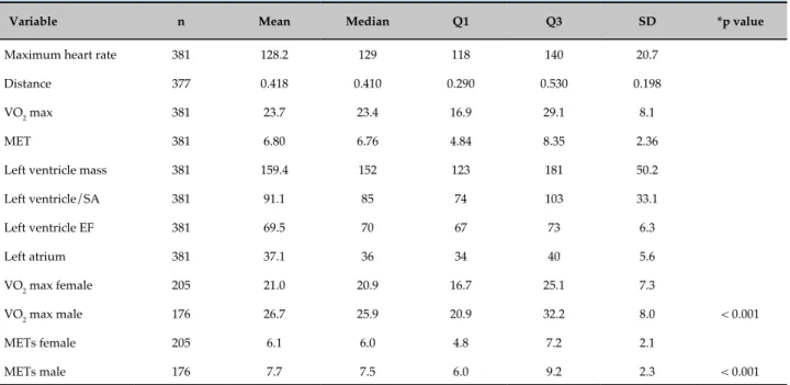
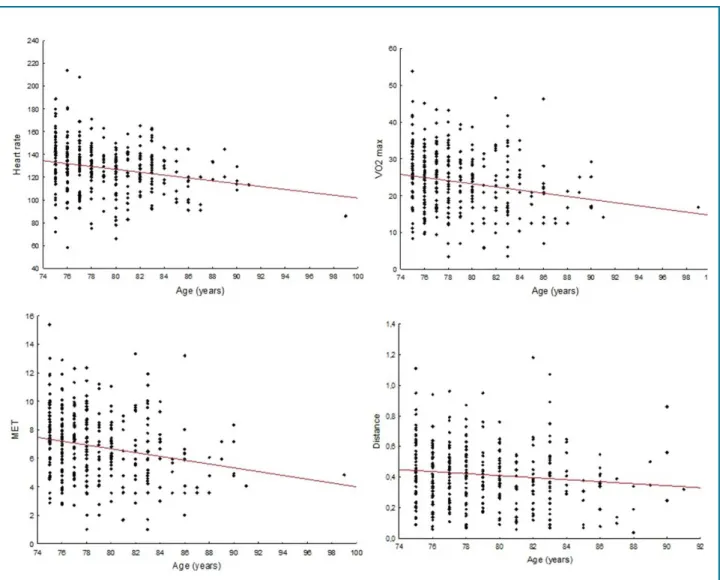
branch block and ST segment alterations. Forty (10.5%) of the patients tested positive for myocardial ischemia and 79 (21.8%) showed abnormal heart rate response in the first minute. As age increased, the distance covered by participants decreased (p = 0.021), as well the expected increase in HR (p < 0.001), VO2 max (p < 0.001) and METs (p < 0.001) (Tables 3 and 4; Figure 1) in men and women.
Women showed lower values of VO2 max and METs
when compared to men (Table 2). Inverse correlation
was noted of the distance covered, VO2 max and METs
with the BMI (Table 3 and 4). Only 4 patients (1%) showed systolic pressure in the pulmonary artery above 40 mmHg in the echocardiogram at rest, which did not influence the distance covered by the subjects, HR at the
first minute (p = 1), VO2 max (p = 0.5), MET (p = 0.5) or
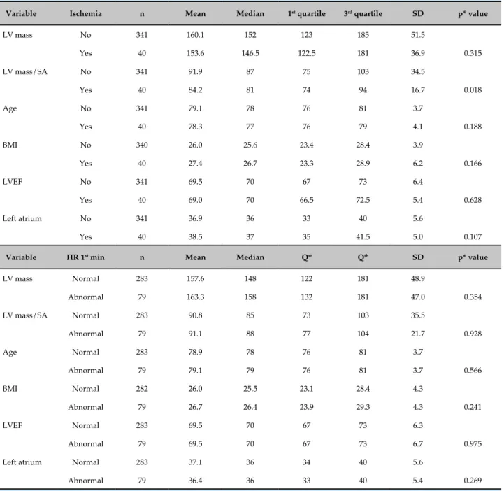
ischemia (p = 1.0) (data not shown). The volume of the left atrium and left ventricular mass had no influence on the ergometric test variables (Table 5). Ischemia at stress test did not correlate with any echocardiographic variable (Table 5). In 198 patients (67.3%), atherosclerotic plaques in the extracranial carotid arteries were detected, which also did not correlate with any of the variables analyzed (data not shown). Severity of stenosis was not considered relevant, only the presence of the atherosclerotic plaque.
Discussion
The present study showed that relatively healthy patients aged 75-81 years, with similar demographic and echocardiographic characteristics, showed a
progressive decrease in METs and VO2 max, associated
with a decrease in the distance covered during ergometric test with increasing age. These findings corroborate
previous studies showing a marked decrease in VO2 max
with aging.15-18 Considering that only individuals with
preserved left ventricular systolic function was studied, we did not expect an influence of this parameter on the results. Similarly, no influence of left ventricular diastolic
function was expected,19 as individuals with grade II and
III diastolic dysfunction were excluded from the study.
Regarding the left atrial volume, since there was no significant variation in its values among the patients, its influence on the ergometric parameters was not expected either, unlike previous studies that reported a worsening of functional capacity due to the increase in left atrial
volume.20-23 The same was observed with left ventricular
mass and left ventricular mass index.24
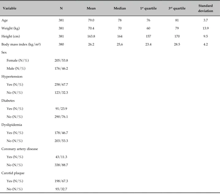
Table 1 - Patients’ baseline characteristics
Variable N Mean Median 1st quartile 3rd quartile Standard
deviation
Age 381 79.0 78 76 81 3.7
Weight (kg) 381 70.4 70 60 79 13.9
Height (cm) 381 163.8 164 157 170 9.5
Body mass index (kg/m²) 380 26.2 25,6 23.4 28.5 4.2
Sex
Female (N/%) 205/53.8
Male (N/%) 176/46.2
Hypertension
Yes (N/%) 258/67.7
No (N/%) 123/32.3
Diabetes
Yes (N/%) 91/23.9
No (N/%) 290/76.1
Dyslipidemia
Yes (N/%) 178/46.7
No (N/%) 203/53.3
Coronary artery disease
Yes (N/%) 43/11.3
No (N/%) 338/88.7
Carotid plaque
Yes (N/%) 198/67.3
No (N/%) 93/32.7
indicates that, in relatively healthy individuals older than 75 years old, the decrease in functional capacity is associated with age, progressive physical deconditioning and comorbidities, which will negatively affect
their independence and daily physical activity.25-28
Nevertheless, comorbidities such as previous stroke, bone and articular diseases, and chronic obstructive pulmonary disease were not analyzed in the present study. In regard to HR at the first minute after the test, it is known that its restoration to baseline values reflects the integrity of the vagal system, which is compromised in older ages, in patients with diabetes,
cardiac failures, and increased BMI.29-31 In this regard,
in the present study, only 21.8% of the individuals
showed an abnormal HR response at the first minute after the exercise test. Also, there was no correlation of this variable with echocardiographic parameters, age, sex or BMI. This finding was expected, as the studied cohort comprised an aged population with similar clinical and echocardiographic characteristics. Another relevant finding was the fact that only 10% of the individuals in the present study showed positive for myocardial ischemia. Sensitivity of the ergometric test was similar
to that documented by Vacanti et al.,6 using myocardial
Table 2 - Echocardiographic and ergometric results
Variable n Mean Median Q1 Q3 SD *p value
Maximum heart rate 381 128.2 129 118 140 20.7
Distance 377 0.418 0.410 0.290 0.530 0.198
VO2 max 381 23.7 23.4 16.9 29.1 8.1
MET 381 6.80 6.76 4.84 8.35 2.36
Left ventricle mass 381 159.4 152 123 181 50.2
Left ventricle/SA 381 91.1 85 74 103 33.1
Left ventricle EF 381 69.5 70 67 73 6.3
Left atrium 381 37.1 36 34 40 5.6
VO2 max female 205 21.0 20.9 16.7 25.1 7.3
VO2 max male 176 26.7 25.9 20.9 32.2 8.0 < 0.001
METs female 205 6.1 6.0 4.8 7.2 2.1
METs male 176 7.7 7.5 6.0 9.2 2.3 < 0.001
* Student’s t- test for independent samples, p < 0.05. VO2 max: maximum oxygen consumption; MET: metabolic equivalent; SA: surface area; EF: ejection fraction.
Table 3 - Correlation of heart rate (HR) and distance covered in exercise stress ECG test with echocardiographic data, age, surface area (SA) and body mass index (BMI) of patients
Variable n Spearman
coefficient p value
HR max x left ventricle mass 381 -0.09 0.082
HR max x left ventricle mass/
SA 381 -0.03 0.556
HR max x age 381 -0.23 < 0.001
HR max x BMI 380 -0.06 0.249
HR max x left ventricle ejection
fraction 381 0.13 0.010
HR max x left atrium 381 -0.05 0.325
Distance x left ventricle mass 377 0.03 0.520
Distance x left ventricle mass/
SA 377 0.08 0.101
Distance x age 377 -0.12 0.021
Distance x BMI 376 -0.17 0.001
Distance x left ventricle ejection
fraction 377 0.04 0.450
Distance x left atrium 377 0.01 0.833
Table 4 - Correlation of metabolic equivalent (MET) and maximum oxygen consumption (VO2 max) with
echocardiographic data, age, body mass index (BMI) and surface area (SA) of patients
Variable N Spearman
coefficient p value
MET x left ventricle 381 0.03 0.581
MET x left ventricle/SA 381 0.06 0.217
MET x age 381 -0.21 <0.001
MET x BMI 380 -0.13 0.011
MET x ejection fraction 381 0.05 0.327
MET x left atrium 381 0.00 0.959
VO2 max x left ventricular
mass 381 0.04 0.488
VO2 max x left ventricular
mass/SA 381 0.07 0.167
VO2 max x age 381 -0.19 <0.001
Figure 1 - Correlation of age with heart rate, VO2 max, metabolic equivalent (MET), and distance (m) covered in exercise stress
ECG test.
this population must be interpreted differently than in younger individuals, since even in patients classified as low risk by risk stratification scores, an annual cardiac mortality rate of 2% was found in patients aged 75
years or older.32,33 These findings confirm the need for
specific protocols and instruments for elderly patients,34
considering the great heterogeneity in aging process and
its biological consequences.33
In addition, considering the presence of atherosclerotic plaques in the extracranial carotid arteries in our patients, we expected its correlation with the other variables analyzed, which did not happen. In fact, its presence was previously shown to be correlated with systolic functions and left filling ventricular pressures,which revealed to be similar in all patients of this study.
An additional important finding was the inverse correlation of the distance covered, functional capacity
(METs) and VO2 max with BMI. There is a progressive
BMI increase as age advances and the prevalence of
obesity has considerably increased in the elderly.36
This has a direct impact on individuals’ health and life quality, since weight gain is associated with a decrease in functional capacity and vitality, body pain, emotional and physical problems, and increased risk for morbidity and disability.37,38
Table 5 - Echocardiographic variables, presence of ischemia and heart rate in the first minute of the exercise stress ECG test (HR 1st min) of the patients
Variable Ischemia n Mean Median 1st quartile 3rd quartile SD p* value
LV mass No 341 160.1 152 123 185 51.5
Yes 40 153.6 146.5 122.5 181 36.9 0.315
LV mass/SA No 341 91.9 87 75 103 34.5
Yes 40 84.2 81 74 94 16.7 0.018
Age No 341 79.1 78 76 81 3.7
Yes 40 78.3 77 76 79 4.1 0.188
BMI No 340 26.0 25.6 23.4 28.4 3.9
Yes 40 27.4 26.7 23.3 28.9 6.2 0.166
LVEF No 341 69.5 70 67 73 6.4
Yes 40 69.0 70 66.5 72.5 5.4 0.628
Left atrium No 341 36.9 36 33 40 5.6
Yes 40 38.5 37 35 41.5 5.0 0.107
Variable HR 1st min n Mean Median Qst Qth SD p* value
LV mass Normal 283 157.6 148 122 181 48.9
Abnormal 79 163.3 158 132 181 47.0 0.354
LV mass/SA Normal 283 90.8 85 73 103 35.5
Abnormal 79 91.1 88 77 104 21.7 0.928
Age Normal 283 78.9 78 76 81 3.7
Abnormal 79 79.1 79 76 81 3.7 0.566
BMI Normal 282 26.0 25.5 23.1 28.4 4.3
Abnormal 79 26.7 26.4 23.9 29.3 4.3 0.241
LVEF Normal 283 69.5 70 67 73 6.3
Abnormal 79 69.5 70 67 73 6.7 0.975
Left atrium Normal 283 37.1 36 34 40 5.6
Abnormal 79 36.4 36 33 40 5.4 0.269
* Student’s t- test for independent samples, p < 0.05. LV: left ventricular; SA: surface area; EF: ejection fraction; BMI: body mass index.
it difficult to accurately analyze and compare the ergometric variables between the subjects. Second, since only patients with preserved left ventricular systolic and diastolic functions were selected, no significant
difference was expected in VO2 max, METs, HR, and
distance covered. Thus, further studies including patients with different degrees of left ventricular dysfunction in the elderly are necessary.
Conclusions
Individuals aged 75 years or older, of both genders, relatively healthy, with preserved left ventricular systolic and diastolic functions, showed progressive decrease in
the distance covered, VO2 max, METS and at the expected
1. Priebe H.-J. The aged cardiovascular risk patient. Br J Anaesth. 2000;85(5):763-78.
2. Sicari R, Nihoyannopoulos P, Evangelista A, KasprzaK J, Lancellotti P, Poldermans D, et al, on behalf of the European Association of Echocardiography. Stress echocardiography expert consensus statement-executive summary. European Association of Echocardiography (EAE) (a registered branch of the ESC). Eur Heart J. 2009;30(30):278-89.
3. Pellikka PA, Naguch SF, Elhendy AA, Kuchl CA, Sawada SG. American Society of Echocardiography recommendations for performance, interpretation, and application of stress echocardiography. J Am Soc Echocardiogr. 2007;20(9):1021-41.
4. Bayliss EA, Bayliss MS, Ware Jr JE, Steiner JF. Predicting declines in physical function in person with multiple chronic medical conditions: what we can learn from the medical problem list. Health and Qual Llife Outcomes 2004 Sep 7;2:47.
5. Jeger RV, Zellweger MJ, Kaiser C, Grise L, Osswald S, Buser PT, et al. Prognostic value of stress testing in patients over 75 years of age with chronic angina. Chest. 2004;125(3):1124-31.
6. Vacanti LJ, Sespedes LBH, Sarpi MO. Exercise stress testing is useful, safe, and efficient even in patients aged 75 years or older. Arq Bras Cardiol. 2004;82(2):151-4.
7. Elveback LE, Lie JT. Continued high incidence of coronary artery disease at autopsy in Olmsted Country. Circulation .1984;70(3):345-9.
8. Jonas MA, Oates JA, Ockene JK, Hennekens CH. Statement on smoking and cardiovascular disease for health care professionals. Circulation .1992;86(5):1664-9.
9. Mancia G, Fagard R, Narkiewicz K, Redón J, Zanchetti A, Böhm M, et al. 2013 ESH/ESC Guidelines for the management of arterial hypertension. The task force for the management of arterial hypertension of the European Society of Hypertension (ESH) and of the European Society of Cardiology. J Hypertens. 2013;31(7):1281-357.
10. Stone NJ, Robinson JG, Lichtenstein AH, Merz CNB, Blum CB, Eckel RH, et al. 2013 ACC/AHA Guideline on the treatment of blood cholesterol to reduce atherosclerotic cardiovascular risk in adults. A report of the American College of Cardiology/American Heart Association task force on practice guidelines. Circulation. 2014;129[ 25 Suppl2]:S46-8.
11. Fletcher GF, Ades PA, Kligfield P, Arena R, Balady GJ, Bittner VA, et al.; on behalf of the American Heart Association Exercise,
Cardiac Rehabilitation, and Prevention Committee of the Council on Clinical Cardiology, Council on Nutrition, Physical Activity and Metabolism, Council on Cardiovascular and Stroke Nursing, and Council on Epidemiology and Prevention. Exercise standards for testing and training: a scientific statement from the AHA. Circulation. 2013;128(8):873-934.
12. Sociedade Brasileira Cardiologia. III Diretrizes da Sociedade Brasileira de Cardiologia sobre Teste Ergométrico. Arq Bras Cardiol. 2010;95(5 supl.1):1-26.
13. Lang RM, Badano LP, Mor-Avi V, Afilalo J, Armstrong A, Ernande L, et al. Recommendations for cardiac chambre quantification by echocardiography in adults: na update from American Society of Echocardiography and the European Association of Cardiovascular Imaging. J Am Soc Echocardiogr. 2015;28(1):1-39.
14. Nagueh SF, Smiseth OA, Appleton CP, Byrd BF, Dokainish H, Edvardsen T, et al. Recommendations for the evaluation of left ventricular diastolic function by echocardiography: na update from American Society of Echocardiography and the European Association ofn Cardiovascular Imaging. J Am Soc Echocardiogr. 2016;29(4):277-314.
15. Roh J, Rhee J, Chaudhari V, Rosenzweig A. The role of exercise in cardiac aging. From physiologic to molecular mechanisms. Circ Res. 2016;118(2):279-95.
16. Fleg JL, Morrell CH, Bos AG, Brant LJ, Talbot LA, Wright JG, Lakatta EG. Accelerated longitudinal decline of aerobic capacity in healthy older adults. Circulation. 2005;112(5):674-82.
17. Cress ML, Buchner DM, Questad KA, Esselman PC, de Lateur BJ, Schwartz RS. Continous-scale physical functional performance in healthy older adults: a validation study. Arch Phys Med Rehabil. 1996;77(12):1243-50.
18. Spin JM, Prakash M, Frielicher VF, Partington S, Marcus R, Do D, Myers J. The prognostic value of exercise testing in elderly men. Am J Med. 2002;112(6):453-9.
19. Otto MEB, Pereira MM, Beck ALS, Milani M. Correlação da função diastólica com a capacidade máxima de exercício ao teste ergométrico. Arq Bras Cardiol. 2011;96(2):107-13.
20. Kusunose K, Motoki H, Popovic ZB, Thomas JD, Klein AL, Marwick TH. Independent association of left atrial function with exercise capacity in patients with preserved ejection fraction. Heart.2012;98(17):1311-7.
References
Author contributions
Conception and design of the research: Baroncini LAV, Baroncini CV, Leal JF. Acquisition of data: Baroncini LAV, Baroncini CV, Leal JF. Analysis and interpretation of the data: Baroncini LAV, Baroncini CV, Leal JF. Statistical analysis: Baroncini LAV. Writing of the manuscript: Baroncini LAV. Critical revision of the manuscript for intellectual content: Baroncini LAV, Baroncini CV, Leal JF. Supervision / as the major investigador: Baroncini LAV.
Potential Conflict of Interest
No potential conflict of interest relevant to this article was reported.
Sources of Funding
There were no external funding sources for this study.
Study Association
This study is not associated with any thesis or dissertation work.
Ethics approval and consent to participate
21. Pellett AA, Myers L, Welsch M, Jazwinski SM, Welsh DA. Left atrial enlargement and reduced physical function during age. J Aging Phys Act 2013;21(4):417-32.
22. Acarturk E, Koc M, Bozkurt A, Unal I. Left atrial size may predict exercise capacity and cardiovascular events. Tex Heart Inst J. 2008;35(2):136-43.
23. Aurigemma GP, Gottdiener JS, Arnold AM, Chinali M, Hill JC, Kitzman D. Left atrial volume and geometry in healthy aging. The cardiovascular health study. Circ Cardiovasc Imaging. 2009;2(4):282-9.
24. Yoneyama K, Donekal S, Venkatesh BA, Wu CO, Lui C-Y, Nacif MS, et al. Natural history of myocardial function in adult human population. J Am Coll Cardiol Imaging. 2016;9(10):1164-73.
25. Alves LC, Leimann BCQ, Vasconcelos MEL, Carvalho MS, Vasconcelos AGG, Fonseca TCO,et al. A influência das doenças crônicas na capacidade funcional dos idosos do município de São Paulo. Cad Saúde Pública. 2007;23(8):1924-30.
26. Noonan V, Dean E. Submaximal exercise testing: clinical application and interpretation. Phys Ther. 2000;80(8)782-807.
27. Freitas RS, Fernandes MH, Coqueiro RS, Reis Junior WM, Rocha SV, Brito TA. Capacidade funcional e fatores associados em idosos: estudo populacional. Acta Paul Enferm. 2012;25:933-9.
28. Pedrazzi EC, Rodrigues RAP, Schiaveto FV. Morbidade referida e capacidade funcional de idosos. Cienc Cuid Saúde. 2007;6:407-413.
29. Lind L, Bertil A. Heart rate recovery after exercise in related to the insulin resistance syndrome and heart rate variability in elderly men. Am Heart J. 2002;144(4):666-72.
30. Barbosa Lins TC, Valente LM, Sobral Filho DC, Barbosa Silva O. Relation between rate heart recovery after exercise testing and body mass index. Rev Port Cardiol. 2015;34(1):27-33.
31. Lindemberg S, Chermont S, Quintão M, Derossi M, Guilhon S, Bernardez S, et al. Heart rate recovery in the first minute at the six-minute walk test in patients with heart rate. Arq Bras Cardiol. 2014;102(3):279-87.
32. Kwok JMF, Miller TD, Hodge DO, Gibbons RJ. Prognostic value of the Duke treadmill score in the elderly. J Am Coll Cardiol. 2002;39(9):1475-81.
33. Freitas WM, Carvalho LSF, Moura FA, Sposito AC. Atherosclerotic disease in octogenarians: a challenge for science and clinical practice. Atherosclerosis. 2012;225(2):281-9.
34. Huggett DL, Connelly DM, Overend TJ. Maximal aerobic capacity testing of older adults: a critical review. J Gerontol Med Sciences. 2005;60(1):57-66.
35. Chahal NS, Lim TK, Jain P, Chambers JC, Kooner JS, Senior R. The distinct relationships of carotid plaque disease and carotid intima-media thickness with left ventricular function. J Am Soc Echocardiogr. 2010;23(12):1303-9.
36. Arterburn DE, Crane PK, Sullivan SD. The coming epidemic of obesity in elderly americans. J Am Geriatr Soc. 2004;52(11):1907-12.
37. Fine JT, Colditz GA, Coakley EH, Moseley G, Manson JE, Willett WC, Kawachi I. A prospective study of weight change and health-related quality of life in women. JAMA. 1999;282(22):2136-42.
38. Stenholm S, Solovieva S, Viikari-Juntura E, Aalto V, Kivimäki M, Vahtera J. Change in body mass index during transition to statutory retirement: an occupational cohort study. Int J Behavioral Nutr Phys Act. 2017;14(1):85.