Effect of Diaphragmatic Breathing on Heart Rate Variability in
Ischemic Heart Disease with Diabetes
Anupama Bangra Kulur
1, Nagaraja Haleagrahara
2, Prabha Adhikary
3, Jeganathan P. S.
3Faculty of Medicine, University College of Sedaya International1, School of Medicine, International Medical University2, Kuala Lumpur, Malaysia; Faculty of Medicine, Kasturba Medical College3, Mangalore - India
Summary
Background: Reduced heart rate variability is associated with an unfavorable prognosis in patients with ischemic heart disease and diabetes. Whether change in breathing pattern can modify the risk factor in these patients has not been definitely proved.
Objective: To evaluate the effect of diaphragmatic breathing on heart rate variability (HRV) in ischemic heart disease patients with diabetes.
Methods: Study population consisted of 145 randomly selected male patients of which 45 had ischemic heart disease (IHD), 52 had IHD and diabetes (IHD-DM) and the remaining 48 had IHD and diabetic neuropathy (IHD-DN). HRV was assessed by 5 minute-electrocardiogram using the time domain method. The intervention group was divided into compliant and non-compliant groups and follow-up recording was carried out after three months and one year.
Results: Baseline recordings showed a significant decrease in HRV in ischemic heart disease (IHD) patients with or without diabetes (p<0.01). IHD patients had higher HRV than IHD patients with diabetes (p<0.01) or diabetic neuropathy (p<0.01). Increase in HRV was observed in patients who practiced diaphragmatic breathing for three months (IHD-DM: p<0.01; IHD-DN: p<0.05) and for one year (IHD-DM: p<0.01; IHD-DN: p<0.01). The HRV significantly decreased after one year in non-compliant patients. The regular practice of diaphragmatic breathing also improved the glycemic index in these patients.
Conclusion: The regular practice of diaphragmatic breathing significantly improves heart rate variability with a favorable prognostic picture in ischemic heart disease patients who have diabetes. These effects seem to be potentially beneficial in the management of IHD patients with diabetes. (Arq Bras Cardiol 2009; 92(6) : 423-429)
Key Words: Respiration; heart rate; heart failure; diabetes mellitus; myocardial ischemia.
Mailing address: Nagaraja H S •
School of Medicine, International Medical University, Plaza Komanwel, Bukit Jalil, 57000 Kuala Lumpur - Malaysia
E-mail: [email protected]
Manuscript received January 13, 2008; revised manuscript received April 26, 2008; accepted May 8, 2008.
Introduction
Heart rate variability (HRV) is the natural rise and fall of the heart rate in response to breathing, blood pressure, hormones and emotions1. It is seen as reflective and predictive of general
health and overall psychological illness. HRV represents one of the most promising markers of autonomic activity2. HRV
impairment has been observed in many clinical scenarios, including autonomic neuropathy, heart transplantation, congestive heart failure, myocardial infarction and other cardiac and non-cardiac diseases3. Several studies have
demonstrated reduced heart rate variability in patients with ischemic heart disease4,5. The mechanism of HRV reduction
in ischemic heart disease is not known5,6. Intermittent or
chronic ischemia may play an important role, as it has been demonstrated that ischemic episodes are associated with acutely reduced vagal tone7.
Diabetic autonomic neuropathy affects about 40% of all diabetic patients and in its severe form offers a very poor prognosis8. As HRV reflects the degree of autonomic control of
the heart, it is widely used for the diagnosis of the autonomic dysfunctions in non-cardiac diseases on the assumption that, if such a dysfunction in the heart is identified, it is a sign of a more widespread autonomic neuropathy affecting all organs9.
It has been observed that cardiovascular autonomic diabetic neuropathy is associated with the loss of heart rate variability. In diabetes mellitus-associated neuropathy, characterized by the alteration of the small nerve fibers, a reduction in time domain parameters of the HRV seems not only to carry a negative prognostic value, but also to precede the clinical expression of autonomic neuropathy10,11.
Because decreased HRV is associated with an adverse outcome, researchers thought that increasing HRV would improve survival rates. Many of the interventions associated with decreased mortality such as exercise practice, cessation of smoking and drug therapy are also associated with increased HRV10-12. Respiratory activity is known to influence heart rate
through both anatomical and physiological mechanisms13.
is a useful adjunct therapy for cardiorespiratory control14.
According to Chacko et al15, slow breathing at 6 cycles per
minute increases the baroreflex sensitivity in normal subjects and patients with heart failure15. Slow abdominal breathing is
known to decrease the sympathetic nervous system activity16.
Abdominal breathing has been shown to reduce the occurrence of coronary events in individuals that have already suffered a heart attack. This type of breathing is known to reduce the tension in the respiratory muscle, to decrease functional symptomatology and to reduce anxiety and promote a feeling of relaxation17. Recent evidence suggests that slow breathing
lowers blood pressure in patients with mild and moderate hypertension and those with resistant hypertension, without changes in medication18.There have been hardly any studies
on the effect of diaphragmatic breathing maneuvers on heart rate variability in diabetic patients. Hence, the aim of this study was to determine the HRV in ischemic heart disease patients who have diabetes with or without autonomic neuropathy. Our research hypothesis was that the regular practice of diaphragmatic breathing would improve heart rate variability in ischemic heart disease patients with type 2 diabetes mellitus.
Methods
The study group consisted of 145 randomly selected male subjects from Kasturba Medical College Hospital, of Manipal University, India, with an age range of 40 -70 years. The patients’ group consisted of 45 ischemic heart disease patients without diabetes, 52 patients with IHD and type 2 diabetes mellitus and the remaining 48 patients with IHD and diabetic autonomic neuropathy. After the inclusion and exclusion criteria were applied, patients were interviewed and general physical examination was carried out and the subjects’ data on the clinical profile, pharmacological treatment and epidemiological profile were collected. Height, weight, waist-hip ratio, blood pressure, heart rate, respiratory rate and complete systemic examination were performed. A 12-lead electrocardiogram (ECG) was recorded and the findings were recorded. The glucose dehydrogenase method was used to measured glucose levels in the fasting blood samples. Glycosylated hemoglobin (HbA1C) was measured by the immunoturbidimetric method.
Patients diagnosed with angina, myocardial infarction or myocardial infarctions with cardiac failure that had diabetes mellitus were included in the study group. Exclusion criteria were the following; patients with valvular heart disease, obstructive lung disease, asthma, cardiomyopathies, atrial fibrillation, and bundle branch block. The diagnosis of ischemic heart disease was documented by; ECG evidence of prior myocardial infarction, ECG evidence of ST-depression, chest pain on a treadmill test within the past six months, coronary angiography showing >60% stenosis of one major coronary artery. Diabetes mellitus patients were classified as having diabetes on the basis of history, regardless of disease duration or need for anti-diabetic agents. Diabetes was defined
as fasting glucose ≥ 7.0 mmol/L. Diabetic neuropathy was
established if two or more standard cardiovascular reflex tests of autonomic function were abnormal19,20. The autonomic
function tests included
I) beat-to-beat measurement of heart rate variation during timed ventilation,
II) postural heart rate response (30:15 ratio),
III) heart rate variation during a standardized Valsalva maneuver and
IV) hemodynamic response to assumption of upright posture. Sixty age-matched healthy subjects were used as the control group. They had no symptoms of any disease, were drug-free and had normal electrocardiogram results at rest. All the study groups were well balanced with regard to the demographic data and pharmacological treatment. All the subjects were instructed about the protocol and gave their informed consent to participate in the study. The study was approved by the Institutional Ethical Committee and complies with all the principles postulated in the Declaration of Helsinki in 1983.
Analysis of heart rate variability (HRV)
Deep breathing heart rate test21-23 was conducted
with the subject in the supine position during standard electrocardiogram recording. The subjects were taught to breathe at a rate of 6 respiratory cycles per minute; 5s for each inhalation and 5s for each exhalation. The electrocardiogram was recorded continuously at 25mm/second for five minutes while the patients breathed as instructed. Beat-to-beat alterations in heart rate were evaluated by the time-domain method. The R-R interval was measured in each respiratory cycle and an average R-R interval was considered for the measurement of HRV. The variability in the heart rate was calculated as the difference between the shortest and the longest R-R intervals. Deep breathing HRV test was chosen as a short bedside test based on the experience achieved in testing autonomic nervous control of the heart in patients with diabetes mellitus. A test result was pre-specified as normal if there was a difference of ten beats or more per minute between the slowest and the fastest heart rate.
Diaphragmatic Breathing
Patients and the control subjects were taught diaphragmatic breathing (abdominal breathing) by a hospital physiotherapist. They were asked to perform abdominal breathing while lying in the supine position. They were asked to inhale slowly and deeply through the nose into the bottom of the lungs, i.e., sending the air, as deep into the lungs as possible. The chest moves slightly while the abdomen expands and the diaphragm moves downwards. After taking a full breath, the patients were asked to hold it for a moment and then to exhale slowly through controlled expiration.
to practice this breathing technique for one year and report for observations. Patients and the control subjects were followed on an outpatient basis for a period of 12 months, with visits at one-month intervals. During the visit, the physiotherapist interviewed the subjects and recorded the details about their breathing exercise regimen. They were regularly motivated to continue the breathing exercise. The subjects were instructed at the hospital by a clinician on the management of ischemic heart disease and type 2 diabetes mellitus and the role of breathing exercises in the management of diseases. Each subject received a notebook with instructions and benefits of breathing exercises and illustrations of abdominal breathing. Subjects were advised to note down their drug therapy, diet and other physical activities during the study period. During the entire 12 months of follow up period all the subjects included in the study were closely supervised by a clinician and a physiotherapist. Both compliant and non-compliant group subjects were reported for the follow up recordings. During the follow-up period of three months and twelve months, heart rate variability, fasting blood glucose, body weight and glycated hemoglobin data were recorded. The subjects’ physical activities, body weight, drug therapy, diets etc., were checked and any problems observed during the breathing exercises were discussed.
All the results were expressed as mean ± standard error of mean or numbers and percentages. The statistical package for social sciences (SPSS, version 11) was used for the statistical analysis. The Mann-Whitney U test was used to compare various parameters between different groups. The Wilcoxon signed-rank test was employed to compare the results between baseline and follow-up recordings. To study whether the diaphragmatic breathing alone influenced HRV, a stepwise multiple regression test was used. P values less than 0.05 were considered significant in all the statistical analysis.
Results
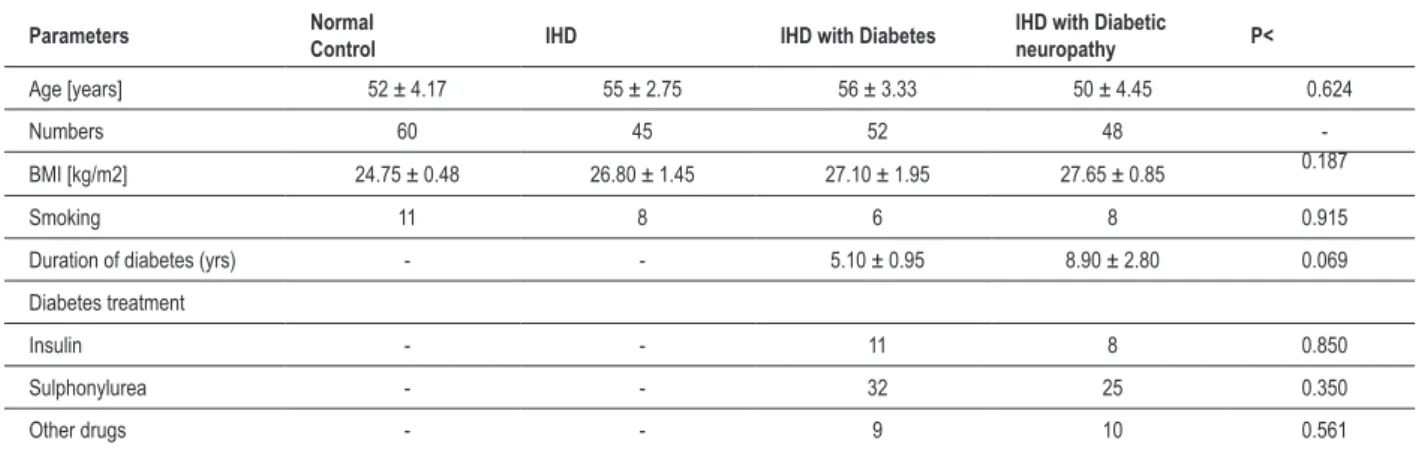
Clinical characteristics of the patients are shown in Table 1. The three study groups and the control group were well-balanced concerning demographic data. The study groups did not significantly differ regarding age, duration of diabetes and medication use. There was a significant decrease in the
Table 1 - Demographic and clinical characteristics of the patients and control subjects; Results are expressed as Mean ± SEM; *p<0.05 – between groups
Parameters Normal Control IHD IHD with Diabetes IHD with Diabetic neuropathy P<
Age [years] 52 ± 4.17 55 ± 2.75 56 ± 3.33 50 ± 4.45 0.624
Numbers 60 45 52 48
-BMI [kg/m2] 24.75 ± 0.48 26.80 ± 1.45 27.10 ± 1.95 27.65 ± 0.85 0.187
Smoking 11 8 6 8 0.915
Duration of diabetes (yrs) - - 5.10 ± 0.95 8.90 ± 2.80 0.069 Diabetes treatment
Insulin - - 11 8 0.850
Sulphonylurea - - 32 25 0.350
Other drugs - - 9 10 0.561
heart rate variability in IHD patients without diabetes (p<0.01) when compared to the healthy controls. IHD patients with diabetes mellitus (p<0.01) and with neuropathy also had a significantly reduced (p<0.01) heart rate variability compared to controls. Ischemic heart disease patients without diabetes had a significantly higher HRV (p<0.05) than IHD patients with diabetes (DM) or diabetic neuropathy (DN). There was no significant difference in HRV between IHD with DM or with DN groups. HbA1C and fasting blood glucose levels were significantly higher in IHD with diabetes (p<0.01) and IHD with diabetic neuropathy (p<0.01) groups when compared to control and IHD groups (table 2).
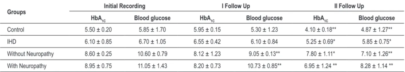
As shown in Table 3, patients with IHD without diabetes and those with diabetes with or without neuropathy, all showed a significant increase in HRV (p<0.01) at the three -month and one-year follow-up after diaphragmatic breathing exercises. A more statistically significant increase in HRV was recorded (p<0.01- for all groups) after one year of follow-up, when compared to their baseline recordings in all the study groups. Increase in HRV after the follow-up was significantly higher (p<0.01) in IHD with diabetes group than IHD with diabetic neuropathy group. The regular practice of diaphragmatic breathing increased heart rate variability in control subjects also after one year (p<0.01) (Table 3). A significant decrease in HbA1C (p<0.01) and blood glucose level (p<0.01) was seen after 3 and 12 month-follow up recording in the compliant group. Even though the levels of HbA1C and
blood glucose were significantly higher (p<0.01) than in the control and IHD groups, the regular practice of diaphragmatic breathing significantly decreased these metabolic parameters after the 12-month follow-up, when compared to baseline levels (Table 4).
Table 3 - HRV (beats/min) in diabetes (without neuropathy & with neuropathy) in the compliant group.Values are expressed as Mean ± SEM, ** p< 0.01 – Initial recording with I and II follow up recording
Groups RecordingInitial I Follow Up II Follow Up
Control 22.85 ± 0.63 23. 90 ± 0.16 25.22 ± 1.37** IHD 17.14 ± 0.18 19.21 ± 0.11** 20.05 ± 0.24 ** Diabetes
without neuropathy
14.68 ± 0.49 17.00 ± 1.23 ** 17.69 ± 1.21 **
Diabetes with
neuropathy 13.50 ± 0.78 14.69 ± 1.11 ** 15.00 ± 1.14 **
Table 4 - HbA1C (%) and blood glucose levels (mmol/l) in IHD and Diabetes (without Neuropathy & with Neuropathy) in the Compliant Group. Values are expressed as Mean ± SEM; * p< 0.05; ** p< 0.01 – Initial recording with I and II follow up recording
Groups Initial Recording I Follow Up II Follow Up
HbA1C Blood glucose HbA1C Blood glucose HbA1C Blood glucose
Control 5.50 ± 0.20 5.85 ± 1.70 5.95 ± 0.15 5.30 ± 1.23 4.10 ± 0.18** 4.87 ± 1.27** IHD 6.10 ± 0.85 6.70 ± 1.05 6.55 ± 0.42 6.10 ± 0.84 5.25 ± 0.69* 5.85 ± 0.75* Without Neuropathy 8.60 ± 0.25 10.60 ± 0.79 8.12 ± 1.23 9.05 ± 0.13** 7.80 ± 1.11* 7.10 ± 1.26** With Neuropathy 8.95 ± 0.75 11.05 ± 1.43 8.20 ± 0.73 10.73 ± 0.85** 6.95 ± 1.24 ** 8.28 ± 1.14 **
Table 2 - HbA1C, (%), blood glucose (mmol/l) and heart rate variability in controls and IHD patients with diabetes and diabetic neuropathy. Values are expressed as Mean ± SEM, ♦p< 0.05; ♦♦p<0.01- control
with other groups, * p< 0.05 – without diabetes, with diabetes and diabetic Neuropathy
Groups HbA1C (%)
Blood glucose
(mmol/l) (beats/minute)HRV
Control 5.50 ± 0.20 5.85 ± 1.70 22.85 ± 0.63 IHD 6.10 ± 0.85 6.70 ± 1.05 17.14 ± 0.18 ♦♦ IHD with
diabetes 8.60 ± 0.25 **♦♦ 10.60 ± 0.79**♦♦ 14.68 ± 0.49 * ♦♦ IHD with
diabetic
neuropathy 8.95 ± 0.75**
♦♦ 11.05 ± 1.43**♦♦ 13.50 ± 0.78 * ♦♦
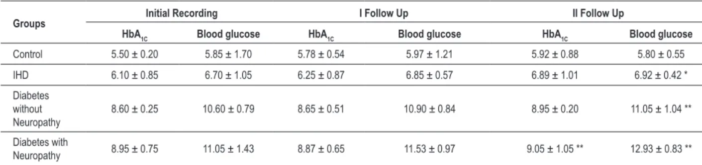
increased significantly after one year of follow-up in IHD patients with diabetic neuropathy (p<0.01). Blood glucose levels increased significantly in non-compliant IHD patients without DM (p<0.05), IHD with DM (p<0.01) and IHD with DN (p<0.01), indicating that the disease condition worsened without diaphragmatic breathing (table 6).
When heart rate variability was compared between the compliant and non-compliant groups after one-year follow-up, a more significant increase was recorded in all the groups including controls (p<0.01) (Table 7). In the non-compliant groups, there was a significant decrease in HRV among IHD patients with DM and DN (p<0.01), when compared to compliant patients.
Multiple regression analysis indicated that breathing alone accounted for a 58.8% (R=0.588) variation in HRV after one year follow-up. When other predictive variables were included, the contribution of breathing increased to 72%
(R=0.725). Diaphragmatic breathing alone showed the greater contribution for the observed changes in HRV. The coefficient of diaphragmatic breathing was the highest, showing the maximum contribution when compared to other predictors such as medication, duration of diabetes, age etc.
Discussion
The present study showed that there was a significant decrease in heart rate variability in ischemic heart disease patients and IHD patients who had diabetes. Few studies have reported the reduction of HRV in diabetes. In diabetic patients without evidence of autonomic neuropathy, the reduction in heart rate variability during controlled conditions has also been reported11,24,25. Abnormal heart rate variability in diabetes
represents an increased risk for ventricular arrhythmias, as well as total cardiovascular morbidity and mortality12. The decrease
in HRV in IHD patients with diabetes reflects the reductions in both sympathetic and parasympathetic modulation of the heart. The observed decrease in heart rate variability in IHD patients could be due to the autonomic dysfunction as a result of ischemia. Imbalanced regulation of the cardiac autonomic nervous system is one of the important pathophysiological changes in IHD26,27. There was a more significant decrease
in HRV in IHD patients with neuropathy than the patients without neuropathy. The heart rate variability may be diminished in patients with diabetes due to cardiovascular autonomic neuropathy. It has been observed that in adult patients with diabetes, there is dysfunction of the autonomic nervous system with the long duration of the disease26-29.
Diabetic autonomic neuropathy is accompanied at first by disorders of the parasympathetic and later, of the sympathetic system28. Our assessment of the HRV changes supports the
view that there is a decrease in autonomic function early in the development of diabetes and that diabetes leads to a progressive decline in autonomic function and that heart rate variability may be an efficient method for early detection of diabetic autonomic neuropathy.
In this study, the practice of abdominal breathing improved the heart rate variability measures. The present study showed that HRV improved in non-DM IHD patients and DM –IHD patients who practiced regular breathing exercises. There was a significant increase in HRV in normal control subjects who practiced diaphragmatic breathing for one year. The observed improvement in HRV with the modified breathing technique could be due to the direct effect of breathing on the autonomic nervous system controlling the heart30. In the human body,
Table 5 - HRV (beats/min) in IHD and diabetes (without neuropathy & with neuropathy) in the non- compliant group. Values are expressed as Mean ± SEM; **p< 0.01 – Initial recording with I and II follow up recording
Groups Initial Recording I Follow Up II Follow Up
Control 22.85 ± 0.63 22.04 ± 1.13 21.05 ± 0.86 IHD 17.14 ± 0.18 16.01 ± 0.18 15.08 ± 0.20 ** Diabetes
without Neuropathy
14.68 ± 0.49 13.33 ± 1.45 11.44 ± 1.35 ** Diabetes
with Neuropathy
13.50 ± 0.78 12.22 ± 0.99 10.78 ± 0.98 **
Table 6 - HbA1C (%) blood glucose levels (mmol/l) in IHD and diabetes (without neuropathy and with neuropathy) in the non- compliant group. Values are expressed as Mean ± SEM; * p< 0.05; ** p< 0.01 – Initial recording with I and II follow up recording
Groups Initial Recording I Follow Up II Follow Up
HbA1C Blood glucose HbA1C Blood glucose HbA1C Blood glucose
Control 5.50 ± 0.20 5.85 ± 1.70 5.78 ± 0.54 5.97 ± 1.21 5.92 ± 0.88 5.80 ± 0.55 IHD 6.10 ± 0.85 6.70 ± 1.05 6.25 ± 0.87 6.85 ± 0.57 6.89 ± 1.01 6.92 ± 0.42 * Diabetes
without Neuropathy
8.60 ± 0.25 10.60 ± 0.79 8.65 ± 0.51 10.90 ± 0.84 8.95 ± 0.20 11.05 ± 1.04 ** Diabetes with
Neuropathy 8.95 ± 0.75 11.05 ± 1.43 8.87 ± 0.65 11.53 ± 0.97 9.05 ± 1.05 ** 12.93 ± 0.83 **
mode-locked to breathing. Proper breathing could influence even subtle cardiac parameters such as ejection fraction, aortic pressure, and pulmonary arterial pressure, preload and afterload and even tissue oxygenation31,32. Diaphragmatic
breathing may reduce sympathetic activity by enhancing central inhibitory rhythm33. Due to increased tidal volume
during deep diaphragmatic breathing, there is the activation of the Hering-Breur reflex34 which reduces the chemoreflex
sensitivity and might enhance the baroreflex and reduce the sympathetic activity34-36. It seems that deep breathing induces
generalized decrease in the excitatory pathways regulating respiratory and cardiovascular system. The respiratory and cardiovascular systems share similar control mechanisms and alterations in one system will modify the functioning of the other15,35. In ischemic heart disease, there is increased
sympathetic and chemoreflex activation and diaphragmatic breathing induced a reduction in the sympathetic activation, which might have increased the HRV. There was a significant improvement in the metabolic parameters studied after the regular practice of diaphragmatic breathing in the present study. The decrease in HbA1C is in agreement with the earlier reports37,38. The decrease in glycated hemoglobin and blood
glucose recorded in IHD patients with diabetes and diabetic neuropathy indicates that regular prolonged breathing exercises can significantly improve the blood glucose homeostasis and the much-needed glycemic control among these patients. Hyperglycemia and its related metabolic consequences leads to the pathogenesis of autonomic neuropathy in diabetes11,24.
The observed improvement in metabolic parameters could be attributed to the autonomic changes brought about by
diaphragmatic breathing, which decreased the sympathetic activity15,35,39,40.
Stress may have negative effects on health and the fact that patients with type 2 diabetes may be at increased risk for the experience of stress is associated with the release of counter-regulatory hormones that will result in elevated blood glucose levels41. In addition, stress can disrupt diabetes
control indirectly through effects on diet, exercise, and other self-care behaviors. Stress can be managed through the use of behavioral stress management programs or through medication use. Both types of interventions have been reported to improve glycemic control in patients with type 2 diabetes38,42,43. Stress
management training typically includes progressive muscle relaxation with or without electromyography biofeedback, mental imagery, diaphragmatic breathing , yoga and instructions on how to modify the physiological, cognitive and behavioral responses to stress. Reports from researchers investigating the effect of stress management on glycemic control have been inconsistent44,45, but several of these studies
were underpowered, and others had design problems. Our study supports the view that that intervention in the form of deep diaphragmatic breathing practice would improve the glycemic control and also decrease the cardiac autonomic impairment in IHD patients with diabetes mellitus.
Physical activity, as a non-pharmacological intervention, is very important in patients with the metabolic syndromes in whom the simultaneous occurrence of diabetes is accompanied by an increased activity in the sympathetic nervous system. Thus, the effect of diaphragmatic breathing on the heart rate variability is mainly due to the alteration in the balance between the sympathetic and parasympathetic nervous systems activity on the heart. Long-term practice of diaphragmatic breathing lead to stable modifications in the autonomic control of the heart and resulted in increased heart rate variability. Abdominal breathing is known to evoke internal quieting, relaxation and peripheral warming. Diaphragmatic breathing is considered as the healthiest form of breathing and it is one of the simplest, yet most powerful, stress management techniques16,32. Diaphragmatic breathing tries to reverse the
autonomic balance in favor of the parasympathetic stimulation from the sympathetic stimulation in diabetic patients, reducing the functional symptomatology and promoting a positive impact on individual health40. The study clearly indicates a
Table 7 - Changes in HRV (beats/min) in IHD and diabetes (without neuropathy & with neuropathy) in the compliant and non-compliant groups after 1-year follow up.
Groups Compliant Non-Compliant
Control 25.22 ± 1.37 21.05 ± 0.86 ** IHD 20.05 ± 0.24 15.08 ± 0.20 ** Diabetes without neuropathy 17.69 ± 1.21 11.44 ± 1.35 ** Diabetes with neuropathy 15.00 ± 1.14 10.78 ± 0.98 **
Values are Mean ± SEM; ** p< 0.01 – Compliant with non-compliant groups
patients with ischemic heart disease and IHD patients with diabetes and diabetes autonomic neuropathy.
Study limitations
There are several limitations in this study. The absence of metabolic data, lack of accompanying cardiovascular measures, the effect of different meal compositions on HRV, measurement of exercise capacity (peak VO2), and absence of echocardiographic data are the major limitations. One of the difficulties faced by our research group is the lack of frequency-domain HRV analysis. Our laboratory was not specifically equipped for this purpose; hence, we could not conduct the HRV analysis by spectral analysis, which has higher sensitivity and specificity. However, the clinical usefulness of the heart rate variability findings from short-term ECG recordings is well-established46,48.The influence of drug treatment and
its withdrawal on heart rate variability should be carefully considered. For ethical reasons, we did not discontinue the treatment for an extended period of time.
In conclusion, the study confirms that heart rate variability
decreases in patients with ischemic heart disease who have diabetes and that this decrease is higher in patients with diabetic neuropathy, indicating severe autonomic imbalance. The regular practice of diaphragmatic breathing increases heart rate variability in normal control subjects and in ischemic heart disease patients with diabetes. The observed data should encourage the regular practice of diaphragmatic breathing as a non-pharmacological therapy in patients with ischemic heart disease with or without diabetes and diabetic neuropathy, along with the regular treatment. The aim of the approach would be to maintain an optimal autonomic nervous system balance and physiological heart rate regulation. As the diaphragmatic breathing is an easy, safe and psychologically acceptable physical activity, we consider this as a valuable finding and considering that the socio-demographic profile of the patients in this study were similar to other international studies, we believe the results of the present study can be applied to other populations. Further studies may be required to establish the long-term benefits of diaphragmatic breathing on other established methods of HRV measurements in larger populations.
Potential Conflict of Interest
No potential conflict of interest relevant to this article was reported.
Sources of Funding
There were no external funding sources for this study.
Study Association
This study is not associated with any post-graduation program.
References
1. Kristal Boneh E, Raifel M, Froom P, Ribak J. Heart rate variability in health and disease. Scan J Work Environ Health. 1995; 21:85-95.
2. Pomeranz B, Macauley RJ, Caudill MA, Kutz I, Adam D, Gordon D. Assessment of autonomic function in humans by heart rate spectral analysis. Am J Physiol. 1985; 248: H151-H153.
3. Tsuji H, Venditti FJ, Manders ES. Reduced heart rate variability and mortality risk in an elderly cohort. The Framingham Heart Study. Circulation. 1994; 90: 878-83.
4. Huikuri HV, Makikallio TH. Heart rate variability in ischemic heart disease. Auton Neurosci. 2001; 90 (1-2): 95-101.
5. Rich MW, Saini JS, Kleiger RE, Carney RM, Te Velde A, Freedland KE. Correlation of heart rate variability with clinical and angiographic variables and late mortality after coronary angiography. Am J Cardiol. 1988; 62: 714-7.
6. Hayano J, Sakakibara Y, Yamada M, Ohte N, Fuginami T, Yokoyama K, et al. Decreased magnitude of heart rate spectral components in coronary artery disease: its relation to angiographic severity. Circulation. 1990; 81 (4): 1217-24.
7. Vardas PE, Kochiadakis GE, Manios EG, Kanoupakis EM, Zouridakis EG, Chlouverakis G. Spectral analysis of heart rate variability before and during episodes of nocturnal ischemia in patients with extensive coronary artery disease. Eur Heart J. 1996; 17: 383-93.
8. Al Hazimi A, Al Ama N, Syiamic A, Qosti R, Abdel Galil K. Time domain analysis of heart rate variability in diabetic patients with and without autonomic neuropathy. Ann Saudi Med. 2002; 22 (5-6): 400-3.
9. Masaoka S, Lev-Ran A, Hill LR, Vakil G, Hon EH. Heart rate variability in diabetes; relationship to age and duration of the disease. Diabetes Care. 1985; 8: 64-8.
10. Kamath MV, Fallen EL. Power spectral analysis of heart rate variability; a non invasive signature of cardiac autonomic function. Crit Rev Biomed Eng. 1993; 1: 245-311.
11. Kitney RI, Byrne S, Edmonds ME, Watkins PJ, Roberts VC. Heart rate variability in the assessment of autonomic diabetic neuropathy. Automedica. 1982; 4: 155-67.
13. Sipinakova J, Hahn G. Effect of respiration and posture on heart rate variability. Physiol Res. 1997; 46: 173-9.
14. Pinheiro CHJ, Medeiros RAR, Pinheiro DGM, Marinho MJF. Spontaneous respiratory modulation improves cardiovascular control in essential hypertension. Arq Bras Cardiol. 2007; 88 (6): 576-83.
15. Joseph CN, Porta C, Casucci G, Casiraghi N, Maffeis M, Rossi M, et al. Slow breathing improves arterial baroreflex sensitivity and decreases blood pressure in essential hypertension. Hypertension. 2005; 46: 714-8.
16. Pepper E, Crane-Gockley V. Towards effortless breathing . Medical Psychotherapy. 1990; 3: 135-40.
17. Boyer BA, Poppen R. Effects of abdominal and thoracic breathing on multiple site electromyography and peripheral skin temperature. Percept Mot Skills. 1995; 81: 3-14.
18. Grossman E, Grossman A, Schein MH, Zimlichman R, Gavish B. Breathing control lowers blood pressure. J Hum Hypertens. 2001; 15: 263-9.
19. American Diabetes Association and American Academy of Neurology. Proceedings of a consensus development conference on standardized measures in diabetic neuropathy. Diabetes Care. 1992; 15 (Suppl. 3): 1080-107.
20. May O, Arildsen H. Assessing cardiovascular autonomic neuropathy in diabetes mellitus. How many tests to use? Journal of Diabetes and Its Complications. 2000; 14: 7-12.
21. Katz A, Liberty IF, Porath A, Ovsyshcher I, Prystowsky EN. A simple bedside test of one minute heart rate variability during deep breathing as a prognostic index after myocardial infarction. Am Heart J. 1999; 138 (1 Part 1): 32-8.
22. Jagomagi K, Raamat R, Talts J, Lansimies E, Jurvelin J. Portapres and differential oscillometric finger blood pressure changes during deep breathing test in the assessment of BRS index. Clin Physiol Function Imag. 2003; 23: 9-13.
23. Tamosiunaite M, Urbonaviciene G, Vainoras A, Gargasas G, Kaminskiene S, Bluzaite I, et al. Influence of deep breathing on heart rate variability in patients with ischemic heart disease. Elektronika ir Elektrotechnika. 2005; 59: 33-6.
24. Takase B, Kurita A, Noritake M, Uehata A, Maruyama T, Nagayoshi H, et al. Heart rate variability in patients with diabetes mellitus, ischemic heart disease and congestive heart failure. J Electrocardiol. 1992; 25: 79-8.
25. Pfeifer MA, Weinberg CR, Cook CL, Reenan A, Halter JB, Ensinck JW, et al. Autonomic neural dysfunction in recently diagnosed diabetic subjects. Diabetes Care. 1984; 7: 447-53.
26. Yan W, Zuo W, Lin Q. Evaluation of autonomic nervous function and heart rate variability and cardiovascular reflex tests in type II diabetes mellitus patients. Zhonghua Nei Ke Za Zhi. 2000; 39: 670-3.
27. Pagani M, Malfatto G, Pierini S, Casati R, Masu AM, Poli M, et al. Spectral analysis of heart rate variability in the assessment of autonomic diabetic neuropathy. J Auton Nerv Syst. 1988; 23: 143-53.
28. Barkai L, Madacsy L. Cardiovascular autonomic dysfunction in diabetes mellitus. Arch Dis Child. 1995; 73 (6): 515-8.
29. Malliani A, Pagani M, Lombardi F, Cerutti S. Cardiovascular neural regulation explored in the frequency domain. Circulation. 1991; 84: 1482-92.
30. Menezes Jr AS, Moreira HG, Daher MT. Analise da variabilidade da
frequência cardíaca em pacientes hipertensos, antes e depois do tratamento com inibidores da enzima conversora da angiotensina II. Arq Bras Cardiol. 2004; 83 (2):165-8.
31. Bernardi L, Spadacini G, Bellwon J. Effect of breathing rate on oxygen saturation and exercise performance in chronic heart failure. Lancet. 1998; 351: 1308-11.
32. Van Dixhoorn J. Favorable effects of breathing and relaxation instructions in heart rehabilitation: a randomized 5-year follow-up study. Ned Tijdschr Geneeskd. 1997; 141: 530-4.
33. Montano N, Cogliati C, Porta A, Pagani M, Malliani A, Narkiewicz K, et al. Central vagotonic effects of atropine modulate spectral oscillations of sympathetic nerve activity. Circulation. 1998; 98: 1394-9.
34. Bernardi L, Gabutti A, Porta C, Spicuzza L. Slow breathing reduces chemoreflex response to hypoxia and hypercapnia, and increases baroreflex sensitivity. J Hypertens. 2001; 19: 2221-9.
35. Spicuzza L, Gabutti A, Porta C, Montano N, Bernardi L. Yoga and chemoreflex response to hypoxia and hypercapnia. Lancet. 2000; 356: 1495-6.
36. Francis DP, Ponikowski P, Coats AJS. Chemoreflex-baroreflex interactions in cardiovascular disease. In: Bradley DT, Floras JS (eds). Sleep apnea: implications in cardiovascular disease. New York (NY): Dekker; 2000. p. 33-56.
37. VanRooijen AJ, Rheedes P, Eales CJ, Becker PJ. Effect of exercise vs relaxation on HbA1C in black females with type 2 diabetes mellitus. Q J Med. 2004; 97: 343-51.
38. Surwit RS, Feinglos MN, Van Tilburg MAL, Edwards CL, Zucker N, Williams P. Stress management improves long-term glycemic control in type 2 diabetes. Diabetes Care. 2002; 25: 30-4.
39. Goso Y, Asanoi H, Ishise H, Kameyama T, Hirai T, Nozawa T. Respiratory modulation of muscle sympathetic nerve activity in patients with chronic heart failure. Circulation. 2001; 104: 418-23.
40. Pepper E, Tibbetts V. Fifteen month follow up with asthmatics utilizing EMG incentive inspirometer feedback. Biofeedback and Self Regulation. 1992; 17: 143-51.
41. Surwit RS, Schneider MS. Role of stress in the etiology and treatment of diabetes mellitus. Psychosom Med. 1993; 55: 380-93.
42. Lane JD, McCaskill CC, Ross SL, Feinglos MN, Surwit R. Relaxation training for NIDDM: predicting who may benefit. Diabetes Care. 1993; 16: 1087-94.
43. Lustman PJ, Griffith LS, Clouse RE, Freedland KE, Eisen SA, Rubin EH, et al. Effects of alprazolam on glucose regulation in diabetes: results of double-blind, placebo-controlled trial. Diabetes Care. 1995; 18: 1133-9.
44. Bradley C. Contributions of psychology to diabetes management. Br J Clin Psychol. 1994; 33: 11-21.
45. Aikens J, Kiolbasa TA, Sobel R. Psychological predictors of glycemic change with relaxation training in non-insulin-dependent diabetes mellitus. Psychother Psychosom. 1997; 66: 302-6.
46. Vinik AI, Maser RE, Mitchell BD, Freeman R. Diabetic autonomic neuropathy. Diabetes Care. 2003; 26: 1553-9.