Universidade de Lisboa
Faculdade de Farmácia
Anticancer prodrugs for targeted therapy
Débora Reis Veiga de Almeida
Mestrado Integrado em Ciências Farmacêuticas
Universidade de Lisboa
Faculdade de Farmácia
Anticancer prodrugs for targeted therapy
Débora Reis Veiga de Almeida
Monografia de Mestrado Integrado em Ciências Farmacêuticas
apresentada à Universidade de Lisboa através da Faculdade de Farmácia
Orientador: Professora Doutora Ana Paula Francisco
II
Resumo
Nos tempos que correm, o cancro é a segunda maior causa de morte globalmente, sendo uma doença com uma grande expressão e fisiopatologia complexa.
Existem diversos tipos de tratamentos anti cancro como, por exemplo, cirurgia e radioterapia porém, quando o tumor se encontra distribuído e com desenvolvimento de metástases, surge a quimioterapia. Apesar da sua ação ser focada no decréscimo da proliferação de células cancerígenas, a maioria dos fármacos citotóxicos não está apta a localizar, seletivamente, o local do tumor, o que leva a variados efeitos adversos indesejados. Por este motivo, tornou-se urgente a procura de novas soluções que possam otimizar o tratamento anti cancro e a terapia localizada, com a utilização de pró fármacos, é uma das potenciais estratégias.
Em comparação com os tecidos ditos normais, as células cancerígenas são caracterizadas por únicos e anormais marcadores e, por isso, a estratégia baseada no uso de pró fármacos irá explorar essas diferenças, de modo a afetar somente o tumor, sem causar dano aos tecidos saudáveis.
As células cancerígenas são, então, caracterizadas pelo seu microambiente específico com baixos valores de pH, elevada concentração de espécies reativas de oxigénio e glutationa e, ainda, por uma elevada expressão de certas enzimas e antigénios específicos. Esta última característica está relacionada com abordagens mais experimentais focando-se em anticorpos monoclonais ou terapia genómica.
Deste modo, estão a ser desenvolvidas estratégias centradas nos mecanismos e singularidades do cancro e alguns exemplos já se encontram disponíveis, incluindo não só pró fármacos que já se encontram no mercado, mas também aqueles que ainda se encontram em estados mais primordiais do seu desenvolvimento.
Consequentemente, esta monografia irá focar-se não só no design de pro fármacos mas na tentativa de ter uma maior perceção de todos estes métodos através de vários exemplos detalhados de alguns dos pro fármacos já comercializados ou ainda em desenvolvimento.
Palavras-chave: pró fármacos, cancro, terapia localizada, pH, microambiente
III
Abstract
In the current times, cancer is the second leading cause of death globally, being a wide spread disease with a complex physiopathology.
There are various types of cancer treatment, such as surgery and radiotherapy but, when the tumor is well spread with the development of metastases, chemotherapy comes to picture. Although its action is focused on decreasing the proliferation of cancer cells, the majority of antitumor drugs cannot selectively localize the cancer site, leading to several undesired side effects. So, it has become urgent to find new solutions that can optimize the anticancer treatment and targeted therapy, using prodrugs, is one potential strategy.
In comparison with normal tissues, cancer cells are characterized by unique abnormal markers, thus the prodrug strategy will exploit these differences, in order to kill solely the cancer tissues without damaging the healthy ones.
Cancer cells are, then, characterized of its specific microenvironment with low pH levels, elevated ROS or high levels of GSH, unique overexpressed enzymes and also specific antigens. This last characteristic is related to more experimental approaches focusing on mAb or gene therapy.
Therefore, strategies are being developed focusing on the cancer mechanisms and singularities and some proven examples are already coming to light, regarding non only prodrugs that are already in the market, but also the ones that are still in earlier stages of development.
Hence, this review will be focused not only on the prodrug design but also in trying to have a better understanding of all these methods with given detailed examples of some prodrugs already on the market or still in development.
Key-words: prodrugs, cancer, targeted therapy, pH, cancer microenvironment,
IV
Acknowledgements
There is no successful journey if travelled alone so, I would like to thank all the people that, in a way or another, have been part of these last years.
First, I would like to thank my family for all the support during the course of this Master’s Degree in the Faculty of Pharmacy of University of Lisboa (FFULisboa). Without their patience and care I would not have achieved so much in terms of personal and academic conquests.
To all my friends, I would like to thank them for all the great college days and the long study nights. For all the given advices, all the memories and all the stories that we will always share, Bárbara Cochicho, Mariana Chaves, Laura Moura, Gonçalo Chasqueira, Inês Lima and Leonor Baptista, I owe all of those to you.
To Tuna A Feminina, for allowing me to be myself from North to South of the country, always reminding me that everything can be accomplished. I also would like to address a special thank you note to my music team, you will always have a special place in my heart.
To my loving boyfriend, João Lourenço, I would like to thank him for being the most wonderful and kind person I have ever met. To have stuck with me in all the days and nights this paper and this course made obligatory, always with a motivational smile to share with me. Without you this journey would certainly have been much harder.
Finally, I would like to thank my teacher, Ana Paula Francisco, for accepting me in this project, always helping me every step of the way. This is yours as well.
V
Abbreviations
5-FC – 5-fluorocytosine 5-FU – 5-fluouracyl 5-FudR – 5-fluorodeoxyuridine 6-MP – 6-mercaptopurine 6-TG – 6-thioguanine 𝛾CL – 𝛾-glutamylcysteine ligase 𝛾GT – 𝛾-glutamyl-transpeptidase ADC – Antibody drug conjugateADEPT – Antibody-directed enzyme prodrug therapy ADME – Absorption, distribution, metabolism, excretion ADRs – Adverse drug reactions
AELs – Antitumor ether lipids ATP – Adenosine triphosphate AVTG – 6-(2-acetylvinylthio)guanine AVTP – 6-(2-acetylvinylthio)purine
BQC – 5,6-dihydro-4H-benzo[de]quinoline-camptothecin BSA – Bovine serum albumin
CPA – Cyclophosphamide CPT – Camptothecin CYP – Cytochrome P450
DCM – Dicyanomethylene-4H-pyran DNA – Deoxyribonucleic acid
DNR – Daunorubicin
DPPC – 1,2-dipalmitoyl-sn-glycero-3-phosphocholine DOX – Doxorubicin
EPR – Enhanced permeation and retention FA – Folic acid
GCV – Ganciclovir
GDEPT - Gene-directed enzyme prodrug therapy GI – Gastrointestinal
GSH – Glutathione
GSSH – Glutathione dissulfide GST – Glutathione-S-transferases HIF – Hypoxia-inducible factor HDACs – Histone deacetylases
VI HPV – Human papilloma virus
HAS – Human serum albumin
HSV-TK – Herpes Simplex Virus Thymidine Kinase IFA - Ifosfamide
IgG – Immunoglobulin
INNO-2016 – 6-malimidocaproyl hydrazone
IUPAC – International Union of Pure and Applied Chemistry IV – Intravenous
mAb – Monoclonal antibody MMAE – Monomethyl auristatin E MMAF – Monomethyl auristatins F MMC – Mitomycin 2
MMP-2 – Matrix metalloproteinase MTX – Methotrexate
NAC – N-acetyl-C-cysteine NIR – Near infracted NO – Nitric oxide
NQO1 - NAD(P) H:quinone oxidoreductase 1 NTR – Nitroreductase
PEG-PAA – Poly(ethylene glycol)-b-poly (acrylic acid) PEG-PLA - Polyethylene glycol−polylactic acid
PLA2 – Phospholipase A2
PSA – Prostate specific antigen
PTA – Peptide-bridged twin-acylhydrazone PTX – Paclitaxel
RGD - Arg-Gly-Asp
ROS – Reactive oxygen species scFv – Single-chain fragment TOS – Vitamin E succinate TPG - Thapsigargin
TPGS - D-alpha-tocopherol polyethylene glycol 1000 succinate VBL – Vinblastine
VDEPT – Virus-directed enzyme prodrug therapy VRL – Vinorelbine
VII
Index
1. Cancer ... 1 1.1. Epidemiology ... 1 1.2. Pharmacotherapy ... 3 2. Methods ... 5 3. Prodrugs ... 6 3.1. Types of prodrugs ... 6 4.2. Benefits of prodrugs ... 74.2.1. Enhancement of ADME properties ... 8
4.2.2 Targeted delivery ... 8
5. Prodrugs based on intracellular cancer microenvironment ... 10
5.1. pH-sensitive prodrugs ... 10
5.2. ROS-activated prodrugs ... 14
5.3. Glutathione-responsive prodrugs ... 18
6. Prodrugs activated by cancer specific enzymes ... 22
6.1. Prodrugs activated by hydrolases ... 22
6.1.1. Proteases ... 23 6.1.2. Esterases ... 25 6.1.3. Glycoside hydrolases ... 27 6.1.4. Protein deacetylases ... 29 6.2. Oxidoreductases ... 29 6.3. Transferases ... 30
7. Antibody prodrugs based on cancer specific antigens ... 31
7.1 Antibody-drug conjugates (ADCs) ... 31
7.2 Antibody-directed enzyme prodrug therapy (ADEPT) ... 37
8. Gene-directed enzyme prodrug therapy (GDEPT) ... 39
VIII
List of Figures
Figure 1 - 2018 Worldwide Incidence and Mortality rates ... 2
Figure 2 - Number of new cases and deaths in 2018, both sexes, all ages ... 3
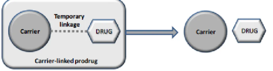
Figure 3 - Schematic representation of a carrier-linked prodrug ... 7
Figure 4 - INNO-206 and pH-sensitive DOX release ... 11
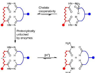
Figure 5 - Proteolytic unlocking of PTA-linkers ... 12
Figure 6 - MMP-2 unlocking and pH-sensitive release of MMAE ... 12
Figure 7 - Acetal-linked pH-sensitive PTX prodrug nanoparticles ... 13
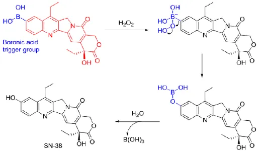
Figure 8 - ROS-activated SN-38 with a boronic acid trigger ... 15
Figure 9 - ROS-activated SN-38 with a boronate ester trigger and a coumarin fluorophore ... 16
Figure 10 - Nitrogen mustards with boronate ester triggers ... 17
Figure 11 - ROS-activated thiazolidinone prodrug ... 17
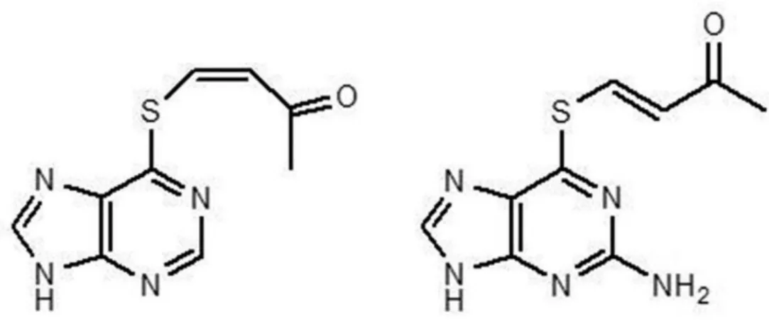
Figure 12 - AVTP and AVTG ... 19
Figure 13 - GSH-responsive JS-K activation ... 20
Figure 14 - GSH-responsive FA-CPT activation ... 20
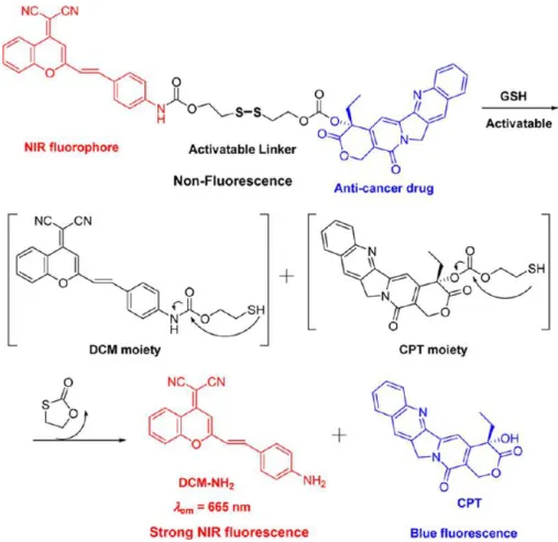
Figure 15 - GSH-responsive DCM-S-CPT activation with strong NIR fluorescence ... 21
Figure 16 - MMP-activated prodrug Cap-ProCitGly ~ HofTyrLeu-DOX ... 25
Figure 17 – sPLA2 action site in a C6-RAR prodrug ... 26
Figure 18 – PLA2 mediated AELs release ... 26
Figure 19 - Prodrugs activated by β-glucuronidase ... 27
Figure 20 - β-glucuronidase-responsive albumine-binding prodrug ... 28
Figure 21 - NAC protecting and NQO1 responsive release of β-lapachone ... 30
Figure 22 - SGN-35 oteolysis and subsequent MMAE release ... 33
Figure 23 - Transtuzumab Emtansine ... 34
Figure 24 - Inotuzumab Ozogamin ... 35
Figure 25 - SYD985 ... 36
Figure 26 - Triazene prodrug and CPG2 mediated conversion into MMT ... 38
Figure 27 – β-glucuronidase activation of BQC-G ... 39
Figure 28 - TK activation of GCV ... 41
Figure 29 - 5-FC ... 42
Figure 30 - BSA nanoparticles of 5-FC ... 42
Figure 31 - CYP450 activation of CPA ... 43
Figure 32 - CYP450 activation of IFA ... 43
1
1. Cancer
Cancer is a general term for a large group of diseases with various causes and different characteristics that make every type unique and unlike the other. (1)
There are several types of cancer, depending on its origin. The most common, carcinoma, starts in the epithelial tissue, usually diffusing through the lymphatic circulation. Leukaemia starts in immature blood cells produced by the bone marrow which tend to accumulate in the bloodstream, lymphoma originates in the lymph nodes and immune system tissues and sarcoma starts in the connective, muscular or adipose tissue, bone and cartilage, diffuses through bloodstream and usually metastasizes to lungs. Given this information, it is possible to conclude how the cancer’s nomenclature system works, since the prefix is directly related to where it is located. (2)
This disease, triggered by deoxyribonucleic acid (DNA) mutation, causes normal genes involved in cell growth to become oncogenes and inactivates tumour supressing genes, which can prevent cancer by slowing or stopping cell growth. So, it leads to an alteration of the regular growing and proliferating cell mechanisms, shown by an uncontrolled cell growth, local tissue invasion and distant metastases, the pathogenic spread of neoplasic cells from an initial or primary tumour site to a secondary site.
Tumour growth is strictly related to the cell cycle duration and the growing cells fraction. Once formed, the tumour rapidly acquires a specific microenvironment, characterized by a high redox homeostasis, a low pH and a particular enzyme metabolism. (3)
About the carcinogenic process, the one responsible for cancer’s entire evolution, it is multifactorial, since the etiology can vary greatly with various agents involved. Cancer’s precipitating factors can be divided in two branches, endogenous factors, for example, genetic modifications and exogenous factors that are passible of change, such as tobacco, alcohol, lack of physical activity, low vegetables/fruit intake, radiations, some drugs and virus. In addition to being multifactorial, the carcinogenic process have multiple stages, comprising initiation, promotion and, at last, progression, the phase when all modifications become more and more profound. (4)
1.1. Epidemiology
Nowadays, cancer is one of the most impacting diseases, being the second leading cause of death globally, responsible for an estimated 9.6 million deaths just in 2018. So,
2 worldwide, 1 in 6 deaths is due to cancer, 70% of those occurring in low and middle income countries.
With this numbers, is crucial to explore more key facts concerning this disease, in order to fully understand its influence in the current times. Around one third of deaths caused by cancer are due to behavioural and dietary risks, with tobacco being the most important exogenous cause, responsible for approximately 22% of cancer deaths.
Late-stage presentation and inaccessible diagnosis are pretty common, especially in low-income countries (in 2017, only 26% of this group reported having oncology services available in the public sector, in contrast with high-income countries, whose numbers are much higher, around 90%), where infections such as hepatitis and human papilloma virus (HPV) represent 25% of the cancer causing factors. Adding to that, only 1 in 5 of these countries have the necessary data to drive cancer policy.
About economics, the impact of cancer is increasing day by day and the latest collected data, from 2010, estimated that the total annual economic cost of cancer was close to 1.16 trillion of American Dollars. (1)
As seen in Figure 1, cancer has an enormous expression all around the world but every continent has its own numbers, Asia being the one with the greatest incidence and mortality rates.
3 Regarding all cancer types, in 2018, there was a total of 18078957 new cases with lung, breast and colorectal cancers taking the major part of them (11.6%, 11.6% and 10.2% of the new cases, respectively). On the deaths subject, the distribution was quite different but lung cancer still took the first place with 18.4%, followed by colorectal (9.2%) and stomach cancer (8.2%). (5)
Although it is important to have a global understanding of this disease, numbers concerning Portugal also require our attention, whose will follow in Figure 2. In 2018, with the total population of 10291198, there were 581999 new cases but only 28960 deaths. The risk of dying from cancer before the age of 75 years was about 10.6%, with the male population being more susceptible than the female population (14.7% versus 6.9%) and the top 5 most frequent cancers, excluding non-melanoma skin cancer, were colorectal, breast, prostate, lung and stomach. Prostate cancer was the most common in the male population (20.4% of the cases) and breast cancer (27.1%), the most incident in women. (6)
1.2. Pharmacotherapy
Over the last years, not only our cancer knowledge has improved but various were the advances regarding its treatment. Depending on the patient in question, his symptoms, co-morbidities and the medication he is taking, every process is different but the main therapeutic goals remain the same, starting with the cure, characterized by the eradication of every neoplasic cells. (7) Other aims include having control over the disease by stopping its progression; prophylaxis – preventing cancer’s proliferation or
4 metastization after surgery or radiotherapy, and palliative care – providing symptomatic relief from pain or physical and mental stress, at any stage of illness, in order to improve the patient’s quality of life, preventing any further infections. (8)
There are many sorts of therapeutic approaches, depending on a huge variety of factors such as the cancer type and its stage of progression, for example, if it is just a carcinoma in situ – stage 0, or if we are, already, in the presence of metastases – stage IV. Surgery and radiotherapy are intimately related to localized and isolated tumours and chemotherapy, a systemic treatment, is utilized in widespread tumours. Adding to those, adjuvant therapy is required in order to maximise the main methods, culminating in a major reduction of the tumoral volume and a greatest destruction of the metastases. Biologic or immunotherapy can also be utilized, stimulating the immune system and, in the end, contributing for the total eradication of the disease. (7,9)
Surgery and radiotherapy, if possible, tend to solve the problem nonetheless, when the tumour’s progression does not allow it, chemotherapy comes to picture, with the use of cytotoxic drugs. Their action focuses on decreasing the proliferation of cells, therefore they cannot selectively localize the cancer site (10), which may lead to a wide variety of side effects on non-affected tissues. It is also important to refer that they destroy a constant fraction of cells, making their toxicity proportional to the dose administrated.
Cytotoxic drugs can be used in monotherapy with limited clinical results due to tumour heterogeneity and drug resistances, caused by changes in molecular targets, the cell’s inability to repair any side damage the drug might have caused, the cytotoxic extrusion by efflux systems, lack of activation of drugs and so on. So, in order to avoid that, combined therapies can be an option, leading to a synergistic increase in antitumor activity, obtained with a low dosage of each drug and fewer side effects. (11)
This drugs are divided in many classes, here follow the main ones. Alkylating drugs induce alterations in the DNA, interfering with the cell replication and they are divided in mustards, nitrosureias and platinum agents. (12) Anti-metabolites act by compromising the cell division. They can be pyrimidine compounds like 5-flurouracil (5-FU), used in breast and gastrointestinal (GI) cancers, purine compounds, per example, fludarabine and 6-mercaptopurine (6-MP) and folate antagonists such as methotrexate (MTX) (13). Plants alkaloids are divided in vinca alkaloids like vinblastine (VBL), vinorelbine (VRL) and vincristine (14), topoisomerases I and II inhibitors and taxanes such as paclitaxel (PTX) and docetaxel used for advanced ovarian and breast cancers. (15) In addition, some antibiotics are also used as cytotoxic drugs with special emphasis to doxorubicin (DOX). (16)
5 As seen, the drugs used in chemotherapy have a direct influence in DNA and cell cycle mechanisms what leads to serious risks associated with their high toxicity profile. Hence, we are towards complex therapeutic regimens with a narrow therapeutic index and serious adverse drug reactions (ADRs), decreasing patient compliance and quality of life. Some of these induced ADRs are hematologic, cardiovascular, GI, neurologic, genitourinary, renal and dermatologic (like alopecia) changes, compromise of the immune system, teratogenicity and infertility. (17)
Because of that, has become more and more urgent to find solutions that can improve the selectivity of cytotoxic drugs and decrease their acquired multiple resistances and one potential strategy is the application of targeted prodrugs, which will be explored in this review.
2. Methods
In order to write the current monography, various were the consulted search platforms, including Pubmed (www.ncbi/nlm.nih.gov/pubmed), Google Scholar (scholar.google.pt) and Sciencedirect (www.sciencedirect.com), each one with really useful information about the topic that is about to be explored. The search was made using English terms and the following keywords: cancer, physiopathology, therapy, prodrug, targeted therapy, tumor targeting, nanotechnology, cancer microenvironment, cancer specific enzymes, antibody prodrugs and suicide gene therapy. The articles are dated from 1980to 2019.
Even though the major part of the searching process was based in articles and systematic reviews, fonts such as the sites of the World Health Organization (www.who.int) and American Cancer Society (www.cancer.org) were also crucial, proving cancer statistics, insights on the disease and its treatment and some of the latest news regarding this subject, keeping this monography as updated as possible.
6
3. Prodrugs
The concept of prodrug is not a new one, having been introduced around 1951. According to the definition that is accepted by the International Union of Pure and Applied Chemistry (IUPAC), a prodrug is any compound derivative of a drug molecule that undergoes enzymatic or chemical transformation in vivo before exhibiting pharmacological effects, leading to the release of the active drug. In general, the metabolic transformation is catalysed by specific enzymes with most focus to hydrolases and it should take place at a targeted tissue, in order to avoid non-desirable side effects (18).
So, their main purpose is to overcome flaws of viable drug candidates or even clinically approved drugs but they are only taken in consideration after lead optimization if the selected drug candidate faced any type of limitations.
Although this is true, based on the success of recent marketed prodrugs, it is clear that they should be considered in earlier stages of lead optimization, what is becoming more evident with prodrugs having accounted for about 10% of small molecular weight drugs that have come to the market in the last five years. There are also a great number of prodrugs undergoing late stage clinical trials and, consequently it is in the Pharmaceutical Industry biggest interests to pursue the development of these drugs in new projects and a foreseeable future (19).
3.1. Types of prodrugs
Prodrugs are divided into two classes, carrier-linked and bioprecursor prodrugs. In carrier-linked prodrugs, the drug is linked to a carrier moiety by a temporary linkage whose cleavage generates a molecule with increased pharmacokinetic or physicochemical properties and a side product which may be biologically inert or have targeting properties (18). Figure 3
To be perfectly designed they must follow some rules. To begin with, the linkage should be a covalent bond which is broken in vivo, the prodrug or the carrier itself cannot be toxic and the reaction that frees the active drug should have fast kinetics in order to ensure effective drug levels at the site of action and minimize any sort of prodrug metabolism or drug inactivation. (20)
There is, at least, one functional group responsible for the attachment of the drug to the carrier moiety, preferably hydroxyl or amino groups but carboxylic acids or carbonyl groups are also possible to be found and the hydrolysis conditions vary, depending on
7 the implied functional group. About the carrier, it is generally lipophilic and can be small or a macromolecule.
The activation can be enzymatic, non-enzymatic or a sequential combination of both. (18)
Bioprecursor prodrugs, in another hand, are the result of a molecular modification of an inactive compound that generates a new one capable of being a substrate of an enzymatic reaction with the metabolite being the expected active compound.
The bioprecursor is intimately related to the functional groups of the drug. For instance, if the drug contains a carboxylic acid group, the bioprecursor would probably be an alcohol that is metabolized by oxidation to the aldehyde and then to the carboxylic acid drug (21).
The active drug is most of the times formed by Phase I reactions, such as oxidation, reduction or phosphorylation but it can also be developed by Phase II reactions like methylation, sulphation (with, per example, glutathione-S-transferases (GST) which catalyses the nucleophilic conjugation of GSH, a thiol-containing endogenous tripeptide involved in the antioxidant cellular defence, whose concentration is elevated in the cancer microenvironment), acetylation and glucuronidation (Paclitaxel, an anticancer prodrug which will be further explored is activated by human’s β-glucuronidase, a good target for cancer specific prodrug conversion). (10,21)
4.2. Benefits of prodrugs
During the course of its research and development, prodrugs have proved to possess a great number of applications, enhancing the absorption, distribution, metabolism and excretion (ADME) properties of active parent molecules. They also have an important role in targeted drug release and reducing metabolism and/or side effects of some drugs. (18,22)
8
4.2.1. Enhancement of ADME properties
Many active drugs with proven therapeutic benefit do not have excellent ADME properties. Per example, they could have low bioavailability after per os administration due to factors like poor absorption or susceptibility to first-pass metabolism, leading to drug inactivation or production of toxic metabolites which will cause side effects. (18)
One solution to overcome this problem is the formulation of a solution that improves oral bioavailability through excipients that increase intestinal membrane permeability but it is not perfect because sometimes these excipients can cause serious damage to the intestinal epithelium. (23) So, prodrugs become the most viable solution, being great in the delivery of drugs to their site of action by modulating the properties that affect, in this specific case, absorption.
To give drugs the ability of becoming better absorbed through crossing cell membranes, they should become more lipophilic, accomplished, for example, by protecting an acid group like a carboxylic acid in the form of a less polar ester.
Other strategy are nanostructured delivery systems like liposomes, micelles and nanoparticles, which are becoming more and more popular. (24)
4.2.2 Targeted delivery
Sometimes, the chosen active drug cannot selectively localise the action site, what leads to undesired side effects and cytotoxic agents are known for having this problem. As said above, multidrug resistance is another issue concerning this drugs. So, in this case, targeted therapy with the application of anticancer prodrugs which can be activated selectively in the cancer tissue is becoming a strategy worth exploring. (25) Here, conventional chemotherapeutic agents are rationally modified into prodrugs to improve their selectivity in the targeted delivery cancer cells. (10)
Entering the cancer subject, to fully understand how targeted therapy works for this specific disease, is crucial to acknowledge that cancer formation is a complex and highly regulated multi-step process, highly dependent on its particular environment. (26)
Cancer tissues have certain markers that are absent in normal tissues. Therefore, the prodrug strategy relies on the biological differences between cancer and normal cells and the cancer specific markers.
There are plenty of strategies, each one related to a cancer specific marker. Some prodrugs are based on the intracellular cancer microenvironment, becoming active
9 because of hypoxia conditions that leads to low pH values and elevated ROS or high levels of GSH. Enzymes like oxidoreductases and hydrolases, which are usually overexpressed in cancer cells are also important, since as soon as prodrugs reach these neoplasic sites they are activated, leading to an enzymatic triggered release. (27)
Although more experimental, antibody prodrugs based on cancer specific antigens are becoming a valuable tool. The rationale behind this approach is the binding between the monoclonal antibody (mAb) and an antigen with amplified expression on cancer tissues but, although antibodies have therapeutic efficacy, their activity is not enough what lead to the development of antibody drug conjugates (ADCs) and antibody-directed enzyme prodrug therapy (ADEPT). In addition, there is gene-directed enzyme prodrug therapy (GDEPT), also known as suicide gene therapy. This one, like ADEPT, attempts to localize non-endogenous activating enzymes into specific cancer sites before the administration of the prodrug, which is made possible by specific genes inducted into cancer cells. (10,18,22)
Further in this review, all of these strategies will be explored in detailed focusing on its singularities and with proven examples of prodrugs already in the market or some who are still part of clinical trials.
10
5. Prodrugs based on intracellular cancer microenvironment
5.1. pH-sensitive prodrugs
The difference of pH between normal and cancer tissues is one of the many that can provide a basis for the selective treatment of cancer. As it was early stated, cancer tissues have some markers that cannot be found in normal ones and hypoxia-inducible factor (HIF) is an example of those. HIF is responsible for the activation of carbonic anhydrase IX and XII that catalyse the transformation of carbon dioxide and water into carbonic acid, which diffuses out of the cell membrane, leading to the accumulation of H+ in the cancer microenvironment. These hypoxia conditions also activate the glycolytic
pathway that leads to the overproduction of lactic acid and carbonic acid, keeping the pH acidic. (28)
Another thing we have to keep in mind about cancer cells is that their energy and regulation metabolisms are aberrant, therefore, these tissues can maintain the acidic intracellular microenvironment, what would not happen in normal tissues.
So, based on the stated several differences, pH-sensitive prodrugs are developed to, assuming these hypoxia conditions exist, target cancer cells, what will lead to an increase of the concentration of drugs in the site of action and, in conclusion, an improvement of the drug efficacy.
One of the approaches related to the cancer’s microenvironment acidic conditions is related to the chemical bounds (they have to be acid-labile) between a chosen drug and its carrier. In this case, the bound is hydrolysed at acidic pH, allowing the drug’s release. There are some linkers that satisfy this premise such as hydrazone, carboxylic hydrazone, acylhydrazone, acetal, ketal, cisaconityl and trityl bounds. (26)
In this list, hydrazone is one of the most commonly used linkers to conjugate with anticancer drugs to achieve specific targeting. 6-malimidodocaproyl hydrazone (INNO-206), a derivative of DOX, is an albumin-binding prodrug that contains an acid-labile hydrazone linker and a thiol-reactive group maleimide moiety (29). First, it is administrated by intravenous via and then, the maleimide moiety of INNO-206 reacts selectively with the cysteine-34 position of endogenous human serum albumin (HSA) via Michael addition (30). This complex is stable at physiological pH but since cancer cells are the target, after that, DOX is released from its albumin carrier because of the low pH values, which break the hydrazone bond. (31) Figure 4
If we compare DOX with INNO-206, in phase I clinical trials, the prodrug has demonstrated high efficiency and reduced toxicity, what would allow for the
11 administration higher doses without any compromise to other tissues. Then, in phase II, no survival benefit was evident and in phase III it has become clear that INNO-260 presented a cardiotoxicity profile. Although the results were not perfect, this prodrug is still being studied for the treatment of soft tissue sarcoma, small-cell lung cancer, glioblastoma and HIV-related Kaposi’s sarcoma with the development of a new formulation who provides the elimination or decrease of the excess of free DOX and its metabolites, which are responsible of the cardiotoxicity. (26,31)
Acylhydrazone linkers are also particular interesting due to the fact that the carbonyl group is readily introduced into various drug molecules and the conjugation of drugs through C=N bond formation is of high efficacy and have no side effects. Adding to that, carbonyl group is a functional moiety of low polarity and, therefore, it will not cause serious changes to the hydrophobicity of drugs.
To be successful, a prodrug should rely on stability and responsiveness of cleavable linkers so, in order to achieve that, a new class of peptide-bridged twin-acylhydrazone linkers was developed (PTA-linkers). They display an ultrahigh stability in neutral and acidic conditions but when the peptide chain is proteolytically cleaved by enzymes, the acylhydrazone linkers can be cleaved under acidic pH, as it is supposed to. Cytotoxic drugs with carbonyl groups are more efficiently delivered into cells through PTA-linkers.
Figure 5
12
Figure 5 - Proteolytic unlocking of PTA-linkers, adapted from (32)
An example of these acid-labile linkers is accomplished with targeted prodrugs which exploit a cyclic Arg-Gly-Asp (RGD) ligand as a targeting agent, monomethyl auristatin E (MMAE) as a cytotoxic drug, peptide substrates specific to be cleaved by extracellular matrix metalloproteinase 2 (MMP-2) and lysosomal cathepsin B as peptide bridges. This design enables a site-specific and acid-triggered release of active drugs with two carbonyl groups in lysosomes, after a proteolytic unlocking of the linkers, and in the end, shows improved activity. Figure 6 (32)
13 Beyond of the chemical bounds approach, pH-sensitive nanocarriers are also been exploited. This group includes polymer nanoparticles, liposomes and micelles, which utilize the cancer cells low pH to deliver drugs. In this case, the drug is attached to the chosen nanocarrier via acid-labile chemical bonds that are stable at a neutral pH but broken in an acidic environment. (10,33–35) One example of this method can be seen with acetal-linked pH-sensitive PTX prodrug nanoparticles, set by the conjugation, via this bond, of PTX and water-soluble poly(ethylene glycol)-b-poly (acrylic acid) (PEG-PAA) block copolymers. Here, in low pH, the oxygen atom of the acetal gets protonated and activates the neighbouring carbon, leading to the release of PTX. Figure 7
This prodrug is responsible for a fast release of the active drug, showing a good in vitro antitumor activity. (36)
One example of the utilization of micelles are the pH-sensitive TOS-H-DOX prodrug-loaded D-alpha-tocopherol polyethylene glycol 1000 succinate (TPGS) nanomicelles for co-delivery of vitamin E succinate (TOS) and DOX, in order to reduce the known cardiotoxicity of the drug. So, DOX is conjugated to TOS through a hydrazone bond and then the complex is encapsulated in the core of TPGS via hydrophobic effects. In recent studies, the pH-sensitive nanomicelles have exhibited a potent release of DOX and an excellent synergistic anti-tumor efficacy in MCF-7 tumor-bearing nude mice model has been confirmed. Furthermore, cardiotoxicity and hepatotoxicity were drastically lower. (35)
Still about DOX, a polymer-prodrug conjugate has been developed, conjugating, via hydrazone bond, the drug and a polyphosphoester containing a group of 2,3-dimethylmaleic anhydride. The conjugate is negatively charged and self-assembled into nanoparticles and, when it arrives to the extracellular acidic environment, the bond
14 between an amino group and 2,3-dimethylmaleic anhydride suffers hydrolysis, leading to a charged reversal from negative to positive and facilitating cell internalization. At last, due to an increased acidity, the hydrazone bond is cleaved and the drug is released from the endocytosed drug carriers. Here, we are towards a dual pH-sensitive nanoparticle that respond to pH gradients to enhance cellular uptake and promote acid-triggered intercellular release of cytotoxic drugs. (37)
5.2. ROS-activated prodrugs
Low pH values are not the only thing that makes the cancer’s microenvironment so unique. Beyond that, cancer tissues are also characterized by the production of great quantities of ROS, which keep them under a higher redox state, leading to DNA alterations, oxidative damage, metastasis and even apoptosis. (38,39)
ROS are chemically reactive molecules generated, in its majority, in the mitochondria, as products of aerobic metabolism. This group of molecules includes the superoxide anion (O2.-), hydrogen peroxide (H2O2) and hydroxyl radicals (HO-). (40)
So, assuming the biochemical and metabolic characteristics of cancer cells, it is possible to design ROS-activated prodrugs which will convert to active agents when ROS levels are high and then, selectively target these cells.
ROS-activated prodrugs comprise two separate functional domains, a ROS accepting moiety which acts as trigger and an effector, linked by a linker system so that the reaction of the trigger causes an increase in the cytotoxic potency of the effector. The trigger units should be non-toxic and ROS acceptors that can supress the effector’s toxicity, releasing the active drug by a ROS-reaction.
Aryl boronic acids and their esters can selectively react with H2O2 forming a boronate
intermediate that, post-hydrolysis, leads to the release of the leaving group, resulting in phenol plus borate ester or boric acid, which are non-toxic. Additionally, the selective reactivity of boronic acids and esters towards H2O2 provides a specific method for its
detection and this, coupled with their known stability makes them good triggers for the development of ROS-activated prodrugs. (41)
In this matter, 7-ethyl-10-boronic acid camptothecin was developed as a prodrug of SN-38. It contains a H2O2-induced cleavable boronic acid moiety as a trigger, linked to
SN-38, and responsible for its release. First, the boron suffers an electrophilic attack from H2O2, following that, the aryl group migrates from boron to oxygen and the borate
ester is generated. The borate ester, after that, is hydrolysed into boronic acid and the active drug. Figure 8
15 Recent studies have shown that the described prodrug was equally or more effective in inhibiting the growth of six different cancer cells than SN-38. It also has displayed more Topo I inhibitory activity what led to the conclusion that the prodrug can be seen as a typical Topo I inhibitor. (42)
Using the same active drug, SN-38, another prodrug was developed, this time, using a boronate ester as a trigger and a coumarin unit as a fluorophore to spot the release of the drug after the reaction with H2O2. This prodrug has demonstrated a clear florescence
activity, indicating the successful release of SN-38. Figure 9
In vivo studies performed on mice confirmed that the prodrug accumulated in
metastasized lung tumors, realising, there, the active drug. So, targeted therapeutic activity was proved and, since the prodrug reacts with H2O2, it can also be used as a
diagnostic agent for the intracellular detection of this ROS. (43)
16 Nitrogen mustards are not only another example of cytotoxic agents, but they also can be incorporated in ROS-responsive prodrugs. In this case, the same trigger is used and the nitrogen mustards act as the effector, being released in response to high concentrations of H2O2 in situ. This prodrug has shown good anticancer activity, with a
range between 60 and 90% of cancer cells inhibition and beyond that, the normal tissues have not been affected by the drug. Figure 10 (44)
In this field, there are more nitrogen mustards synthesized with several leaving groups. In the boron-containing aromatic mustard prodrugs they have two linker systems and several leaving groups.
The boronate ester trigger masks the mustard activity, which is just restored after interacting with H2O2. Upon this interaction, the carbon-boron bond suffers oxidation,
leaving the prodrug with a hydroxyl group which, as it is an electron-donating group, will release an electron to the nitrogen of the mustard. Then, an aziridinium ring is formed and cytotoxicity is achieved, leading to DNA alkylation.
Figure 9 - ROS-activated SN-38 with a boronate ester trigger and a coumarin fluorophore, adapted from (43)
17 It also important to acknowledge that compounds with halogens as leaving groups have demonstrated the lowest potency in inducing DNA cross-linking, in comparison with the ones having a methyl mesylate group.
This prodrug is only responsive to high H2O2, therefore, in normal tissues, the
above-mentioned mechanism does not apply but it has shown good results in several in vivo studies performed in several cell lines. (45)
Figure 10 - Nitrogen mustards with boronate ester triggers, adapted from (10)
Beyond aryl boronic acids and their esters, thiazolidinone is also been developed as a promoiety by the activation of H2O2. Studies have found that this promoiety possess
high stability and is resistant to the attack of common biological nucleophiles. So, prodrugs based in thiazolidinone have no activity in normal tissues but only in the presence of high levels of H2O2, in which the promoiety is hydrolysed to generate an
active compound with a free carboxylic acid. Figure 11
Based on this, it is possible to conclude that this approach is useful for derivatizing carboxylic acid therapeutics to H2O2-targeted release. (46)
18
5.3. Glutathione-responsive prodrugs
GSH is a thiol-containing endogenous tripeptide involved, in its majority, in the antioxidant cellular defence. As it was explained in the previous topic, cancer cells are characterized for having high concentrations of ROS, due to the unbalance of its metabolism and frequent genetic mutations. Therefore, it is essential that cells have some control mechanisms, such as ROS-scavenging molecules and GSH is one of many examples of those. For that reason, GSH is usually augmented in cancer tissues due to the oxidative stress they have to endure.
Under physiological conditions, the reduced GSH concentration is very high in comparison with ROS but, in a pathological situation of oxidative stress like cancer, it is converted by GSH-dependent peroxidases into glutathione dissulfide (GSSG). So, the redox status of the cell is directly related to the GSH/GSSG ratio. (47)
So, it is possible to observe elevated GSH concentrations in various types of tumors, being also important to add that the content of GSH in some cancer tissues is associated with higher levels of GSH-related enzymes, per example, 𝛾-glutamylcysteine ligase (𝛾CL) and 𝛾-glutamyl-transpeptidase (𝛾GT), which can be taken in consideration for targeted therapy as well. (48) Still related to this topic is the superfamily of dimeric enzymes glutathione-S-transferases (GSTs), responsible of catalysing the cellular biotransformation of electrophilic compounds and the conjugation of GSH to its electrophiles as well.
About this tripeptide, GSH is a soft nucleophile, what makes its molecular structural elements like the thiol functionality and other electron-rich sites capable to react with electrophilic agents. Because of that, this cancer’s microenvironment attribute can be seen as a good target of GSH-responsive prodrugs. (49)
Using this information, various were the studies that have tried to prove this statement. In one of them, 6-mercaptopurine (6-MP) and 6-thioguanine (6-TG), two anticancer drugs with high systemic toxicity due their lack of target specificity were chosen with the purpose of increasing their selectivity towards cancer tissues.
The design of the prodrugs was inspired by the toxicity of trichloroethylene (TCE), a human carcinogen, which undergoes the mercapturic acid pathway to form S-(1,2-dichlorovinyl)-L-cysteine (DCVC). DCVC, in its turn, can experience an enzymatic reaction forming a reactive thiol or be oxidized by flavin-containing monooxygenase 3 to form DCVC sulfoxide, a good Michael acceptor that can react with GSH. (50,51)
So, based in this reaction, an analogy was developed, culminating in 6-MP and 6-TG derivatives that can release the active drugs through a Michael addition-elimination
19 mechanism. They carried vinyl carboxylic acid- or methyl vinyl ketone-moieties and were able to respond to high levels of GSH and, among them, here follow the most successful ones, cis-6-(2-acetylvinylthio)purine (AVTP) and trans-6-(2-acetylvinylthio)guanine (AVTG). Figure 12
They have exhibited excellent anticancer activity against almost 50 tumor cell lines from different tissues, delivering more thiopurines to tumor cells in vivo than 6-MP and 6-TG. Furthermore, they have showed less toxicity in vivo than the active drugs. (52)
Nitric oxide (NO), another drug with significant anti-tumor activity, also causes unwanted and serious toxic side effects. In order to change that and, taking advantage of GSH-responsive prodrugs, a class of O2 arylated diazeniumdiolate NO-generating
agents has been studied, being just activated to release NO upon nucleophilic attack by reduced thiols, particularly when GSH levels are high. Therefore, this will target NO specifically to cancer cells. (53)
JS-K is one of the prodrugs within this group and upon contact with GSH, a reaction of dearylation of the diazeniumdiolate takes place, involving a nucleophilic aromatic substitution. Then, the product suffers hydrolysis, resulting in the release of the active drug. In comparison with NO, it shows great toxicity in various cancer models, being related to apoptosis and angiogenesis inhibition, which leads to the conclusion that can be a great candidate for anticancer targeted therapy. Figure 13 (54)
20 To date, the most commonly used trigger for GSH-responsive prodrugs has been the disulfide bond, known for being easily cleaved by thiol-containing species, like GSH and providing several advantages like connecting two different functional moieties. (55)
Various are the prodrugs that use this triggered self-immolation, per example, FA-CPT, conjugates folic acid (FA) and camptothecin (CPT) and, once in an aqueous solution, this conjugate self-assembles into nanoaggregates. Then, in the presence of high levels of GSH, it disintegrates, releasing CPT selectively. Figure 14 (56)
Figure 14 - GSH-responsive FA-CPT activation, adapted from (10) Figure 13 - GSH-responsive JS-K activation, adapted from (54)
21 Another application of this disulfide linker can be seen in the prodrug DCM-S-CPT. In this case, the researchers have conjugated a dicyanomethylene-4H-pyran derivative (DCM) as a near infracted (NIR) fluorophore – in order to monitor the activation of the prodrug in vivo, assessing its efficiency – to the active drug CPT.
It was verified that the high GSH concentrations typical of tumor cells were responsible for the cleavage of the trigger, releasing not only CPT but also showing a significant NIR fluorescence turn-on, which makes us conclude that the prodrug DCM-S-CPT is, in fact, GSH-responsive. Figure 15
Adding to that, DCM-S-CPT has been used in vivo with a great level of success, exhibing excellent therapeutic results and being directly delivered to the target with less side effects than CPT.
The conjugate prodrug can be loaded as well to polyethylene glycol−polylactic acid (PEG-PLA) nanoparticles and, it this situation, they have shown even higher antitumor activity than free CPT, with no side toxicity.
In conclusion, DCM-S-CPT and PEG-PLA/DCM-CPT are promising prodrugs and studies are required to reach more advances in this targeted delivery subject. (57)
Figure 15 - GSH-responsive DCM-S-CPT activation with strong NIR fluorescence,
22 As seen above, nanoparticles are a great alternative to improve chemotherapy, being used to encapsulate some drugs. One fact about chemotherapy is that it can induce the development of drug resistances, which can be solved by drug combination therapy. Following this two thoughts, another prodrug was synthetized by conjugating disulfide-containing CPT to poly(L-glutamic acid)-graft-methoxy poly(ethylene glycol) (PLG-g-mPEG) through a esterification reaction. Then, the prodrug would self-assemble into nanoparticles, encapsulating another drug, DOX and, after that, due to high concentrations of GSH, both CPT and DOX would be released.
Subsequently, in this case, we are towards a GSH-responsive dual release drug delivery, a promising strategy to targeted cancer therapy that mediates effective cancer cells killing due to the synergy effect between the two active drugs. (58)
6. Prodrugs activated by cancer specific enzymes
Enzymes have a great variety of purposes, being associated, mostly, with their catalytic properties which play a big part in the metabolism of living organisms, and their biorecognition skills as well.
Their composition, quantity and expression can vary, depending on the tissue they are related to. For instance, in cancer cells, the concentration of enzymes like hydrolases, oxidoreductases and transferases is higher than in normal tissues and, because of that, this abnormal expression of enzymes can be explored as an instrument for targeted therapy through the design of enzyme-triggered delivery systems, per example, in the form of anticancer prodrugs.
These systems are responsible for enhancing the efficacy of anticancer drugs not only by increasing their local therapeutic concentrations and cellular uptake but also by reducing the toxic side effects usually caused by them, all because of the controlled-release of the active drug to the specific target. (59,60)
6.1. Prodrugs activated by hydrolases
Starting by hydrolases, they are divided in proteases, esterases and glycoside hydrolases.
23
6.1.1. Proteases
Proteases, having the ability to degrade extracellular matrices and proteins, play a huge role in cancer progression. In association, this group of enzymes can also perform reactions of hydrolysis, recognizing and degrading specific substrates, which makes them the ultimate effector biomolecules used in specific targeted therapy. (59)
Usually, a delivery system passible of enzymatic catabolism is composed of an active drug and a specific peptide sequence that will be degraded and, although it is already functional, the addiction of a third element, a nanoparticle carrier, can really enhance the drug release. So, it is clear that protease-responsive prodrugs comprising this elements are, in fact, a strategy that deserves attention and an extensive development.
Among proteases, cathepsins are well known for being upregulated in several tumor tissues, being important markers for targeted therapy. (61)
The first studies regarding overexpressed cathepsins focused on the conjugation between single amino acids or dipeptides and cancer drugs like DOX or daunorubicin (DNR). L-Leu-DNR, Val-DNR, Ile-DNR, Ala-Leu-DNR and Leu-Leu-DNR were some of these conjugates and all of them have demonstrated great results both concerning tumor suppression and survival rate, in an experiment involving the intravenous (IV) administration of the prodrug into L1210 leukaemia xenografts of murines. Because of the decrease of the drug’s accumulation in several non-carcinogenic tissues, only possible by this means, they also were responsible for the decline of cardiotoxicity, a major side effect of anthracycline derivatives. (61,62)
The same results were found when conjugating cathepsins to DOX, with the side-toxicity being much lower and the antitumor efficacy much superior than with free DOX. It was also suggested that the superior antitumor efficacy was due to the conjugate’s enhanced hydrophobicity and the high proteolysis whose cathepsins are responsible for. (61)
The studies that followed this one, investigated other peptide subtracts with different proteolysis mechanisms like albumin and other macromolecules. DNR and DNR derivatives were, then, conjugated with albumin establishing new compounds such as Leu-Ala-Leu-DNR, Ala-Leu-Ala-Leu-DNR, DNR, Leu-DNR or ALB-Ala-Leu-DNR and incubated with lysosomal enzymes and, in this case, the active drugs was only released in the ALB-tri-/tetra-peptide. It was also proved that their anticancer activity in L1210 leukaemia xenografts was superior in comparison to mono-peptide linkers, which lead to the conclusion that tri/tetra peptide linkers are responsible for a
24 higher internalization of the prodrug, releasing DNR inside the cancer cells due to the proteolysis of the subtract by cathepsins, majorly by cathepsin B. (61)
One of the main things we can take from the last experiment result is that the bigger the amino acidic chain is, the better is the anticancer activity, which happens because of the macromolecules enhanced permeable and retention (EPR) effect - they accumulate in tumor tissue and remain there for a long period of time. (63) An example of this can be seen in the new prodrug of DOX, Ac-Phe-Lys-(para-aminobenzyloxycarbonyl) (PABC)-DOX, which shows a great anticancer efficacy and low rates of toxicity. In this prodrug, Phe-Lys is the specific dipeptide for cathepsin B, which is cleaved due to proteolysis, followed by the self-hydrolysis of PABC and the consequent release of DOX. Here, the active drug will just target cancer cells with success, resulting in is damage and later apoptosis. (64)
Prostate specific antigen (PSA) is another protease, commonly found in prostate cancer. To target this specific organ, a peptide sequence highly specific for PSA (Ser-Ser-Lys-Leu-Gln) was coupled with DOX, which resulted in the PSA-responsive prodrug (morpholinocarbonyl (Mu)-Se-Ser-Lys-Leu-Gln-DOX. This peptide prodrug showed severe cytotoxicity to both PC-82 and LNCaP human prostate cancer cells and it was not detected in PSA-nonproducing cells, indicating great targeting sensitivity.
Other drugs like thapsigargin (TPG) and 5-fluorodeoxyuridine (5-FudR) were also associated with the same peptide substate specific to PSA and the obtained results were really satisfactory as well. (61)
In one more successful application of prodrugs activated by enzymes, DOX can be conjugated with albumin via (Ɛ-maleimidocaproic acid (EMC)-Arg-Arg-Ser-SerTyr-Tyr-Ser-Gly. In this example, the conjugate increased suppression of tumor growth by 62% in comparison with free DOX and the metastatic burden in lungs was reduced by over 50%. (65)
Although there are lots of different proteases, MMPs are, probably, the ones with most representation within this group. They are a family of zinc and calcium-dependent proteolytic enzymes and can be found in the extracellular area of the cell, serving as excellent protease targets. (66)
One example of prodrugs cleaved by MMPs can be seen in Cap-ProCitGly ~ HofTyrLeu-DOX, in which the terminal carboxyl group of the peptide sequence is linked through an amide bond to the amino group of DOX and the N-termini of it is capped to prevent aminopeptidase degradation and increase the drug’s solubility. In this case, first, the prodrug is cleaved by MMP-2, MMP-9 and MMP-14, generating HofTryLeu-DOX that
25 is degraded by extracellular proteases, becoming Leu-DOX which will suffer other reaction of proteolysis, releasing DOX at last. Figure 16
In a preclinical trial using HT1080 xenografts, results have concluded that the prodrug had a higher therapeutic index with less toxicity. (67)
Although MMPs overexpression is linked to various cancers, they have the disadvantage of cleaving many subtracts due to the existence of numerous MMPs types. So, one peptide sequence can target various MMPs, like it was stated before but, even if this is a limitation regarding targeting a specific MMP type, multiple-MMP targeted prodrugs can still be used, especially in metastasized forms of cancer. (61)
Figure 16 - MMP-activated prodrug Cap-ProCitGly ~ HofTyrLeu-DOX, adapted from
(10)
6.1.2. Esterases
Esterases are known to be up-regulated in infectious or inflammatory diseases, being found in abnormally high concentrations in cancer tissues. One representative of this group is the extracellular enzyme phospholipase A2 (PLA2) and it is usually increased in
prostate, breast and pancreatic cancers. (24)
Therefore, it is logical to investigate biomaterials like nanoparticles that would encapsulate the chosen drug and release it, upon degradation by PLA2, becoming the
object of targeted cancer therapy. (60)
PLA2 is a Ca2+ - dependent esterase that hydrolyses phospholipids at the SN2-fatty
acyl ester position, producing free fatty acid and lysophospholipid. So, it can mediate the hydrolysis of liposomes, through the disruption of the lipid bilayer, leading to the release of the encapsulated drug.
26 So, to develop a PLA2-sensitive prodrug, first, the acyl chain at a SN2-position in the
phospholipid is replaced for a lipophilic drug that can be cleaved for this esterase. Then, this drugs aggregate in liposomes and, when the level of PLA2 is enhanced, the drug is
released in its active form. (68)
In one example, prepared liposomes contained 1-O-stearyl-2-RAR-C6-sn-glycero-3-phosphoglycerol as C6-RAR prodrug and 1,2-dipalmitoyl-sn-glycero-3-phosphocholine (DPPC); RAR, 4-(4-octylphenyl)-benzoic acid being the active drug, a selective antagonist for the retinoic acid receptor β2 which inhibits cell growth and were add to a MT-3 breast carcinoma cell line. (69)
In the presence of PLA2, the conjugate was hydrolysed to C6-RAR and it was also
concluded that DPPC have accelerated the hydrolysis rate. Figure 17
Although the results in vitro were excellent, the effectiveness in vivo was not studied yet but it is expected that esterases-responsive prodrugs will be a promising model to deliver toxic lipophilic drugs. (24)
Figure 17 – sPLA2 action site in a C6-RAR prodrug, adapted from (24)
Liposomes can not only encapsulate prodrugs but act as them if they release cytotoxic lysolipids, per example antitumor ether lipids (AELs) that inhibit cell growth. Here, AELs are hydrolysed by PLA2 as well, exhibiting great toxicity in situ. Figure 18 (59)
27 In conclusion, liposomes responsive to esterases are good carriers for anticancer targeted therapy, not only converting the prodrug in its active form in the specific site, but also protecting non-cancer cells against the harmful side effects they cause.
6.1.3. Glycoside hydrolases
Although there are various glycoside hydrolases, the focus will be on β-glucuronidase, secreted extracellularly in necrotic tissues and specially increased in breast, lung and GI tract tumors. The prodrugs targeted for this enzyme can contain several classes of cytotoxics like anthracyclines, taxanes, CPT derivatives, nitrogen mustards, histone deacetylase inhibitors, auristatins and duocarmycins and include a self-immolative linker between the carbohydrate trigger of the glucuronic acid (which will target β-glucuronidase) and the chosen drug. (70,71)
The release of the active drug is comprised of two steps, the first being the hydrolysis of the glycosidic bound, followed by the degradation of the linker which will culminate in the release of the drug. About the linker, its main purpose is to avoid the non-specific delivery of the drug so, it has a huge impact on the prodrug’s toxicity, pharmacokinetics, distribution and bioavailability. Figure 19
Figure 19 - Prodrugs activated by β-glucuronidase, adapted from (70)
Many are the glucuronide prodrugs that already have been subject of studies in vitro and almost all of them have had superior efficacy than their relative free drugs. Per example, prodrugs of duocarmycins and auristatins have showed great therapeutic efficacy, even when they were administrated in low doses, which proved that they were responsive to the saturation of β-glucuronidase.
Although this enzyme is usually high in tumors, its activity is diminished in some forms of cancer what can be solved by antiangiogenic agents that synergise the anticancer activity of β-glucuronidase responsive prodrugs by enhancing the concentration of the enzyme, selectively in the tumor microenvironment. (70)
28 As seen above, the targeted therapy with less toxicity of non-cancer tissues was accomplished with this prodrugs, however they faced an issue of rapid renal clearance that lead to a decrease of their action along the time (72). Responding to this matter, researchers have started developing β-glucuronidase-responsive albumin-binding prodrugs. The first documented example with an improved half-life consists on DOX and a glucuronide trigger, linked by a self-immolative linker bearing a poly(ethylene glycol) side chain terminated by a maleimide functional group.
First, the prodrug is administrated via IV and then it will bind to plasmatic albumin, producing the macromolecular drug carrier which will prevent the rapid renal clearance due to its size. After that, when the prodrug arrives at the tumor site where the concentration of β-glucuronidase is highly increased, the enzyme catalyses the cleavage of the glycosidic bond, leading to the release of DOX after the disintegration of the self-immolative linker. In studies in vivo, this prodrug has shown great efficacy, being responsible for greater pharmacokinetics regarding this model of drug delivery. Figure
20 (73)
Figure 20 - β-glucuronidase-responsive albumine-binding prodrug,
29
6.1.4. Protein deacetylases
Within this class, histone deacetylases (HDACs) which have an abnormal expression in cancer tissues, are responsible for the regulation of the initiation and progression mechanisms in the carcinogenic process. Using this enzyme’s activity, a new strategy for targeted release of anticancer agents has been on development through the combination between HDACs and proteases. (60)
This approach that led to the conception of the prodrug Boc-Lys(Ac)-Puro consists on coupling an Ɛ-acetylated lysine group to the anticancer drug Puromicyn, an inhibitor of the protein synthesis, masking its cytotoxic effect. Then, HDAC deacetylates the connection, exposing an amide bond that is soon hydrolysed by the protease cathepsin L, culminating in the activation of Puromycin.
Upon the development of the prodrug, in vitro studies in colon cell lines indicated a high selectivity of Boc-Lys(Ac)-Puro towards high concentrations of HDACs and cathepcin L and the in vivo studies proved that the prodrug-treated mice bearing human cancer xenografts showed a good tumor growth inhibition, with no relevant side effects. Yet again, it has been proved that hydrolases-responsive prodrugs are a promising strategy, especially in this case with the application of the cancer-selective cleavage of the masking group. (74)
6.2. Oxidoreductases
The cancer’s microenvironment is well known for its constant state of oxidative stress, caused not only by ROS but also by the presence of oxidoreductases which have a primary role in this process. (75)
One of these oxidoreductases is NAD(P) H:quinone oxidoreductase 1 (NQO1) and is highly expressed in a wide variety of tumors. To become activated by NQO1, the prodrug has to possess a quinone pharmacophore passible of undergoing bioreductive activation to generate cytotoxic hydroquinones. (76)
Per example, mitomycin C (MMC), being an indolequinone, fulfils this requirement and, after being activated, a hydroquinone intermediate is formed which then is rearranged to form alkylating species. Nevertheless, MMC is not a specific subtract to NQO1, having the big disadvantage of being activated by other oxidoreductases. (77)
Unlike MMC, β-lapachone is an anticancer agent activated specifically by NQO1. This activation, in its turn, does not produce alkylating species and it causes the auto-oxidation of the drug back to its original state, what have a huge impact on the redox homeostasis. To prevent the prodrug’s return to β-lapachone, one solution is to associate
30 it with N-acetyl-L-cysteine (NAC) that will keep the integrity of the prodrug during the circulation until it reaches the cancer site in which the NQO1 levels are really high. Figure
21 (78)
Figure 21 - NAC protecting and NQO1 responsive release of β-lapachone,
adapted from (10)
Examples of this approach can be seen in prodrugs using cytotoxic agents like CPT which have shown high efficacy in cancer cells with over-expressed NQO1 and SN-38 whose prodrug consists on the agent, a self-immolative linker and a trigger activated by high levels of NQO1 and biotin as well. (10)
6.3. Transferases
Cancer’s physiopathology is a multifactorial process where phosphorylation is essential. Therefore, transferases like kinase are associated with human cancer initiation and progression, being a promising target in cancer therapy.
31 Most of the recent kinases-responsive prodrugs target the adenosine triphosphate (ATP) binding site of kinase enzymes but, in order to reduce possible drug resistances, a possible approach is to induce and stabilize inactive kinase conformations. In the future, scientific advances may allow to combine mutagenesis screens through next generation sequencing and proteomic techniques with the computational modelling of compound interactions with all possible mutant variants of a targeted kinase what will lead to the development of well-tolerated kinase inhibitors compared to traditional chemotherapeutic treatments. (79)
7. Antibody prodrugs based on cancer specific antigens
The cancer’s microenvironment is not only characterized by very low levels of pH, abnormal concentrations of ROS and GSH or specific enzymes but also by particular antigens highly related with the carcinogenic process. Therefore, therapy in the form of prodrugs based on cancer specific antigens has become more and more a valuable tool which has brought several advances in delivery of anticancer of drugs directly to the target, even when the tumor is metastasised. (80)
In this approach, mAb can be either linked to anticancer drugs and bind to their correspondent antigen – ADCs or an activating enzyme can be linked to a cancer-specific antibody, producing antibody-enzyme conjugates that will become integral in the drug’s release – ADEPT. So, both strategies have the induction of an immunological response against the target cancer cells as their main requirement and, although mAbs cannot always exert therapeutic effect themselves, they are crucial for the development of these kind of prodrugs.
7.1 Antibody-drug conjugates (ADCs)
One of the most promising approaches to achieve a selective anticancer treatment is to associate mAbs to cytotoxic agents, establishing a conjugate that, once administrated, will recognize and specially bind to tumor-associated antigens. (80)
There are three essential components of an ADC, the mAb (which usually does not have therapeutic effect), the linker and the active drug – payload.
The mAb, highly specific for cancer antigens, will bind to them due to their enhanced expression in tumors and, because of that the drug is delivered directly to the target. So, because of that specificity, drugs whose side toxicity is too high can be administrated in a much safer way. (81)