UNIVERSIDADE DA BEIRA INTERIOR
Desenvolvimento de matrizes poliméricas para a
aplicação na regeneração da pele
Patrícia Esteves Fernandes
Dissertação para obtenção do Grau de Mestre em
Biotecnologia
(2º ciclo de estudos)
Orientador: Prof. Doutor Ilídio Joaquim Sobreira Correia, Ph.D.
Co-orientador: Sónia Miguel
Co-orientador: Elisabete Costa
iv
“Tudo aquilo que o homem ignora, não existe para ele. Por isso o universo de cada um, resume-se ao tamanho do seu saber.” ―Albert Einstein
vi
Acknowledgments
I would like to thank to my supervisor Professor Ilídio Correia for supporting the developing of my work.
I would like thank Sónia Miguel for all guindance along my practical work.
I would like to thank Elisabete Costa for all the help in the most important and final phase of my thesis.
I also would like to thank Lino Cipriano for all the support and patience over the last seven years.
I would like to thank my group colleagues for their support and to my closest friends that have always be on my side during these last years.
Lastly, and most importantly, I would like thank to my family, especially my parents and my sister for their support.
viii
Abstract
Skin is the largest organ of the human body, and it is involved in the preservation of homeostasis of the body fluids, temperature and protection against infectious agents. When skin is injured, a complex process of regeneration begins. To promote skin regeneration it may be coated with proper biomaterials that contribute for the restoration of skin structure and functions, by reducing the risk of infection, avoiding dehydration, pain and reducing the formation of scar. Herein, a new sponges (S) aimed to promote skin regeneration ware developed. The materials used of the production this sponge were: Chitosan and Gelatin.
Sponges were coated with a membrane (M), the materials used were deacetylated Chitosan, Poly (ethylene oxide) (PEO) and Poly (ε-caprolactone) (PCL), mimicking the natural anatomy and physiology of skin. The coated sponge (CS) produced is biocompatible, biodegradable, anti-inflammatory and antimicrobial porous structure that allow the difusion of nutrients and waste products. Furthermore, Ibuprofen was also loaded at sponges to improve the skin regenenation, by decreasing wound edema and decreased production of inflammatory mediators.
The structure of the biomaterials developed here (S, M and CS) were initially characterized by Fourier transform infrared spectroscopy (FTIR). Ther morphology was characterized by scanning electron microscopy (SEM). Cellular adhesion and internalization into the porous structures of the biomaterials were visualized by confocal laser scanning microscopy (CLSM). The cytotoxicity profile of the biomaterials were characterized through 3-(4,5-Dimethyl-2-thiazolyl)-2,5-diphenyl-2H-tetrazolium bromide (MTT) assays and the results obtained confirmed their biocompatibility. The antimicrobial activity of the sponges were also evaluated and the results showed that they were able to inhibit the growth, at the surface, of the most common microorganism in skin infection (Staphylococcus aureus). In conclusion, the produced porous sponges has suitable properties for improving the healing process of cutaneous wounds.
ix
Resumo
A pele é o maior órgão do corpo humano e este órgão está envolvido na preservação da homeostase dos fluidos corporais, manutenção da temperatura e protecção contra agentes infecciosos. Quando a estrutura da pele é comprometida inicia-se um complexo processo de regeneração. Para promover este processo a pele pode ser revestida com biomateriais com o objectivo de reduzir o risco de infecção, desidratação, dor e a formação de cicatriz.
No presente estudo foram desenvolvidas novas esponjas (S) para a regeneração da pele. Os materiais utilizados na sua produção foram: O Quitosano e a Gelatina.
Por outro lado as esponjas também foram revestidas com uma membrana (M), em que os materiais usados foram o Quitosano desacetilado, Óxido de polietileno (PEO) e policaprolactona (PCL), imitando a anatomia e fisiologia natural da pele. A esponja revestida (CS) é biocompatível, biodegradável, possi uma estrutura porosa com propriedades antimicrobianas, que permite a difusão de nutrientes e produtos residuais. Além disso, no interior do CS as células permacem viáveis e ocorre a proliferação. O Ibuprofeno foi também incorporado nas esponjas para acelarar a regeneração da pele, ao diminuir o edema da ferida por diminuição da produção de mediadores inflamatórios.
A estrutura dos biomateriais produzidos, foram analisadas por espectroscopia de infravermelho com transformada de Fourier (FTIR). A morfologia da superfície e do interior das esponjas foi caracterizada por microscopia eletrónica de varrimento (SEM). A adesão celular e internalização das células nas estruturas porosas foram visualizadas através de imagens de microscopia confocal. Os perfis citotoxidos dos biomateriais foram caracterizados por meio de ensaios de viabilidade celular, e os resultados obtidos confirmaram a sua biocompatibilidade. A actividade antimicrobiana dos biomateriais foi também avaliada e os resultados mostraram que as esponjas inibem o crescimento na sua superfície, do microrganismo mais comum das infecções de pele (Staphylococcus aureus). As estruturas porosas têm propriedades adequadas para melhorar o processo de cicatrização de feridas cutâneas.
x
Resumo alargado
A pele é o maior órgão do corpo humano, representando cerca de 7 % da massa corporal, chegando a atingir uma extensão de 2m2 num adulto. Este tecido tem como principal função
servir de barreira protetora do organismo, protegendo contra infeções. Por outro lado, este orgão também preserva a hemóstase dos fluidos do corpo humano e ajuda na regulação da temperatura corporal. A pele é constituída por três diferentes camadas: Epiderme (camada externa), Derme (camada intermédia) e Hipoderme (camada interna).
Diariamente a pele está sujeita a lesões, que podem ser causadas por queimaduras, cirurgias, traumas, contusões e hematomas. Estas lesões levam à disrupção do tecido quer a nível anatómico quer a nível funcional.
Após ocorrer uma lesão de pele, inicia-se um processo de cicatrização que tem por objectivo restabelecer as propriedades e funções nativas da pele. Este processo é complexo e envolve fases que incluem: Hemostase, Inflamação, Migração celular, Proliferação e Maturação. Com o intuito de restabelecer o mais rapidamente possível a estrutura e função da pele têm sido usados auto-, alo- e xeno- enxertos. Até ao presente foram desenvolvidos um grande número de substitutos de pele. No entanto, estas abordagens terapêuticas apresentam algumas limitações tais como, a rejeição por parte do paciente, risco de transmissão de doenças e, ainda uma disponibilidade limitada. Devido a este facto tem-se procurado desenvolver novos substitutos de pele que permitam acelerar o processo de cicatrização.
No presente estudo foi desenvolvido um novo substituto de pele, que consiste numa esponja revestida. A esponja foi produzida por um processo de congelação/descongelação. Posteriormente, esta foi revestida pelo método de electrospinning com uma membrana fibrosa, contendo um agente anti-inflamatório (Ibuprofeno). Os biomateriais escolhidos na produção da esponja revestida foram o Quitosano, Gelatina, PEO e PCL, que são conhecidos por possuírem as propriedades requeridas para serem aplicados na regeneração do tecido, como sejam, a biocompatibilidade, a actividade antimicrobiana, degradabilidade, e ainda uma porosidade que permite a internalização e a proliferação das células dentro da sua estrutura e ainda permitem a difusão de gases, nutrientes e produtos residuais. O Ibuprofeno foi ainda incorporado nas esponjas para reduzir a inflamação associada à lesão.
A estrutura dos biomateriais produzidos, foram analisadas por espectroscopia de infravermelho com transformada de Fourier (FTIR). A morfologia da superfície e do interior das esponjas foi caracterizada por microscopia eletrónica de varrimento (SEM). A adesão celular e internalização das células nas estruturas porosas foram visualizadas através de imagens de microscopia confocal. Os perfis citotoxidos dos biomateriais foram caracterizados por meio de ensaios de viabilidade celular, e os resultados obtidos confirmaram a sua biocompatibilidade. A actividade antimicrobiana dos biomateriais foi também avaliada e os resultados mostraram que as esponjas inibem o crescimento na sua superfície, do microrganismo mais comum das
xi
infecções de pele (Staphylococcus aureus). As estruturas porosas têm propriedades adequadas para melhorar o processo de cicatrização de feridas cutâneas.xii
Table of Contents
Chapter I- Introduction
1 Introduction 2
1.1 Skin 2
1.1.1 Functions and structure 2
1.1.1.1 Epidermis 3 1.1.1.2 Dermis 5 1.1.1.3 Hypodermis 5 1.1.1.4 Skin appendages 5 1.2 Skin wounds 6 1.2.1 Skin burns 7 1.3 Wound healing 8
1.3.1 Phases of wound healing 9 1.3.1.1 Haemostasis 9 1.3.1.2 Inflammation 10 1.3.1.3 Cell migration and proliferation 12 1.3.1.4 Remodelling (maturation) 13 1.3.2 Types of wound healing 14 1.4 Tissue engineering 15 1.4.1 Tissue engineering applied to wound healing 16 1.4.1.1 Comercial available skin substitutes 16 1.5 Polymeric sponges for skin regeneration 18 1.5.1 Methods and techniques used for sponge production 19 1.5.2 Coating of sponges 21 1.5.3 Biomaterials used for sponges and coating production 22
1.5.3.1 Chitosan 23
1.5.3.2 Gelatin 24
1.5.3.3 Poly (ethylene oxide) 25 1.5.3.4 Poly (ε-caprolactone) 25 1.5.4. Incorporation of anti-inflammatory drugs in sponges for skin regeration 26 1.5.4.1 Action of Ibuprofen in the wound healing process 27 1.6 Main goals of the present study 28
Chapter II- Materials and methods
2 Materials and methods 30
2.1 Materials 30
2.2 Methods 30
2.2.1 Sponge production 30 2.2.2 Production of membrane and coated sponge 30
xiii
2.2.2.1 Deacetylation of Chitosan 30 2.2.2.2 Electrospinning setup 31 2.2.2.3 Production of Chitosan/PEO/PCL/Ibuprofen electrospun membrane 31 2.2.3 Characterization of the physicochemicals properties of sponge, membrane and coated sponge
31 2.2.3.1 Scanning electron microscopic analysis 31 2.2.3.2 Fourier transform infrared spectroscopicanalysis 32 2.2.3.3 Contact angle determination 32 2.2.3.4 Swelling studies 32 2.2.3.5 Porosity evaluation 32 2.2.4 Characterization of sponges and coated sponges through in vitro assays 33 2.2.4.1 In vitro degradation assays 333 2.2.4.2 Proliferation analysis of NHDF cells and samples biocompatibility 33 2.2.4.3 Scanning electron microscopic analysis of cells adhesion 34 2.2.4.4 Confocal microscopic analysis of the sponges and coated sponges 34 2.2.5. Incorporation of Ibuprofen in sponges 34 2.2.5.1 IC50 determination of the Ibuprofen in NHDF cells 34 2.2.5.2 Characterization of the Ibuprofen release profile 34 2.2.5.3 Characterization of the cytotoxic profile of the samples loaded with Ibuprofen 35 2.2.6 Sponge and coated sponge antimicrobial activity 35 2.2.7 Statistical analysis of the results 35
Chapter III- Results and discussion
3. Results and discussion 37 3.1 Characterization of the properties of sponge, membrane and coated sponge 37 3.2 Morphologic characterization of the samples 38 3.2.1 Membrane morphology 38 3.2.2 Sponges and coated sponges morphology 39 3.3 Fourier transform infrared spectroscopic analysis of the sponge, membrane and coated sponge
40 3.4 Contact angle of the sponge, membrane and coated sponge 42 3.6 Characterization of the swelling profile of the sponge, membrane and coated sponge 42 3.7 In Vitro degradation of the sponge and coated sponge 45 3.8 Evaluation of cellular viability and cell proliferation in contact with sponge, membrane and coated sponge
47 3.9. Characterization of cells adhesion and penetration within produced samples 49 3.10 Determination of the concentration of Ibuprofen that must be used to improve wound healing
50 3.11 Determination of the release profile of Ibuprofen from coated sponges 51 3.12 Determination of cellular viability in contact with coated sponges loaded with Ibuprofen
52 3.13 Evaluation of antimicrobial activity of the sponge and coated sponge 53
xiv
Chapter IV- Coclusion
4. Conclusion 55
Chapter V- Bibliography
xv
List of Figures
Chapter I – Introduction
Figure 1: Structure of the human skin. 3 Figure 2: Representation of skin layered organization 4 Figure 3: Representation of the different degrees of burn. 8 Figure 4: Representation of the phases of the wound healing. 9 Figure 5: Representation of first phase of the wound healing process: Haemostasis. 10 Figure 6: Representation of early inflammatory phase. 11 Figure 7: Representation of late inflammatory phase. 11 Figure 8: Representation of migration and proliferation phase. 12 Figure 9: Representation of the remodelling phase. 14 Figure 10: Schematic diagram of the electrospinning setup. 22 Figure 11: Chemical structure of Chitosan. 24 Figure 12: Representation of Gelatin structure. 25 Figure 13: Structure chemical of PEO. 25 Figure 14: Structure chemical of PCL. 26 Figure 15: Mechanism of action of the COX-1 and COX-2 in the human body. 26
Chapter III – Results and discussion
Figure 16: Macroscopic and microscopic image of M. 38 Figure 17: Macroscopic and microscopic images of S and CS. 39 Figure 18: Determination of the porosity of S, M and CS. 40 Figure 19: FTIR spectra of the produced S, M and CS. 41 Figure 20: Swelling profile of the produced S. 43 Figure 21: Swelling profile of the produced M. 44 Figure 22: Swelling profile of the produced CS. 45 Figure 23: Characterization of the degradation profile of S. 46 Figure 24: Characterization of the degradation profile of CS. 47 Figure 25: Characterization of cellular viability in the presence of the produced materials.
48 Figure 26: SEM images of NHDF in contact with materials. 49 Figure 27: Characterization of cellular internalization in different sponges. 50 Figure 28: Evaluation of the cellular viability in contact with Ibuprofen. Determination of the IC50.
51 Figure 29: Representation of the calibration curves of Ibuprofen. 51 Figure 30: Characterization of release profile of Ibuprofen. 52 Figure 31: Determination of the cellular viability in contact with Ibuprofen loaded on S, M and CS.
52 Figure 32: Evaluation of the antimicrobial properties of the produced sponges. 53
xvi
List of Tables
Chapter I – Introduction
Table 1: Technologies used for the production of 3D constructs. 21
Chapter III – Results and discussion
Table 2: Degree of deacetylation of the Chitosan. 38 Table 3: Contact angles determined for the produced samples. 42
xvii
List of Abbreviations
CFU Colony forming units
CLSM Confocal laser scanning microscopy COX Cyclooxygenase COX-1 Cyclooxygenase-1 COX-2 Cyclooxygenase-2 CO2 Carbon dioxide CS Coated sponge PCL Poly (ε-caprolactone) DD Deacetylation degree DEJ Dermal-epidermal junction
DMEM-F12 Dulbecco’s modified eagle’s medium DMSO Dimethylsulfoxide
ECM Extracellular matrix
EDTA Ethylenediaminetetraacetic acid EGF Epidermal growth factor
EtOH Ethanol
FBS Fetal bovine serum FGF Fibroblast growth factor
FTIR Fourier transform infrared spectroscopy GAGs Glycosaminoglycans
IL-1 Interleukin-1 IL-6 Interleukin-6 K- Negative control K+ Positive control LMW Low molecular weight
M Membrane
MW Molecular weight
MMW Medium molecular weight
MTT 3-(4,5-Dimethyl-2-thiazolyl)-2,5-diphenyl-2H-tetrazolium bromide
NaOH Sodium hydroxide
NHDF Normal human dermal fibroblast NSAIDs Nonsteroidal anti-inflammatory drugs PBS Phosphate buffered saline solution PDGF Platelet derived growth factor PEO Poly (ethylene oxide)
PFA Paraformaldehyde PI Propidium iodide RGD Arg-Gly-Asp RT Room temperature
xviii
SB Stratum basale SC Stratum Corneum
SEM Scanning electron microscopy SG Stratum granulosum
SL Stratum lucidum SS Stratum spinosum
S Sponge
TE Tissue Engineering
TGF-β Transforming growth factor-β TNF-α Tumour necrosis factor-α TPP Tripolyphosphate
VEGF Vascular endothelial growth factor
WHS Wound Healing Society
2
1. Introduction
Skin is the largest organ of the human body and it plays highly specialized functions like at as a protective barrier against external insults that comprise ultraviolet light radiation (1), chemicals and microorganisms. Skin is also involved in fluid homeostasis maintenance, thermoregulation, immune surveillance, sensory detection and self-healing (2).
Skin injuries have a high impact on the life quality of patients. Skin injuries are caused by burns, accidents, diseases, surgery, trauma and bruises. Due to the various functions of the skin, any loss of skin integrity may result in organism disorders and ultimately in a significant patient disability or even death (2, 3). Skin disruption often leads to an increase in fluid loss, infection, scarring, compromised immunity and change in body image (4). For a complete restoring of both skin structure and function, successful wound healing must occur. The healing process of adult skin is complex, requiring the collaborative efforts of cells as well as both extracellular and intracellular signals (5, 6). With the objective to solve the problems associated with re-establishment of the native structure of skin and also the mechanisms responsible for healing, different wound dressings have been developed so for to protect the wound from bacterial infection, dehydration and allow wound exudate absorption (6). Nowadays, skin substitutes have a high demand for clinical uses. Actually, they represent approximately 50 % of Tissue Engineering (TE) and regenerative medicine market revenues.
TE is emerging as an interdisciplinary field in biomedical engineering that integrates many concepts of science and engineering in order to design and develop biological substitutes that restore, maintain or improve organs functions and damaged tissues regeneration (2, 7). It has appeared as a solution to the a number of clinical problems that were not properly treated by the conventional therapeutics in which wound healing of chronic wounds is include (8). In a near future, TE purposes to produce a biodegradable wound dressing that promotes the re-establishment of skin’s native structure (Epidermis, Dermis and skin appendages). Furthermore, these skin substitutes are expected to reduce costs and pain associated with wound healing (Lanza et al., 2007).
1.1 Skin
1.1.1 Functions and structure
Skin provides an essential protective barrier against external environment avoiding pathogens invasion. Morever, this organ is also responsible for several hemostatic/sensory functions that are vital to health, namely the regulation of temperature and the hydration state for the human body (9-11). In detail, the skin functions are:
3
Protection: Confers protection against harmful external agents (ultraviolet light, mechanical, chemical and thermal insults), and also disables microorganisms penetration;
Sensativity: Skin has various sensory receptors, that allow to monitoring of body temperature, touch, pressure, vibration and pain;
Metabolic: Subcutaneous adipose tissue constitutes a major energy reserve, mainly in the form of triglycerides. Epidermis is synthesizes vitamin D, which is responsible for the maintenance of calcium and phosphorus concentration in the blood;
Thermoregulation: Body temperature regulation, in which hair and pores are present at the surface of the skin.
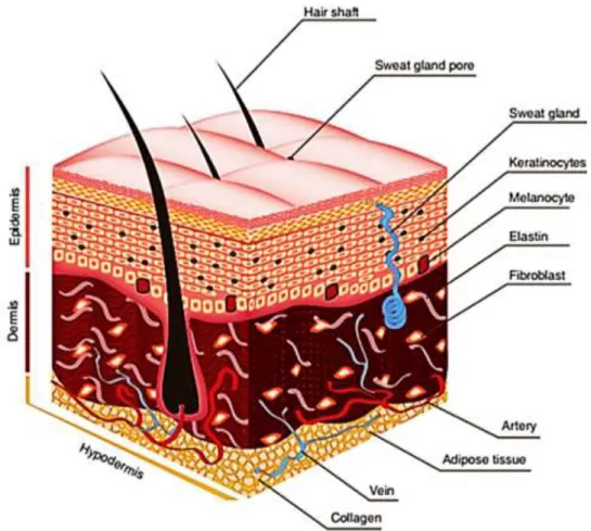
Skin is organized in three anatomical distinct layers known as Epidermis, Dermis and Hypodermis (Figure 1). Between Epidermis and Dermis is a Dermal–epidermal junction (DEJ) that provides mechanical support for the Epidermis and acts as a partial barrier against larger molecules (10).
Figure 1: Structure of the human skin. Adapted from (12).
In the three layers of skin there are skin appendages such as, nails, hair follicles, sweat and sebaceous glands, nerves, lymphatic and blood vessels (13, 14).
4
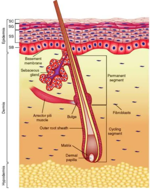
Epidermis is the outermost layer of the skin, acting as a physical barrier against the external environment, preventing the water loss and infections (13). This layer is mainly constituted by keratinocytes (80 % of cellular elements), pigment-producing cells (melanocytes) and specialized dendritic Langerhans cells that have an essential role in the skin immune defense system (15, 16).The Epidermis comprises 4-5 sublayers (17): stratum basale (SB), spinosum (SS), granulosum
(SG), lucidum (SL) and corneum (SC), as can be observed Figure 2.
Figure 2: Representation of skin layered organization. Skin is composed of three layers: Epidermis, Dermis and Hypodermis. Epidermis is a stratified squamous epithelium that is divided into four layers, starting with the outermost layer: stratum corneum (SC), stratum granulosum (SG), stratum spinosum (SS), and stratum basale (SB). Adapted from (18).
5
The SB is comprised mostly by keratinocytes that are in constant division. They are responsible for the continuous self-renewel of Epidermis. The SS also contains keratinocytes that are involved in the process of growth and early keratin synthesis (13, 14). The SG is characterized by the presence of intracellular granules that are involved in the process of keratinization (19-21). The SL represents 3–4 layers of dead and flat cells. This sub-layer is only found in the skin of palms and soles (13, 14). The final result of keratinocyte maturation is found in the SC (Figure 2), which is formed by completely differentiated dead keratinocytes (corneocytes). The resulting structure provides the physical barrier and prevent water loss of the human skin (13, 14, 22).The Epidermis is bound tightly to the underlying Dermis through the basement membrane at the DEJ. The basement membrane can be divided into lamina lucida (the layer closer to the Epidermis that is made of laminin, entactins and dystroglycans) and lamina densa (a sheet-like structure composed mainly by collagen type IV). The whole basement membrane is involved in the mechanical stabilization of the Epidermis (14, 23, 24). Moreover, the basement membrane determines the polarity of the Epidermis and provides a barrier against Epidermal migration, which prevents the direct contact of Epidermal cells with the Dermis (23, 24)
1.1.1.2 Dermis
Dermis lies below the Epidermis and constitutes the main part of the skin (13). Dermis is composed by a high number of fibroblast cells that produce collagen type I and III, elastin and glycosaminoglycans (GAGs). These proteins are the main constituents of the extracellular matrix (ECM) and are responsible for the support and elasticity of the skin (14). Additionally, Dermis confers support to the vascular and lymphatic vessels and nerve bundles (Figure 2) (17, 25).
Dermis is divided in papillary and reticular layers. The superficial papillary Dermis is composed by thin fibers that are loosely arranged and contains blood vessels that supply the Epidermis with nutrients, remove waste products and help in body temperature regulation. The deeper reticular Dermis accounts for 80 % of Dermis and is composed by dense collagen and elastic fiber matrix, conferring strength and flexibility to the skin (17, 24)
1.1.1.3 Hypodermis
The Hypodermis, located below the Dermis, is also known by subcutaneous tissue, is mainly composed by fat and connective tissue (15, 16). This layer is highly vascularized, providing blood vessels and nerves to the skin and also allowing its connection to the underlying bones and muscles. Furthermore, it also contribute for the thermoregulatory and mechanical properties of the skin (12, 16).
6
Skin has a variety of appendages, like, sweat and sebaceous glands, hairs folicules, nails and nerves. The sweat glands, secrete a watery fluid onto the skin surface, by the process of exocrine secretion. These glands play on important role in the thermoregulatory mechanism in humans (26). The main function of the sebaceous glands is to secrete sebum to moisturize the skin and hair and even the hair follicles, which are a source of proliferation of keratinocytes during epithelialization. Hair follicles also play an important role in the wound healing, once the Epidermal basal layer constitutes the outer cell layer of these structures and it has been shown that such basal cells, present in hair follicles, can move out and repopulate the Epidermis after healing. Nails confer protection to the distal phalanx and the fingertip (14). Moreover, skin contains a variety of nerve endings that sense heat and cold, touch, pressure, vibration and tissue injury. All cutaneous nerves have their cell bodies in the dorsal root ganglia, and both myelinated and non-myelinated fibers are found. Free sensory nerve endings lie in the Dermis where they detect pain, itch and temperature. Specialised corpuscular receptors also lie in the Dermis allowing sensations of touch (perceived by Meissner’s corpuscles) pressure and vibration (by Pacinian corpuscles).1.2 Skin wounds
Skin wounds affect millions of people worldwide, being one of the major issues of modern health care (5, 27). According to the Wound Healing Society (WHS), a skin wound can be described as a “disruption of normal anatomic structure and function” of skin, resulting from physical or thermal damage, medical procedures or physiological conditions (28).
Skin wounds can be classified in acute or cronic: Acute wounds:
Acute wounds are usually characterized by a complete healing, with a minimal scar formation. The primary causes of acute wounds include mechanical injuries due to external factors, such as abrasions and tears that are caused by frictional contact between the skin and hard surfaces. Other examples of mechanical injuries include penetrating wounds caused by knives, gun shots or surgical procedures. Burns and chemical injuries (caused by radiation, electricity, corrosive chemicals and thermal sources) are another category of acute wounds (28). Acute wounds heal through a normal, orderly, and timely reparative process that results in a sustained restoration of the anatomic and functional integrity of this organ (28, 29). Chronic wounds:
Chronic wounds are characterized by their slow healing (28). These type of wounds are commonly affected by several factors, such as the absence of clot formation (which reduce the levels of active/vital growth factors in the wound environment) and bacterial colonization that triggers a high immune response for removing the debris, leading to healing time (30, 31).
7
Chronic wounds usually occur in individuals who have underlying comorbidities, including peripheral blood vascular disease, obesity, diabetes, chronic steroid use, or other chronic diseases that impair tissue healing (30, 32). These wounds involve a large surface area and have a high incidence in general population, featuring an enormous medical and economic impact (33). Pressure, venous insufficiency, diabetic foot ulcers and ischemic wounds are the most prevalent types of chronic wounds (28, 33).Additionally, skin wound can also be classified accordingly for the with the depth of injury and the number of layers affected: superficial, superficial partial-thickness, deep partial-thickness and full-thickness wound (28, 34):
Superficial wound:
Results from sunburns, light scalds and abrasions. In this type of wounds only the Epidermis is affected and they are characterized by erythema and minor pain. Such injuries do not require specific surgical treatment, since it regenerates without scaring occurs (34). Superficial partial-thickness wound:
Affects Epidermis and superficial parts of the Dermis. Epidermal blistering and severe pain characterizes this type of injury, especially in the case of thermal trauma. The blood vessel, sweat glands and hair follicles are affected. The cells (keratinocytes) migrate towards each other from the basal layer to surround the wound. The healing occurs purely by epithelialization (28, 34).
Deep partial-thickness wound:
Injuries that involve great Dermal damage take long period to heal. Scaring is more pronounced for depth injuries, as well as the fibroplasia that is more intense, when compared with superficial partial-thickness wounds (5, 34, 35).
Full-thickness wound:
This type of wounds are characterized by a complete destruction of the skin appendages. The healing process involves contraction, and the epithelialization process occurs only from the edge of the wound. All full-thickness skin wounds are more than 1 cm in diameter and require skin grafting, as they cannot epithelialize on their own and may lead to extensive scarring, leading to limitations in joint mobility and severe cosmetic deformities for the patient (5, 34, 35).
1.2.1 Skin burns
Skin burns result from physical, chemical or thermal damages. The severity of the burn is determined by the patient condition (age and health) and it is usualy classified in degrees
8
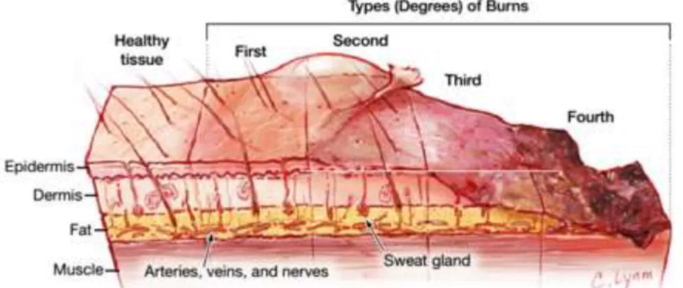
depending on depth, position and size of the burned area (36, 37). Burns are usually divided in four degrees (Figure 3) (38, 39):Figure 3: Representation of the different degrees (first, second, third and fourth) of skin burn severity. Adapted from (40).
First-degree (or superficial):
Burns affect only the top layer of the skin and are the least severe burns. It involves pain and edema formation. The skin usually takes several days to restore its structure, however the formation of scar does not occur. The superficial burns are healed in a period of 2 weeks (34, 40).
Second-degree (or partial-thickness):
Involves the Epidermis and part of the underlying Dermis destruction. Blisters are characteristic of in this type of burns (34, 40).
Third-degree (or full-thickness):
Affect all layers of skin, including the nerves. In these type of burns, skin is restored from the periphery involving the formation of granulation tissue and also scarring. Usually, in this type of burn the necrotic tissue resulting from the burn must be removed (34, 40). Fourth-degree:
Burns extend into the muscle below the skin, including fat tissue, tendons, muscles and bones (40).
1.3 Wound healing
After, a lesion, skin integrity and function must be restored. The main goals of healing is to achieve a rapid wound closure and a functional and aesthetic scar (41). Although, if skin regeneration does not occurs properly significant disability or even death may occur (2, 3).
9
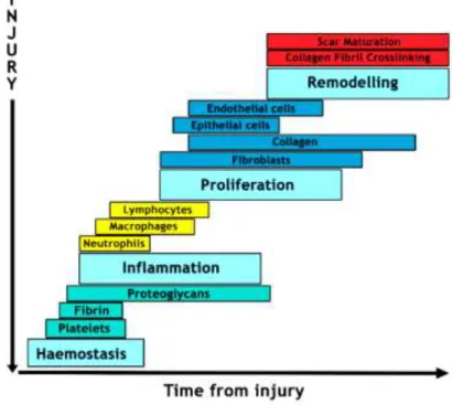
Skin wound healing is a complex process with an orchestrated cascade of events (e.g. coagulation, inflammation, phagocytosis, chemotaxis, mitogenesis, epithelialization and ECM proteins production) (4, 42), where different cellular elements (e.g. platelets, neutrophils, macrophages, keratinocytes and fibroblasts) and soluble factors (e.g. cytokines and growth factors) are involved.Skin healing process can be divided into four overlapping phases: (i) haemostasis, (ii) inflammation, (iii) cell migration and proliferation and (iv) remodeling (43) as displayed in Figure 4.
Figure 4: Representation of the phases of the wound healing. This process involves diferente types of cells and various phases. Phase I - Haemostasis which is characterized by coagulation and platelet activation; Phase II - Inflammatory phase where the cells of the immune system are recruited to injury site; Phase III - Proliferation phase occur the formation of ECM, granulation tissue and also angiogenesis; Phase IV - Remodeling, where the formation and maturation of the scar occurs.
1.3.1 Phases of wound healing
1.3.1.1 Haemostasis
After a tissue injury, the disruption of blood vessels is responsible for the extravasation of blood constituents (5, 35, 44). Bleeding typically occurs when the skin is injured and it allows the removal of bacteria and/or antigens from the wound. In addition, bleeding triggers platelet aggregation, fibrin clot formation and activates the coagulation cascade in order to prevent ongoing fluid losses. Haemostasis is achieved initially by the formation of a platelet plug,
10
followed by the formation of a fibrin matrix that allows cells infiltration. The clot dries to form a scab and provides strength and support to the injured tissue (Figure 5) (28, 45).Figure 5: Representation of first phase of wound healing process: Haemostasis. The red balls represent the platelets releasing several factors, including platelet derived growth factor (PDGF) and transforming growth factor β (TGF-β).
The cytoplasm of platelets contains α-granules filled with growth factors and cytokines, such as PDGF, TGF-β, epidermal growth factor (EGF) and insulin-like growth factors. They also contain dense bodies that store vasoactive amines, like serotonin, which increase the microvascular permeability (35). There is an invasion of inflammatory cells such as leukocytes, macrophages and neutrophils of the wound site. These cells and platelets release cytokines and growth factors in order to activate the inflammatory process (35).
1.3.1.2 Inflammation
The inflammatory phase begins almost simultaneously with haemostasis, sometimes from within a few minutes of injury to 24 hours, and lasts for about 3 days. This phase can be divided into two stages (early and late inflammatory phases) depending either on the time and duration of the response and the type of inflammatory cells involved (35).
11
Figure 6: Representation of early inflammatory phase. The red balls represent the platelets that release several factors, including PDGF and TGF-β, which attract PMNs to the wound, signalling the beginning of inflammation. The blue traces represents fibrin.
In the early inflammatory phase the activation of coagulation and complement system leads to the release of chemoattractants that recruit neutrophils into the wound site (Figure 6) (46). Then, the degranulation of platelets occurs. Moreover, once at the wound site, neutrophils perform their function of killing and phagocyte bacteria and damaged matrix proteins within the wound bed. The role that neutrophils play is crucial within the first days after injury, due to their ability to perform phagocytosis and also secrete proteases, that are involved in killing bacteria and also on degrade the necrotic tissue (42).
Figure 7: Representation of late inflammatory phase. The yellow clots represents the aggregate of macrophages with the PMNs, which are responsible for removing the debris from the wound, release growth factors and begin to reorganize the ECM. The red balls represent the platelets releasing several factors, including PDGF and TGF-β and traces of fibrin.
After 2-3 days in the late inflammatory phase, monocytes appear in the wound area and differentiate into macrophages (5). These macrophages are the most essential inflammatory cells involved in the normal healing response, as can be observed in Figure 7. Once activated
12
they perform phagocytosis of pathogens and of cell debris as well as the secretion of chemokines, inflammatory cytokines (interleukin-6 (IL-6), tumour necrosis factor (TNF-α) that stimulate the epithelialization) and growth factors such as: EGF (that stimulates the re-epithelialization), TGF-β, fibroblast growth factor (FGF), PDGF (which promote cell proliferation and the synthesis of ECM molecules by resident skin cells) and vascular endothelial growth factor (VEGF) (that stimulates the angiogenesis and granulation) (5). Macrophages act as phagocytic cells and secrete growth factors that are responsible for the proliferation of endothelial and smooth muscle cells and also for the production of ECM components by fibroblasts. They also involved in the release of enzymes that help to debride the wound (42). The presence of macrophages at the wound site is a marker that the inflammatory phase is finishing and the proliferative phase is beginning.1.3.1.3 Cell migration and proliferation
The migration phase is the final stage of visible wound healing process (Figure 8). This phase involves the migration of keratinocytes, fibroblasts and endothelial cells to the wound site in order to replace the damaged tissue (47). In this phase, the wound is filled with granulation tissue. The endothelial cells of the adjacent venules initiate the angiogenesis process. These cells also synthesize remodelling enzymes that perform the breakdown of the ECM and thus create defects into which new capillary vessels will form a network and restore the vasculature (5, 28, 35, 39, 44).
The proliferative phase starts three days after injury and lasts for about 2 weeks (35, 47). It is characterized by fibroblasts cells migration and by the deposition of newly synthesized ECM and formation of granulation tissue (35, 48). With progression of the proliferative phase, the provisional fibrin/fibronectin matrix is replaced by the newly formed granulation tissue (48). Epithelialization of the wound represents the final stage of the proliferative phase (35).
Figure 8: Representation of migration and proliferation phase. The proliferation phase begins when fibroblasts are recruited to the wound site through the release of growth factors by inflammatory cells. Then fibroblasts start the synthesis of collagen.
13
Migration of fibroblasts:Fibroblasts appear at the wound site after 2–4 days and endothelial cells come about one day later (5). Following injury, fibroblasts are attracted to the wound by a several growth factors, including PDGF and TGF- β. After that, fibroblasts proliferate and produce the matrix proteins: fibronectin, hyaluronan, collagen and proteoglycans. These components are involved in the production of a new ECM, which supports the migration and proliferation of cells (35, 47).
Production of the new ECM:
The ECM is composed by a network of structural proteins (collagens and elastin) and by an interstitial matrix composed by the adhesive glycoproteins (fibronectin, laminin and thrombospodin) embedded in a proteoglycan and GAGs (5, 49). In wound healing, PDGF, FGF, TGF-β, interleukin-1 (IL-1), TNF induce collagen synthesis during the proliferative and remodeling phases (35, 50).
Formation of the granulation tissue:
After 3–5 days, the development of the granulation tissue occurs, which is characterized by a high density of fibroblasts, granulocytes, macrophages, capillaries and loosely organized collagen bundles (5, 35). Angiogenesis and neovascularization are also processes that occur in to this phase. (5, 35, 42, 49).
Epithelialization:
Within a few hours often skin injury, a single layer of Epidermal cells migrate, from the wound edges, to form a covering over the damaged area. Along this process, a new basement membrane is produced and, thereafter, the growth and differentiation of epithelial cells allows the re-establishment of the stratified epithelium. At the end of this phase the myofibroblasts are responsible for wound contraction, bringing the edges together. The appearance of myofibroblasts corresponds to the initiation of connective-tissue compaction and the contraction of wound (5, 35).
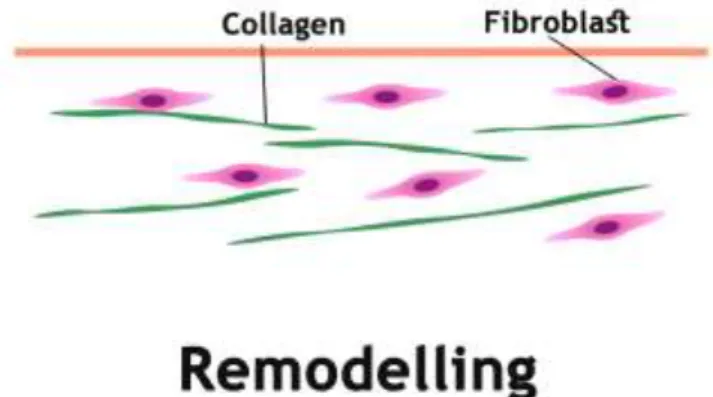
1.3.4 Remodelling (maturation)
Remodelling is the last phase (Figure 9) of the wound healing and occurs from day 21 to up to 1 year after injury. At this stage, the majority of endothelial cells, macrophages and myofibroblasts undergo apoptosis or exit from the wound, leaving a mass that contains few cells and consists mainly of collagen and other ECM proteins (5).
14
Figure 9: Representation of the remodeling phase. The green represents collagen and the purple fibroblasts.
This stage involves the formation of cellular connective tissue and strengthening of the new epithelium. There is a continuous synthesis and breakdown of collagen as well as the remodeling of ECM. Such determines the nature of the final scar (Enoch and Leaper, 2008). Most of the endothelial cells, macrophages and myofibroblasts undergo apoptosis or exit from the wound, leaving a mass that contains few cells and mostly collagen and other ECM proteins (42). Probably, the interactions between the epithelial mesenchymal cells will remain to support the skin integrity and homeostasis. In addition, over 6–12 months, the collagen type III that was produced in the proliferative phase is now replaced by collagen type I. This process is performed by matrix metalloproteinases secreted by fibroblasts, macrophages and endothelial cells (5). Finally, the angiogenic response decreases, the wound blood flow decreases and the acute wound metabolic activity slow down and stop. Subepidermal appendages such as hair follicles or sweat glands are not re-established after a serious injury (5, 42, 51).
1.3.2 Types of wound healing
In each healing process there are several mechanisms involved. The severity of the wound, number of skin layers affected and the occurrence or absence of bacterial infection allows us to classify the wound healing in different categories (5, 35):
Primary healing:
Occurs when a wound, created by laceration or surgical incision, causes only focal disruption of the continuity of the epithelial basement membrane and death of some cells of the underlying connective tissue. The wound is closed within 12-24 hours of its occurrence; In this type of healing, epithelial regeneration predominates over fibrosis (35, 44).
15
Occurs in a contaminated or poorly delineated wound. The closure is performed after the host defenses have helped to debride. After 3-4 days, phagocytic and inflammatory cells are recruited to the wound site to remove the contaminating bacteria. Collagen metabolism is usually unaffected and the wound retains its tensile strength (35, 44).Secondary healing:
Occurs when the wound edges cannot be approximated, due to the extensive loss of soft tissue, caused by a major trauma like severe burns and some surgical procedures. This type of wound healing is common in patients with underlying co-morbidities such as vascular, diabetic and pressure ulcers. The wound is left open and thus more susceptible to infections. The epithelial cells are not capable to restore the skin original architecture, so there is ingrowth of granulation tissue from the wound margins, followed by accumulation of ECM with the laying down of collagen. Myofibroblasts, which have structural properties similar to that of fibroblast and smooth muscle cells, are thought to play a crucial role in the healing of this type of injuries. The secondary healing is slower and may lead to functional defects (35, 44).
Superficial healing:
It is observed in injuries such as superficial burns, split-thickness donor graft sites, and abrasions where the injury involves the epithelium and the superficial (papillary) part of the dermis. The basal layer of cells remains uninjured and the epithelial cells within the Dermal appendages, hair follicles, and sebaceous glands replicate to cover the exposed Dermis; the cells migrate towards each other from the basal layer to surround the wound. Healing occurs purely by epithelialization (35).
1.4 Tissue engineering
TE is a field that applies the principles of biology, engineering and medicine in order to develop the biological substitutes that restore, maintain or improve damaged tissues or organs functions (2). It appeared as a solution for a number of clinical problems that were not properly treated with the use of permanent replacement devices (8).
The underlying concept of TE is to isolate cells from a patient and then produced to their expansion and incorporation in a 3D matrix. The resulting TE construct is then grafted back into the same patient to function as a replacement tissue. In this approach, a highly porous artificial ECM, or scaffold, is required to accommodate mammalian cells and guide their growth in three dimensions.
Major advances in materials science and engineering have contributed for the continuous development of TE and regenerative medicine (7, 52, 53). Nowadays, the tissue engennering is a discipline already applied in a significant number of medical procedures for skin (54), liver (55), pancreas (56), intestines (57), esophagus (58), nerves (59), cartilage (60), bone (61), and tendon (62) replacement.
16
1.4.1 Tissue engineering applied to wound healing
Autografts, allografts and xenografts are the most used therapeutic approachs for skin regeneration. Autografts are obtained from the patient and present a higher healing success rate. However, they have a limited supply and its obtention is associated to morbidity in the donor site (63, 64). Allograft skin is harvested from organisms of the same specie. The use of allograft skin is limited since there is a great risk of disease transmission, eventual immune rejection and other limitatiors associated with its storage (65). The demand for tissues and organs seriously exceeds the supply, creating a substantial waiting list. Moreover, the immune system tends to reject the foreign tissue or organ (14). Xenograft skin is harvested from a different species and the majority of xenograft tissues are rejected by the immune response of the host, that may be caused upon the implantation process, thus leading to a high failure rate (66-68).
In order to overcome the drawbacks associated with the use autografts, allografts and xenografts, different studies have been performed in the area of TE to developed new skin substitutes that can contribute to reduce the mortality and morbidity caused by scarring, changes in pigmentation, reduce the number of surgical procedures and hospitalization period (69).
In the development of materials aimed to produce new skin substitutes it is necessary to take into account three major requirements: the safety of the patient, the clinical efficacy and the convenience of handling and application. Nowadays, skin substitutes are highly porous and some of them can accommodate skin cells and guide their growth in three dimensions (70).
1.4.1.1 Comercial available skin substitutes
Different skin substitutes are already applied in the clinic. Skin substitutes are a heterogeneous group of wound coverage materials that aid in wound closure and help in the reestablishment of the functions of the skin (28, 71, 72). Most of the bioengineered skin devices currently available consist on a combination of sheets of biomaterial matrix (e.g. collagen, hyaluronic acid) containing cultured cells (73-75). Several types of temporary dressings have been designed to provide a bacterial barrier to decrease pain and contribute to an adequate environment for epithelial regeneration.
Wound dressings have been widely used due to their relative low cost, ease use, and effectiveness to clean and protect the wound from the external environment. They act as physical barriers that protect the wound from microorganism invasion and promote moisture environment and allow gases exchanges.
The commercial bioengineered skin equivalent products are classified accordingly to the following parameters (2, 74-76):
17
A. Type of the biomaterial used for their production:a. Biological – e.g. Epicel; b. Synthetic – e.g. MySkin.
B. Composition regarding the cellular components: a. Cellular – e.g. Dermagraft;
b. Acellular – e.g. Integra Dermal regeneration. C. Duration:
a. Temporary – e.g. Dermagraft;
b. Semi-permanent – e.g. Integra Dermal regeneration; c. Permanent – e.g. Epicel.
D. Layer of skin that skin substitutes are able to replace:
Epidermal substitutes - keratinocytes are isolated from a donor and then are cultured
in vitro in order to obtain the necessary number of keratinocytes for therapeutic
purposes. Several Epidermal skin substitutes are already commercially available: a) Epicel ™: is a permanent substitute, which is composed by the in vitro culture
of autologous keratinocytes (confluent cellular sheets) (34, 77).
b) EpiDex ™: in vitro cultured autologous keratinocytes collected from hair bulbs (confluent cellular sheets). It is a permanent substitute (17, 77).
c) MySkin ™: in vitro cultured autologous keratinocytes (subconfluent cellular sheets)which are grown on a silicone support layer with a specially formulated surface coating (34, 77).
Dermal substitutes - Dermal substitutes are usually acellular, based on allogeneic, xenogeneic or synthetic materials (78). Dermal skin replacements present advantages, such as reduced costs, easier manufacture and rigorous quality control. They also add mechanical stability and prevent the wound from contracting (2, 34). However, they can be rejected by the host and be involved in diseases transmission (15). The Dermal substitutes available in include:
a) Dermagraft ™: is composed of polyglactin mesh seeded with living cultured neonatal fibroblasts. It is a temporary substitute (79).
18
b) Alloderm is a freeze-dried human acellular dermal matrix. This type of matrix is ready to be incorporated into the wound, and it does not any immunogenic response from the host due to absence of a cellular component (80);c) Integra®: is composed by two layers: a porous layer of the skin made of bovine collagen type I and shark chondroitin-6-sulphate GAG that is bonded to a silicone pseudo-epidermis Integra®. It is indicated for the treatment of full thickness or deep partial thickness burns (17, 81).
Dermo-epidermal substitutes - These substitutes mimic the Epidermal and Dermal layers. These substitutes are more advanced than the Epidermal and Dermal ones, although they are the most expensive (82).
a) Apligraf ™: is composed by viable allogeneic neonatal fibroblasts grown in a bovine collagen type I gel matrix, combined with viable allogeneic neonatal keratinocytes. It supplies ECM components to the wound, as well as cytokines and growth factors (77, 81).
b) PermaDerm ™: is composed by an Allogenic matrix with bovine collagen. It is a permanent substitute (77).
c) OrCell ™: is a TE skin construct that includes cultured allogenic fibroblasts and keratinocytes from the same neonatal foreskin. Fibroblasts are seeded into a bovine type I collagen sponge (77).
1.5 Polymeric sponges for skin regeneration
Despite the existence of various skin substitutes, none of them is capable of completely replicate the anatomy, physiology, biological stability or aesthetic nature of native skin (15). In addition, they are expensive, require frequent replacement, making the patient susceptible to subsequent secondary bacterial infections. Having this knowledge in mind, there is a huge demand for developing alternative strategies for treating burns or other skin lesions. Researchers from the area of TE have develop new Dermis and Epidermis substitutes using natural or synthetic matrices (83), in order to promote a more rapid and improved healing as well as a reduced scarring (2).
Based on the properties that skin substitutes must have, porous scaffolds emerged as a promising alternative to be used as skin substitutes.
Sponges are three-dimensional (3D) matrices that act as temporary templates for cell adhesion and proliferation, while providing mechanical support, until the new skin tissue is formed at
19
the affected area. Polymeric sponges are potential scaffolds for skin regeneration since they satisfy several requirements (84, 85):a) Protect the wound from fluid and proteins loss; b) Easy to handle and apply at the wound site; c) Present controlled degradation;
d) Enable exudates absorption; e) Minimize scar formation;
f) Large surface area that enables cell adhesion, growth and differenciation;
g) Great porosity that allows cell infiltration, diffusion of nutirents and gases exchange; h) A surface can be easily modified (e.g. with the use of coatings);
i) Can be produced using various techniques;
j) Biocompatible: ability of a biomaterial to perform its desired function, without eliciting any undesirable local or systemic effects in the recipient or beneficiary of that therapy (10);
k) Biodegradable: The by-products of their degradation must be non-toxic and able to exit from the body without interference with other organs. In order to allow degradation at a rate compatible with tissue formation, an inflammatory response combined with controlled infusion of cells such as macrophages is required;
l) Active biomoluces (e.g. growth factors, cell-surface interactive peptides, drugs) can be easily incorporated to the sponge matrix that will facilitate skin regeneration, to stimulate cellular attachment, migration and proliferation.
Based on these properties the development of sponges for skin regeneration may have a huge potential for skin healing.
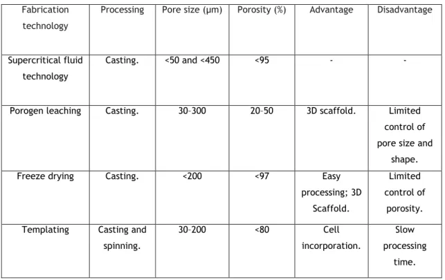
1.5.1 Methods and techniques used for sponge production
Differents methods have been used for the development of sponges for TE, including the supercritical fluid technology, porogen leaching, freeze drying, scaffold templating (see Table 1, for further details).
Supercritical fluid technology:
In this process, high pressures are used to dissolve the polymeric solution with or without a porogen. When a supercritical fluid such as carbon dioxide is used as a nonsolvent, the simple tuning of the processing conditions (pressure and temperature) can tailor the final structure of the sponges. Also, any subsequent drying step is avoided, as the obtained porous structure is a dry product free of any residual solvent (86, 87).
20
Porogen leaching allows the control of pore size and porosity of sponges, allowing the obtention of scaffolds with a more homogeneous pore morphology (86). Porous structures from polymers such as PCL has been produced using this method (88).Freeze drying:
The method is based on the formation of ice crystals that induce porosity through ice sublimation and desorption. The kinetics of the freezing stage controls the porosity and the interconnectivity of the foams (89). 3D structures with values of porosity up to 200 % (86) with different interconnectivities are commonly obtained by freeze-drying. The main difficulty associated with this process is to ensure structural stability and adequate mechanical properties of the porous constructs after subsequent hydration. This limitation hinders its use when the application involves conditions with mechanical stress, even at low-to-moderate levels. Scaffold templating techniques:
Polymeric solutions may be injected into moulds to fabricate scaffolds with various shapes and sizes. Also, the mould template can be design to fabricate a macroporous scaffold (86).
21
Table 1: Technologies used for the production of 3D constructs. Adapted from (86).
1.5.2 Coating of sponges
Electrospinning has been recognized as the simplest technique to produce continuous nanofibers from diverse materials, including polymers (90).
Electrospinning is a process that comprise the application of a needle attached to a syringe filled with polymer solution, a grounded collector plate and a high voltage power supply connected between the capillary and the collector. The feeding rate of the polymer solution is usually controlled using a syringe pump. A charged polymer solution flowing out of the needle is accelerated towards the grounded collector by a strong electrostatic field (91, 92). This field causes the droplet to emerge from the needle to undergo deformation into a conical shape, known as the “Taylor cone”. When a critical value is attained (the repulsive electrostatic force overcomes the surface tension) a fine jet of the solution emerges from the Taylor cone. The jet undergoes twisting instability and a characteristic whipping motion due to the charge-charge repulsion that occurs between the excess charge-charges presented in the jet (Figure 10), and during this phase, the jet is drawn by at least two orders of magnitude, the solvent evaporates, and the dry fibers deposit onto the collector (91, 92).
The properties of the nanofiber mesh depending on fiber diameter, porosity characteristics of the solution and electrospinning equipment processing parameters. The smaller size of the individual fibers, the higher the surface area to volume ratios, which leads to an increase cell proliferation (93). The size, shape, individual fibers, the porosity of the web of fibers obtained and chemical compositions can be easily manipulated (94).
Fabrication technology
Processing Pore size (µm) Porosity (%) Advantage Disadvantage
Supercritical fluid technology
Casting. <50 and <450 <95 - -
Porogen leaching Casting. 30–300 20–50 3D scaffold. Limited control of pore size and
shape. Freeze drying Casting. <200 <97 Easy
processing; 3D Scaffold.
Limited control of
porosity. Templating Casting and
spinning. 30–200 <80 Cell incorporation. Slow processing time.
22
Figure 10: Schematic diagram of the electrospinning setup. Adapted from (25).
In biomedical applications the ultrafine fibrous scaffolds produced by electrospinning have been demonstrated to have suitable properties to promote the adhesion, proliferation and differentiation of several types of cells (94).
Moreover, electrospun fibers can be used to coat scaffolds aimed for tissue regeneration, namely skin regeneration. Several electrospun nanofibrous membranes a contribution of natural/synthetic materials have been already tested for skin regeneration, produced with natural materials or polymers. Electrospun nanofibers reproduce the native topographical features of the natural ECM, promoting the cell’s natural functions (95, 96).
Most of the work performed in this field uses biodegradable synthetic polymers (such as PCL) to produce non-woven membranes for various TE or drug delivery applications (97).
Currently, a variety of natural polymeric-based membranes obtained from Chitin (98), Chitosan (99, 100), Alginate (100), Cellulose (101, 102), Hyaluronic acid (103), Gelatin (104, 105), Collagen (106) and their derivatives have been developed in order to satisfy the hight demand for new materials for the treatment of different wounds. These types of membranes may be composed of dense top layer and underlying porous sponge-like layer. The external layer protects the wound and serves as an artificial Epidermis, while the inner layer is designed for the drainage of wound exudates and attachment of wound tissues (107, 108).
1.5.3 Biomaterials used for sponges production
The first issue with regard to the development of a scaffold for skin TE is the choice of suitable material. Natural polymers can mimic many features of ECM and thus can guide the migration, growth and organization of cells during the wound healing process (109, 110).
These natural polymers include polysaccharides, like Chitosan or proteins-based polymers (Collagen, Fibrin gels, Silk, and Gelatin). Despite their low mechanical strength, these natural
23
polymers have high hydrophilicity, low immune reaction and promote cell adhesion and proliferation (111).1.5.3.1 Chitosan
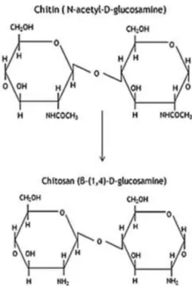
Chitosan is a cationic polysaccharide composed of copolymers of β (1→4)-glucosamine and N-acetyl-D-glucosamine (Figure 11). It presents important characteristics for biomedical applications, such as, biocompatibility, biodegradability, hydrophilicity, hemostatic activity, nonantigenicity, anti-microbial activity and promote wound healing (108, 112). In addition, Chitosan is very abundant, has a low production cost and is environmental friendly.
Chitosan has been applied in the area of TE for a wide variety of applications, like skin regeneration. It induces a faster wound healing and produce smoother scarring possibly due to an enhanced vascularization (113).
Another important property of Chitosan is its antibacterial activity for different strains, such as Enterobacter aerogenes, Salmonellas Typhimurium, Staphylococcus aureus and Escherichia
coli (84, 114, 115). Due to this bactericidal activity Chitosan has been blended with other
polymers (71, 116). Its antimicrobial activity may result from the electrostatic interactions between the positively charged Chitosan with negatively charged molecules at the cell surface, which affects cell permeability (71).This electrostatic attraction promotes cells’ adhesion, proliferation and differentiation (116, 117).
Different studies reported the use of Chitosan for the production of skin substitutes, due to its properties that stimulate haemostasis and fibroblast to synthetize collagen, improving the tissue regeneration (118, 119). In vivo, this polymer stimulates the adhesion of fibroblasts, promoting keratinocytes proliferation and modulate the migration of neutrophils and macrophages, which in turn, modifies the repairing processes such as fibroplasias and reepithelialisation (66, 71).
24
Figure 11: Chemical structure of Chitosan. Adapted from (120, 121).
The deacetylation degree (DD) of commercial available Chitosan is usually between 70 % and 95 %. The different DD are defined in terms of the percentage of primary amino groups in the polymeric matrix (122). Chitosan with higher DD presents a greater number of free amino groups (71). Chitosan is degraded in vivo, through enzymatic hydrolysis. Lysozyme is the primary enzyme responsible for the in vivo degradation of Chitosan.
1.5.3.2 Gelatin
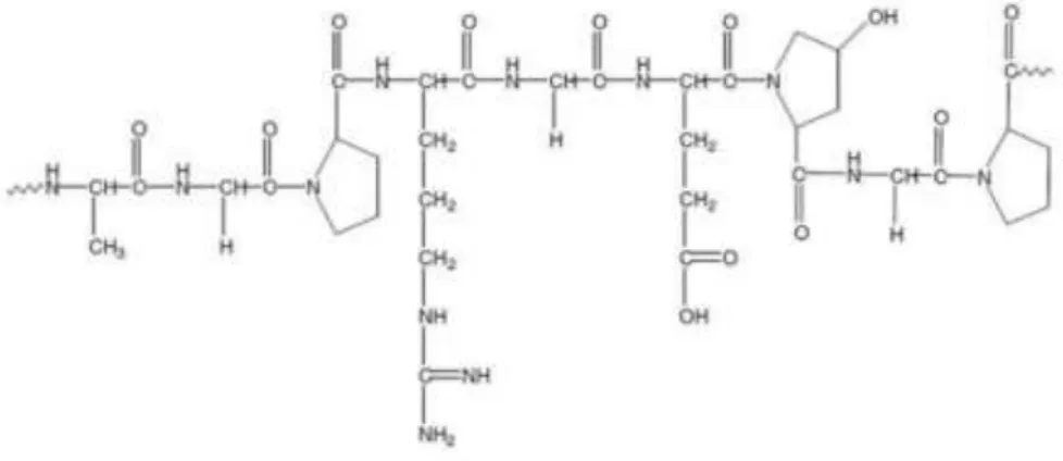
Gelatin is produced by partial hydrolysis of collagen extracted from the boiled bones, connective tissues, organs and some intestines of animals. Gelatin is colorless, brittle (when dry) and a flavorless solid substance. It is commonly used as a gelling agent in food and pharmaceuticals. Gelatin is biodegradable, biocompatible and has low antigenicity (122). Although, it obtained from collagen, it still retains some its properties, such as tripeptide Arg-Gly-Asp (RGD) sequence, that promote cell adhesion, differentiation and proliferation (123). Furthermor it also has a low cost and low immunogenicity. It is soluble at physiological pH and at 40 °C. The large number of amino, carboxyl and hydroxyl groups, allows Gelatin chemical modification, increasing its versatility (Figure 12). Gelatin has been investigated for the production of matrices for skin regeneration (sponge or film) that can promote the epithelialization and granulation tissue formation (124).
25
Figure 12: Representation of Gelatin structure. Adapted from (125).
1.5.3.3 Poly (ethylene oxide)
PEO is a unique class of water-soluble biodegradable biopolymer (Figure 13). Due to its excellent biocompatibility, biodegradability and potential to be used in biomedical applications has attracted a great attention from both the industrial and scientific areas (113, 126). PEO is also used to reduce the viscosity of Chitosan solution, so that the solution is extruded at high polymer concentrations.
Figure 13: Structure chemical of PEO.
1.5.3.4 Poly (ε-caprolactone)
PCL (Figure 14) is a polyester that exhibits good mechanical properties. It a semi-crystalline material. However, due to its hydrophobic character, contains very few cell recognition sites and has a slow degradation rate. This polymer is used for various biomedical applications such as sutures, drug delivery systems and scaffolds in TE, due to its soft- and hard-tissue compatible properties (72).
26
Figure 14: Structure chemical of PCL.
1.5.4. Incorporation of anti-inflammatory drugs in sponges for skin
regeneration
Nonsteroidal anti-inflammatory drugs (NSAIDs) are the most commonly used drugs to treat inflammatory diseases, since they are effective in the management of pain, fever, redness, edema that occur as a consequence of inflammatory mediator release (127, 128). Different studies have shown that both therapeutic and side effects of NSAIDs are dependent of cyclooxygenase (COX) inhibition. COX isoforms have been named constitutive cyclooxygenase-1 (COX-cyclooxygenase-1) and inducible cyclooxygenase-2 (COX-2). COX-cyclooxygenase-1 (such as indomethacin, naproxen, Ibuprofen) catalyzes the formation of cytoprotective prostaglandins in thrombocytes, vascular endothelium, stomach mucosa, kidneys, pancreas, langerhans islets, seminal vesicles and brain (128, 129). Induction of COX-2 by various growth factors, proinflammatory agents, endotoxins, mitogens and tumor agentes (130, 131) indicates that this isoform may have a role in occurence of pathological processes, such as inflammation (132, 133). As a result of studies focused on reduction of the adverse effects of NSAIDs, selective COX-2 inhibitors, such as celecoxib and rofecoxib, have been developed. Today, it is a well-known hypothesis in medicine that COX-1 is constitutive and cytoprotective, while COX-2 is an inducible enzyme in the inflamed tissues (Figure 15).
27
Figure 15: Mechanism of action of the COX-1 and COX-2 in the human body.
NSAID in general in particular Ibuprofen, have been shown to have benefic effects on various acute conditions ranging from sepsis, traumatic induced pulmonary damage and wound healing.
1.5.4.1 Action of Ibuprofen in the wound healing process
Ibuprofen has been shown to have benefic effects on acute episodes of a wide range of tissues. Ibuprofen was the first phenylpropionate to be marketed in the United States. It has analgesic, antipyretic and anti-inflammatory activity and it is well absorbed and well tolerated (134, 135). Ibuprofen has also been shown to improve several aspects of the wound healing ranging from wound edema. As previously described in literature the second degree burn wounds treated with Ibuprofen 5 % from two and five hours after burn showed a significantly reduced lymph drainage and no variation in the wound water content (136).
28
1.6 Main goals of the present study
In this study new skin substitutes were aimed to be produced. The objectives of the workplan comprised:
1) Production of Chitosan-Gelatin sponge using freeze-dried method;
2) Coating of sponges with nanofibers produced with Chitosan deacetylation, PEO, PCL and Ibuprofen);
3) Morphological and physicochemical characterization of the bilayer of the S; 4) Evaluation of biocompatibility of the developed system;