INTRODUCTION
Cyanide is a well known chemical for its properties as suici-dal, homicidal and chemical warfare agent (Hariharakrish-nan et al. 2010). Industries dealing with metal plating and finishing, mining and extraction of metals such as gold and silver, production of synthetic fibers and the processing of coal generate large quantities of cyanide-containing wastes (Rocha-e-Silva et al. 2010). Consequently release of cyani-de containing wastesinto the environment has generated considerable interest. Cyanide is found to be highly toxic to the aquatic organisms, primarily due to the formation of complexes with metal ions that are present as enzyme cofactors. Most notably this occurs with Fe3+ ion in cyto-chrome, thereby inhibiting respiration and hence, oxidative
Effect of exposure to sublethal concentrations of sodium cyanide
on the carbohydrate metabolism of the Indian Major Carp
Labeo
rohita
(Hamilton, 1822)
1Praveen N. Dube2, Shwetha Alavandi2 and Basaling B. Hosetti2*
ABSTRACT.- Dube P.N., Alavandi S. & Hosetti B.B. 2013. Effect of exposure to sublethal con-centrations of sodium cyanide on the carbohydrate metabolism of the Indian Major Carp Labeo rohita (Hamilton, 1822). Pesquisa Veterinária Brasileira 33(7):914-919. Toxico
-logy Laboratory, Department of Applied Zoo-logy, School of Biological Sciences, Jnana Sahya -dri, Shankaraghatta, Shimoga, Karnataka 577 451, India. E-mail: [email protected]
Experiments were designed to study in-vivo effects of sodium cyanide on biochemical
endpoints in the freshwater fish Labeo rohita. Fish were exposed to two sublethal concen -trations (0.106 and 0.064mg/L) for a period of 15 days. Levels of glycogen, pyruvate, lac-tate and the enzymatic activities of laclac-tate dehydrogenase (LDH), succinate dehydrogenase (SDH), glucose-6-phosphate dehydrogenase (G6PDH), phosphorylase, alkaline phosphata-se (ALP), acid phosphataphosphata-se (AcP) were asphosphata-sesphosphata-sed in different tissues (liver, muscle and gills). Result indicated a steady decrease in glycogen, pyruvate, SDH, ALP and AcP activity with a concomitant increase in the lactate, phosphorylase, LDH and G6PD activity in all selected tissues. The alterations in all the above biochemical parameters were significantly (p<0.05) time and dose dependent. In all the above parameters, liver pointing out the intensity of cyanide intoxication compare to muscle and gills. Study revealed change in the metabolic energy by means of altered metabolic profile of the fish. Further, these observations indi -cated that even sublethal concentrations of sodium cyanide might not be fully devoid of deleterious influence on metabolism in L. rohita.
INDEX TERMS: Labeo rohita, metabolism, oxidative enzymes, sodium cyanide, subacute.
1 Received on March 1, 2012.
Accepted for publication on September 10, 2012.
2 Toxicology Laboratory, Department of Applied Zoology, School of Bio
-logical Sciences, Jnana Sahyadri, Shankaraghatta, Shimoga, Karnataka 577
451, India. *Corresponding author: [email protected]
phosphorylation (Daya et al. 2000). Lower concentrations in the range of 50-100µg/l of cyanide have proven even-tually fatal to many sensitive fish species (Dube & Hosetti 2010). Even at concentrations as low as 20 µg/l, chronic or sublethal effects of cyanide have been found in several freshwater species (Heath 1991).
they are present in most tissues. The increase or decrease in their level may be sufficient to provide information of diagnostic values (Begum 2009).
Sodium cyanide is extremely toxic, which might hamper the fish health through the impairment in the metabolism, sometimes leading to death. This is because, most of the cyanide absorbed is detoxified by enzymatic combination with sulfur, and thus the detoxification process imposes a nutritional cost (Prashanth & Neelagund 2007). Concentra -tions of cyanides and lethal and sublethal toxicities of cya -nides to the fishes are well documented (Dube & Hosetti 2010). Further it is also essential to evaluate the sublethal effects of cyanides on the fishes, since they form very im -portant members of the food chain. Consequently it was considered as a matter of warranted interest to elucidate biochemical effects in the fish. Evaluation of biochemical activities in an organism helps to identify disturbances in its metabolism. In this study, we monitored the distur -bances of metabolism in Labeo rohita by exposing them to
different sublethal concentrations of sodium cyanide for a period of 15 days. Labeo rohita constitutes one of the main fish species of economic importance, as an abundant cul -tural fish species in India and as very popular fish among farmers and consumers.
MATERIALS AND METHODS
Studies were conducted on fingerlings of Labeo rohita, obtai
-ned from State Fisheries Department, Bhadra Reservoir Project (BRP), Shimoga, Karnataka, India. Fish weighing 3.5±0.5g with mean body length 6±0.75cm were maintained in large holding tanks following standard fish maintenance procedure during acclimatization and exposure (APHA 2005). Acclimatized fishes were transferred to aquaria (20 L) for subacute studies. Aquarium water was kept oxygen saturated by aeration and its temperatu
-re was maintained at ambient laboratory temperatu-re (25±1°C).
Animals were transferred to a fresh volume of water for every 24
h to minimize contamination from metabolic wastes and the fi
-shes were fed with fish food pellets (Nova Aquatic Pvt Ltd, not less than 3% of body weight). Prior to exposure the fish were exami -ned carefully for any pathological symptoms.
Stock solution of the sodium cyanide (99.9%, Loba Chemicals Mumbai, Maharashtra, India) was prepared in double distilled water. After acclimatization, fish were transferred to aquarium
(20 L capacity) supplied with dechlorinated tap water (total
hardness 23.4±3.4mg as CaCO3/L, phosphate 0.39±0.002μg/L, salinity 0.01ppt, specific gravity 1.001 and conductivity less than 10μS/cm). Experiments were carried out in static renewal system
(APHA 2005). Median lethal concentration (96 h LC50) of sodium cyanide to L. rohita was found out to be 0.32 mg/L (Dube and Ho
-setti 2010). Two sublethal concentrations selected for the present study were based on the 1/3rd and 1/5th of the 96 h LC
50. Accli-matized fish were distributed into seven equal groups with ten fish each. Fish in first three groups were exposed to 0.106mg/L and the next three groups to 0.064mg/L of sodium cyanide, whi -le remaining group was kept in cyanide free tap water, served as
control. Five replicas were maintained for each concentration. The test media was changed daily with fresh addition of toxicant.
At the end of 5, 10 and 15 days of exposure, fish were sacri
-ficed and liver, gill, muscle tissues were collected immediately, washed with distilled water and weighed before homogenization. Tissues were homogenized using a glass homogenizer with res
-pective homogenizer solution and centrifuged. Glycogen content
in the tissue was estimated by using the anthrone reagent as des
-cribed by Caroll et al (1956). The homogenate (5%) was prepared in 5% trichloroacetic acid (TCA). Values were expressed as mg
glycogen/g of tissue. Pyruvate level was measured according to
Friedman & Haugen (1943). Homogenate (50 mg/ml) was prepa -red in 10% TCA. Sodium pyruvate was taken as standard and the
values were expressed as mg pyruvate/g tissue. Lactate content
in the tissues was estimated according to the method of
Hucka-bee (1961). Homogenate (50mg/ml,) was prepared in 10% cold
TCA and centrifuged at 1000 g for 5 minutes. Sodium lactate was
taken as standard. Lactate content was expressed as mg lactate/g
wet weight tissue. LDH (EC 1.1.1.27) was assayed following
Go-vindappa & Swami (1965) and SDH (EC 1.3.99.1) by the method
of Nachilas et al (1960). The formazon extracted was measured at
495nm and the activity of enzyme was represented in µmol form
-azon/mg of tissue. For the estimation of G6PDH (EC 1.1.49)
activi-ty method described by Bergmeyer & Bernt (1965) and Glycogen phosphorylase activity (EC 2.4.1.1.),using; the method described
by Sutherland (1955) was followed and the activity expressed as µM Pi liberated/mg protein/h, using phosphate standards. Acid Phosphatase (AcP; E.C. 3.1.3.2) in the tissues were estimated by using method of Kind & King (1954) and the inorganic phosphate (Pi) liberated was measured by the method of Fiske & Subbarow (1925). The enzyme assays were made after preliminary standar
-dization regarding linearly with respect to time of incubation of enzyme concentration. Enzyme activity was expressed as µmol Pi liberated/mg protein/h.
Each assay was replicated five times, and values thus obtai -ned were converted to percentage of the respective control and
analysis of variance (ANOVA) was employed followed by Duncan
multiple range test as post hoc as test to calculate the significant
difference. The analysis was made between the means of the con
-trol and cyanide treated fishes. In all the cases, differences were considered statistically significant at p<0.05 (Daniel 1987).
RESULTS AND DISCUSSION
No mortality was observed during in the fish exposed to both sublethal concentrations of sodium cyanide. The effect of cyanide in the liver, muscle and gills of Labeo rohita are shown in Table 1 to 4. The biochemical changes observed in the present study were due to disturbed metabolism in the fish. Cyanide has its own target sites of action and most of them are metabolic depressors. They generally affect the activity of biologically active molecules (Prashanth & Nee -lagund 2007).
Effect on glycogen
(Rees et al. 2009). The decrease was greater in liver than in muscle and gills, since liver is the principal organ for glyco-gen storage. Depletion of glycoglyco-gen may also indicate its di-rect utilization for energy generation, a demand caused by cyanide-induced hypoxia (Dube & Hosetti 2010).
Effect on pyruvate
Table 1 show pyruvate levels in all the three tissues. Maximum decrease was recorded in the liver and lowest in the muscle. The percent decrease in the pyruvate con-tent in different tissues was in the order of liver (11.91 to 40.76%) > muscle (15.83 to 36.58%) > gill (9.55 to 28.98%) in 0.106mg/L of sodium cyanide and liver (10.07 to 29.92%) > muscle (7.34 to 29.03%) > gill (8.85 to 22.33%) in 0.064mg/L of sodium cyanide. Decrease in the level of pyruvate suggests the possibility of a shift towards anaerobic dependence due to a remarkable drop in aerobic segment, a characteristic feature of cyanide intoxication (Singh et al. 1989). This might also be due to its conversion to lactate, or due to its mobilization to form amino acids, lipids, triglycerides and glycogen synthesis in addition to its role as a detoxification (De Zwaan et al. 1993).
Effect on lactate
Lactate concentrations reached highest values after subacute exposure compared to control (Table 1). Signi -ficantly elevated lactate concentrations were observed in
the fishes exposed to cyanide. Values ranged from muscle (10.57 to 48.96%) > gills (22.45 to 42.69%) >l iver (26.21 to 39.39%) in 0.106mg/L and in 0.064mg/L sodium cya-nide, muscle (8.54 to 42.03%) > liver (15.95 to 34.59%) > gills (16.21 to 30.13%). The level of lactic acid in the tissue known to act as an index of anaerobiosis (Rees et al. 2009), which might be beneficial to the animal to tolerate hypo -xic conditions (Baud et al. 2002). Observed changes in the lactate level, which was dose depended, may be considered as a symptom of stress induced by the cyanide. Similar ob -servations were noticed in the rats following exposure to cyanide and carbon monoxide (Dodds et al.1992).
Effect on LDH and SDH
In contrast to control, significant inhibition in the acti -vity of SDH and enhancement of LDH levels were noticed at both the concentrations (Table 2). The activity levels sco -red high in liver than the muscles and gills. The LDH activi-ty was in the range of liver (23.27 to 44.16%) > gill (13.79 to 43.34%) > muscle (15.19 to 40.13%) at 0.106mg/L and in 0.064mg/L of sodium cyanide, gill (9.09 to 36.58%) > muscle (12.55 to 33.22%) > liver (14.71 to 33.06%). Si-milarly, SDH activity ranged in liver (16. 12 to 40.32%) > muscle (9.83 to 39.88%) > gill (16.45 to 39.62%) in 0.106 mg/L and in 0.064 mg/L of sodium cyanide, gill (10.65 to 30.70%) > liver (13.09 to 30.08%) > muscle (7.11 to 29.57%) respectively. Any change in the intermediary
me-Table 1. Effect of sublethal concentrations of sodium cyanide on glycogen, pyruvate and lactate levels in different tissues of Labeo rohita
Control 0.106 mg/L 0.064 mg/L
5 10 15 5 10 15
Glycogen(mg / g wet wt)
Liver 7.20 ± 0.47a 5.79 ± 0.34c 5.11 ± 0.22e 4.53 ± 0.65g 6.26 ± 0.11b 5.50 ± 0.32d 4.90 ± 0.18f
Muscle 1.84 ± 0.41a 1.36 ± 0.23e 1.48 ± 0.16c 1.20 ± 0.26g 1.59 ± 0.23b 1.37 ± 0.74d 1.27 ± 0.52f
Gill 1.73 ± 0.12a 1.53 ± 0.37c 1.34 ± 0.12e 1.18 ± 0.32g 1.57 ± 0.54b 1.42 ± 0.18d 1.30 ± 0.65f
Pyruvate(mg / g wet wt)
Liver 12.73 ± 0.43a 11.22 ± 0.32c 10.11 ± 0.67e 7.54 ± 0.54g 10.39 ± 0.44d 11.45 ± 0.68b 8.92 ± 0.84f
Muscle 9.66 ± 0.32a 8.13 ± 0.65c 7.45 ± 0.34e 6.13 ± 0.22g 8.95 ± 0.54b 6.85 ± 0.87f 7.90 ± 0.22d
Gill 10.24 ± 0.78a 8.65 ± 0.44e 9.26 ± 0.93c 7.27 ± 0.42g 9.33 ± 0.65b 8.76 ± 0.65d 7.95 ± 0.12f
Lactate(mg / g wet wt)
Liver 2.55 ± 0.32g 3.22 ± 0.54d 3.55 ± 0.31a 3.35 ± 0.37c 2.95 ± 0.38f 3.18 ±0.24e 3.43 ±0.13b
Muscle 3.47 ± 0.34g 3.84 ± 0.62e 4.23 ± 0.41d 5.18 ± 0.21a 3.77 ± 0.64f 4.64 ± 0.13c 4.94 ± 0.53b
Gill 2.55 ± 0.42g 3.11 ± 0.51e 3.36 ± 0.43b 3.62 ± 0.17a 2.96 ± 0.62f 3.28 ± 0.43d 3.32 ± 0.73c
Data are means ± SD (n=5) for an organ in a row followed by the same letter are significantly different (p<0.05) from each
other according to Duncan’s multiple range test.
Table 2. Effect of sublethal concentrations of sodium cyanide on activities of LDH and SDH in different tissues of Labeo rohita
Control 0.106 mg/L 0.064 mg/L
5 10 15 5 10 15
LDH(mM formazon/mg protein/h)
Liver 5.23 ± 0.44g 6.45 ± 0.34e 6.94 ± 0.64c 7.54 ± 0.35a 5.98 ± 0.32f 6.51 ± 0.44d 6.96 ± 0.44b
Muscle 4.02 ± 0.65g 4.63 ± 0.44e 4.96 ± 0.33c 5.64 ± 0.41a 4.53 ± 0.22f 4.80 ± 0.43d 5.36 ± 0.65b
Gill 3.64 ± 0.44g 4.14 ± 0.31e 4.53 ± 0.24c 5.22 ± 0.11a 3.97 ± 0.85f 4.33 ± 0.77d 4.97 ± 0.93b
SDH(mM formazon/mg protein/h)
Liver 3.62 ± 0.23a 3.04 ± 0.87c 2.74 ± 0.27e 2.16 ± 0.87g 3.15 ± 0.47b 2.85 ± 0.31d 2.53 ± 0.62f
Muscle 1.58 ± 0.62a 1.43 ± 0.98c 1.25 ± 0.86e 0.95 ± 0.34g 1.47 ± 0.11b 1.38 ± 0.60d 1.11 ±0.72f
Gill 0.86 ± 0.62a 0.72 ± 0.46c 0.65 ± 0.13e 0.52 ± 0.87g 0.76 ± 0.62b 0.59 ± 0.45f 0.68 ± 0.11d
Data are means ± SD (n = 5) for an organ in a row followed by the same letter are sig nificantly different (p < 0.05) from each
tabolism due to stress is results in altered activity of oxida -tive enzymes like SDH, LDH, and G6PDH. These may also contribute to the classic signs of cyanide toxicity (Baud et al. 2002). Stimulation of LDH and concurrent inhibition of SDH, in the tissues of the cyanide-intoxicated fish signifies disturbances in the cellular oxidative processes (Daya et al. 2000). The changes appear to favor alternative meta-bolism in response to the environmental stress, probably due to the inability of the tissues of fish to derive sufficient oxygen for the normal metabolic functions. Increased LDH resulted in lactic acidosis, due to an enhanced rate of con-version of pyruvate to lactate (Singh et al. 1989, Rees et al. 2009). Similarly under hypoxic condition augmented relea -se of LDH in to the tissues were reported by Sharma & Jain (2008). The significant decline of LDH activity in L. rohita further suggest the decrease in the glycolytic process due to the lower metabolic rate as a result of cyanide exposure (Bhattacharya et al. 2009). The suppression of SDH activity in subacute conditions indicates derailment of metabolic cycle. This may also be due to the mitochondrial disruption leading to a decrease in activities of oxidative enzymes. The induced decrease of SDH activity can be attributed to the ability of cyanide to inhibit mitochondrial enzymatic acti -vities (Archanakumta & Gaikwad 1998). Ramkritinan et al (2005) reported similar results in the fish Cyprinus carpio
exposed to distillery effluents.
Effect on G6PDH
Glucose-6-Phosphate dehydrogenase (G6PDH), key enzyme that catalyses the oxidative irreversible step PPP (Pentose phosphate pathway) exhibited enhanced acti -vity under cyanide intoxication (Table 3). Acti-vity ranged from liver (21.92 to 49.42%) > gills (16.93 to 41.63%) > muscle (18.77 to 37.79%) in 1/3rd and at 1/5th sublethal concentration, liver (15.72 to 42.93%) > muscle (16.18 to 34.49%) > gills (13.61 to 33.46%) respectively. G6PDH is mainly regulated by the NADPH/NADP ratio (Saraswathi & Govindasamy 2002), needed as reducing power for various detoxification pathways. The effects of different chemical substances on the activity of G6PDH enzyme have been in -vestigated in many in vitro and in vivo studies, performed with various organisms (Murat et al. 2009). In the present study, sodium cyanide significantly stimulated G6PDH acti -vity in the fish indicating mobilization of glucose through pathways other than glycolysis-Krebs cycle. High G6PDH
activity is indicative of increase in the HMP (Hexose mono -phosphate) shunt under stress condition. Since G6PDH can generate NAD+ rapidly, thereby promoting triose phospha -te oxidation which is found to increase during the stress. Similar results were reported by Surendranath et al (1991) and substantiate the present work.
Effect on glycogen phosphorylase
Decrease in the levels of glycogen was accompanied by an increase in the activity of phosphorylase, main glycogen degrading enzyme (Table 3). The activity ranged from liver (20.01 to 39.41%) > muscle (9.65 to 21.95%) > gill (6.75 to 20.11%) in 1/3rd and at 1/5th sublethal concentration muscle (6.45 to 19.40%) > liver (11.54 to 13.71%) > gill (2.13 to 12.31%). Augmentation of phosphorylase activity in tissues of the cyanide exposed fish indicates the active breakdown of glycogen. Therefore the observed decrea -se in the glycogen content of the tissues with concurrent increased efficiency of phosphorylase following exposure to sodium cyanide may be a compensatory mechanism to meet the increased energy demand. Anoxia or malfunction of the respiratory chain could induce the generation of a stimulator of phosphorylase activity (Begum 2009). Hypo-xic conditions in tissues have been reported to increase the catalytic efficiency of phosphorylase (Conaglen et al. 1984). The glycogenolytic rate in hypoxic conditions is about two fold higher than expected from the concentration of the normal phosphorylase (Vandebroeck et al. 1988).
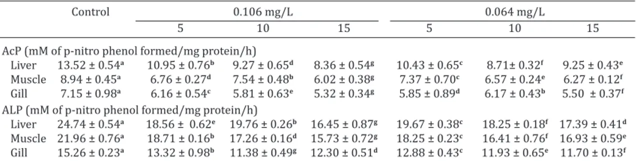
Effect on acid and alkaline phosphatase
Exposure to sodium cyanide induced significant decli -ne in phosphatase activity (Table 4) in the fish. Activity of AcP, ranged from liver (19.06 to 38.21%) > muscle (15.66 to 32.66%) > gill (13.88 to 25.64%) at 1/3rd and at 1/5th sublethal concentration liver (22.91 to 35.57%) > muscle (17.61 to 29.84%) > gill (13.68 to 23.17%). Similarly for ALP, the activity ranged from liver (20.14 to 33.49%) > mus-cle (14.81 to 28.36%) > gill (12.69 to 25.40%) at 1/3rd and at 1/5th sublethal concentration liver (20.48 to 29.72%) > muscle (15.87 to 25.28%) > gill (15.54 to 23.28%) respec-tively. The significant difference in phosphatases activities between the control and experimental groups of fish may be due to the damage of hepatic tissue with disturbed nor -mal liver function. Since, cyanide has anti-phosphatase activity; inhibition of phosphatase activity may be due to Table 3. Effect of sublethal concentrations of sodium cyanide on activities of G6PDH and glycogen
phosphorylase in different tissues of Labeo rohita
Control 0.106 mg/L 0.064 mg/L
5 10 15 5 10 15
G6PDH(mM of Pi formed/mg protein/h)
Liver 5.34 ± 0.44g 6.52 ± 0.15e 7.12 ± 0.54c 7.99 ± 0.32a 6.18 ± 0.35f 6.95 ± 0.12d 7.64 ± 0.31b
Muscle 7.53 ± 0.25g 8.94 ± 0.14e 10.38 ± 0.19a 9.82 ± 0.28c 8.75 ± 0.34f 10.13 ± 0.29b 9.65 ± 0.53d
Gill 2.95 ± 0.22g 3.45 ± 0.16e 3.81 ± 0.33c 4.18 ± 0.21a 3.35 ± 0.28f 3.63 ± 0.63d 3.94 ± 0.72b
Glycogen phosphorylase(mM of Pi formed/mg protein/h)
Liver 7.83 ± 0.01a 9.40 ± 0.08c 10.26 ± 0.01e 10.91 ± 0.02a 6.13 ± 0.05g 5.94 ± 0.08g 5.56 ± 0.01g
Muscle 4.87 ± 0.06a 5.58 ± 0.08c 5.34 ± 0.05e 5.94 ± 0.04g 5.19 ± 0.13g 5.51 ± 0.01g 5.82 ± 0.02g
Gill 1.42 ± 0.03a 1.52 ± 0.15c 1.71 ± 0.01e 1.63 ± 0.10g 1.45 ± 0.01g 1.54 ± 0.04g 1.59 ± 0.02g
Data are means ± SD (n=5) for an organ in a row followed by the same letter are significantly different (p<0.05) from each
reduced protein synthesis, as such phosphatases plays an important role in protein synthesis (Okolie & Osagie 2000). Ogundele et al (2010) illustrated inhibition in ALP and AcP activity in the adult Wistar rats, administered with the cas -sava. Inyang et al (2011) reported inhibition of ALP and AcP activity in the fish Clarias gariepinus resulting from the Diazinon exposure. Das & Mukharjee (2003) made similar observation in the fish Labeo rohita exposed to sublethal concentrations of cypermethrin. Due to the resulting activi-ty of AcP and ALP activiactivi-ty, it may be assumed that the liver tissue of the experimental animal exhibited marked inhibi -tion in the activity of phosphatases by cyanide. As hypoxic cell damages are mediated through increased permeability of cell membrane, the localization of phosphatases within the membrane would make it highly susceptible to release in cyanide-induced tissue lesions (Rees et al. 2009). This may be responsible for the inhibition of phosphatases in the tissue of the cyanide-intoxicated animals.
CONCLUSION
The results of this study show clearly that carbohydrate me -tabolism in the fish disrupted in the presence of the sodium cyanide, and this may lead to a severe energy crisis at the cellular level and altered the intermediary metabolism. De -creased activity of SDH, with concomitant increase in the LDH, G6PDH and phosphorylase in various tissues under cyanide intoxication indicates the switch over of metabolic pathways towards compensatory mechanisms. This clear-ly represents a shift from aerobic to anaerobic metabolism as evidenced by elevated lactate and fall in pyruvate levels. Therefore, alterations in the metabolism of the fish reflects the differential effects of stress and can be considered as a biomonitoring tool in the assessment of environmental pollution by cyanide on non-target organisms.
Acknowledgments.- The authors wish to thank the State Fishery Depart
-ment, BR Project, Shimoga, Karnataka, India, for their timely supplying of the fish fingerlings. The authors wish to express their gratitude to the University Grant Commission, New Delhi, for financial support in the form of a Major Research Project (F.No.34-459/2008 SR) and also to Kuvempu
University for providing necessary infrastructural facilities to carry out
the experiments.
REFERENCES
APHA 2005. Standard methods for the examination of water and was -tewater. 21st ed. American Public Health Association (APHA), American
Water Works Association (AWWA) and Water Environment Federation (WEF). Washington, DC.
Archanakumta A. & Gaikwad S.A. 1998. Effect of nitrite on succinate
dehydrogenase (LDH) and lactate dehydrogenase (LDH) in the
freshwa-ter fish Gambusia affinis. Poll. Res.17:177-179.
Baud F.J., Borron S.W., Mégarbane B., Trout H.D., Lapostolle F., Vicaut E., Debray M. & Bismuth C. 2002. Value of lactic acidosis in the assessment
of the severity of acute cyanide poisoning. Crit. Care. Med. 30(9):2044-2050.
Begum G. 2009. Enzymes as biomarkers of cypermethrin toxicity: respon
-se of Clarias batrachus tissues ATPa-se and glycogen phosphoryla-se as a function of exposure and recovery at sublethal level. Toxicol. Mech.
Methods. 19(1):29-39.
Bergmeyer H.U. & Bernt E. 1965. In: Bergmeyer H.U. (Ed.), Methods of En
-zymatic Analysis. Academic Press, New York.
Bhattacharya R., Satpute R.M., Hariharakrishnan J., Tripathi H. & Saxena P. B. 2009. Acute toxicity of some synthetic cyanogens in rats and their res
-ponse to oral treatment with alpha-ketoglutarate. Food. Chem. Toxicol.
47: 2314-2320.
Caroll N.V., Longley R.W. & Row J.H. 1956. Glycogen determination in liver and muscle by use of anthrone reagent. J. Biol. Chem. 22: 583-594. Conaglen J.V., Malthus R.S., Redshaw-Loten J.C. & Sheyd J.G.T. 1984. The
action of anion and cyanide on glycogen breakdown in the liver of grd/ grd rat. Eur. J. Biochem. 145:323.
Daniel W.W. 1987. Biostatistics: A foundation for analysis in the health
sciences. 4th ed. Wiley, New York, p.276-296.
Das B.K. & Mukherjee S.C. 2003. Toxicity of cypermethrin in Labeo rohita
fingerlings: Biochemical enzymatic and hematological consequences. Comp. Biochem. Physiol. Toxicol. Pharmacol. 137:325- 333.
Daya S., Walker R.B. & Dukie S.A. 2000. Cyanide induced free radical pro
-duction and lipid peroxidation in rat brain homogenate is reduced by aspirin. Metab. Brain. Dis. 5(3):203-210.
De Zwaan A., Cattan O. & Putzer V.M. 1993. Sulfide and cyanide induced mortality and anaerobic metabolism in the arcid blood clam Scaphar-ca inaequiv alvis. Comp. Biochem. Physiol. C. Comp. Pharmacol. 105(1): 49-54.
Dodds R.G., Penney D.G. & Sutariya B.B. 1992. Cardiovascular, metabolic and neurologic effects of carbon monoxide and cyanide in the rat. Toxicol.
Lett. 61: 243–254.
Dube P.N. & Hosetti B.B. 2010. Assessment of acute toxicity of sodium cya -nide to a freshwater teleost, Labeo rohita (Ham). Online. J. Vet. Res. 14 (2):176-187.
Fiske C. & Subbarow Y. 1925. The colorimetric determination of phospho
-rus. J. Biol. Chem. 66:375-400.
Friedman T.E. & Haugen G.F. 1943. Pyruvic acid I. Collection of blood for the determination of pyruvic acid and lactic acid. J. Biol. Chem.
144:67-77.
Govindappa S. & Swami K.S. 1965. Electrophoretic characteristics of sub cellular compounds and their relation to enzyme activities in amphibian muscle. Ind. J. Environ. Biol. 10 (4):349-353.
Table 4. Effect of sublethal concentrations of sodium cyanide on acid and alkaline phosphatase activities in different tissues of Labeo rohita
Control 0.106 mg/L 0.064 mg/L
5 10 15 5 10 15
AcP (mM of p-nitro phenol formed/mg protein/h)
Liver 13.52 ± 0.54a 10.95 ± 0.76b 9.27 ± 0.65d 8.36 ± 0.54g 10.43 ± 0.65c 8.71± 0.32f 9.25 ± 0.43e
Muscle 8.94 ± 0.45a 6.76 ± 0.27d 7.54 ± 0.48b 6.02 ± 0.38g 7.37 ± 0.70c 6.57 ± 0.24e 6.27 ± 0.12f
Gill 7.15 ± 0.98a 6.16 ± 0.54c 5.81 ± 0.63e 5.32 ± 0.34g 5.85 ± 0.89d 6.17 ± 0.43b 5.50 ± 0.37f
ALP(mM of p-nitro phenol formed/mg protein/h)
Liver 24.74 ± 0.54a 18.56 ± 0.62e 19.76 ± 0.26b 16.45 ± 0.87g 19.67 ± 0.38c 18.25 ± 0.18f 17.39 ± 0.41d
Muscle 21.96 ± 0.76a 18.71 ± 0.16b 17.26 ± 0.16d 15.73 ± 0.72g 18.25 ± 0.23c 16.41 ± 0.76f 16.93 ± 0.59e
Gill 15.26 ± 0.23a 13.32 ± 0.98b 11.38 ± 0.49g 12.30 ± 0.51d 12.88 ± 0.43c 11.93 ± 0.65e 11.70 ± 0.13f
Data are means ± SD (n=5) for an organ in a row followed by the same letter are significantly different (p<0.05) from each
Hariharakrishnan J., Satpute R.M. & Bhattacharya R. 2010. Cyanide indu
-ced changes in the levels of neurotransmitters in discrete brain regions of rats and their response to oral treatment with α-ketoglutarate. Ind. J. Exp. Biol. 48:731-736.
Heath A.G. 1991. Water Pollution and Fish Physiology. Lewis Publishers,
Boca, Ranton, Florida.
Huckabee W.E. 1961. Relationship of pyruvate and lactate during anaerobic metabolism. V. Corouary adequacy. Ann. J. Physiol. 200(6):1169-1179. Inyang I.R., Daka E.R. & Ogamba E.N. 2011. Effect of Diazinon on Acid and
Alkaline Phosphatase Activities in Plasma and Organs of Clarias gariepi-nus. Curr. Res. J. Biol. Sci. 3(3):191-194.
Kind P.N.R. & King E.J. 1954. In vitro determination of serum alkaline phosphatase. J. Clin. Pathol. 7:322.
Murat S., Saltuk B.C., Orhan E. & Ömer I.K. 2009. In vitro and in vivo effects of some pesticides on glucose-6-phosphate dehydrogenase enzyme activity from rainbow trout (Oncorhynchus mykiss) erythrocytes. Pestic. Biochem. Phys. 95: 95-99.
Nachilas M.M., Marguiles S.P. & Serigman A.M. 1960. A Calorimetric me
-thod for determination of succinate dehydrogenase activity. J. Biol.
Chem. 235:490-503.
Ogundele O.M., Caxton-Martins E.A., Ghazal O.K. & Jimoh O.R. 2010. Neuro
-toxicity of cassava: Mode of cell death in the visual relay centres of adult Wistar rats. J. Cell. Anim. Biol. 4(8):119-124.
Okolie N.P. & Osagie A.U. 2000. Differential effects of chronic cyanide intoxication on heart, lung and pancreatic tissues. Food. Chem. Toxi -col. 38(6):543-548.
Prashanth M.S. & Neelgund S.E. 2007. Free cyanide-induced Biochemical changes in Nitrogen metabolism of the Indian major carp, Cirrhinus mri-gala. J. Basic Clin. Physiol. Pharmacol. 8(4):77-287.
Ramkritinan C.M., Kumaraguru A.K. & Balasubramanian M.P. 2005. Impact of distillery effluents on carbohydrate metabolism of freshwater fish,
Cyprinus carpio. J. Fish. Biol. 14:693.
Rees B.B., Boily P. & Williamson L.A.C. 2009. Exercise- and hypoxia-indu
-ced anaerobic metabolism and recovery: a student laboratory exercise using teleost fish. Adv. Physiol. Educ.33:72-77.
Rocha-e-Silva R.C., Cordeiro L.A.V. & Soto-Blanco B. 2010. Cyanide toxicity
and interference with diet selection in quail (Coturnix coturnix). Comp. Biochem. Physiol. Part C. 151:294-297.
Saraswathi P.R. & Govindasamy S. 2002. Effect of sodium molybdate on carbohydrate metabolizing enzymes in alloxan induced diabetic rats. J.
Nutr. Biochem. 13:21-26
Sharma M. & Jain K.L. 2008. Alteration in the metabolic enzyme activity of muscle tissues in the fish Cyprinus carpio (L) on chronic exposure to
some pesticides. J. Aqua. 16: 7-12.
Singh B.M., Coles N., Lewis P., Braithwaite R.A., Nattrass M. & FitzGerald M.G. 1989. The metabolic effects of fatal cyanide poisoning. Postgrad. Med. J. 65:923-925.
Surendranath P., Ghouselazam S. & Rama Rao K.V. 1991. Significance of
glucose-6-phosphate dehydrogenase activity in the tissues of penaeid prawn, Metapenaeus monoceros (Fabricius) under acute kelthane expo
-sure. Bull. Environ. Contam. Toxicol. 56:738-744.
Sutherland E.W. 1955. Polysaccharide phosphorylase, liver, p.215-225. In: Colowick S.P. & Kaplan N.O. (Eds), Methods in Enzymology. Vol.I. Acade
-mic Press, New York.
Vandebroeck A., Uyttenhove K., Bollen M. & Stalmans W. 1988. The hepatic glycogenolysis induced by reversible ischaemia or KCN is exclusively ca