http://dx.doi.org/10.1590/s2175-97902017000317095
A
r
*Correspondence: T. Ozen. Department of Chemistry, Faculty of Arts and Sciences, Ondokuz Mayis University, 55139-Samsun, Turkey. Tel: +90 362 312 1919; fax: +90 362 457 6081. E-mail: [email protected]
The impacts of Elaeagnus umbellata Thunb. leaf and fruit aqueous
extracts on mice hepatic, extrahepatic antioxidant and drug
metabolizing enzymes related structures
Tevik Ozen*, Kemal Yildirim, Mehmet Toka
Department of Chemistry, Faculty of Arts and Sciences, Ondokuz Mayis University, 55139-Samsun, Turkey
In this work, the potential chemopreventive activities of Elaeagnus umbellata fruit aqueous (EUFA) and
leaf aqueous (EULA) extracts focusing on the modulatory inluence of xenobiotic metabolizing enzymes
(XMEs), antioxidant enzymes, glucose-6-phosphate dehydrogenase (G6PD), 6-phosphogluconate dehydrogenase (6PGD), lactate dehydrogenase (LDH) activity, lipid peroxidation (LP), sulfhydryl groups were investigated in the hepatic and extrahepatic organs of Swiss albino mice (50 and 100 mg/kg body wt given orally for 14 days) and compared with BHA (0.75 % in diet). The modulatory and chemopreventive
properties of two diferent dosesEUFA and EULA were observed for cytochrome P450, cytochrome b5, sulfhydryl groups, NADPH-cytochrome P450 reductase, NADH-cytochrome b5 reductase,
7-ethoxyresoruin-deethylase and N,N-dimethylaniline N-oxidase activities in the liver and compared
with BHA as a standard. The activities of glutathione S-transferase (GST) and DT-diaphorase (DTD)
showed a signiicant increase in the kidney, forestomach, heart and brain at both doses of EUFA and EULA. The results of EULA-treated groups were found a notable increase in LDH, G6PD, 6PGD, GST and DTD activities. Superoxide dismutase level in liver, kidney and heart exhibited a signiicant increase at both doses of EULA. Glutathione reductase activity was a remarkable level at high dose of EUFA in liver, kidney and EULA in kidney. Both doses of EUFA were efective in inducing glutathione
peroxidase activitiy in heart. The levels of LP at low and high doses of EULA-treated and EUFA-treated
were efective in liver and kidney, respectively. The present results demonstrate that signiicant efects in the level of XMEs and antioxidant enzymes of EUFA and EULA are remarkable for modulating roles and natural chemoprevention properties and therefore is considered for a valuable natural source.
Keywords: Elaeagnus umbellata Thunb. Chemopreventive properties. Xenobiotic metabolizing enzymes. Antioxidant enzymes. Lipid peroxidation. Glutathione. Lactate dehydrogenase. Glucose-6-phosphate dehydrogenase. 6-Phosphogluconate dehydrogenase.
INTRODUCTION
Phenolic compounds found in natural products
of plants and fruits have become important research topics due to their many bioactive properties such
asanticarcinogenic, antiallergic,antimicrobial,
anti-inflammatory and antioxidant. It is known that these natural compounds prevent chain reactions that can lead to many diseases resulting from the deterioration of tissue functions such as cancer, cardiovascular, immunological
system and degenerative diseases by neutralizing
short-lived oxidative damage after metabolism in the human body (Gulcin, 2012).
Elaeagnus umbellata Thunb. (Elaeagnaceae; EU) tree
is the most cultivated medicinal plant in Pakistan, China, Japan, and Korea, Afghanistan, India, USA and Canada,
especially for its fruits because of its high medicinal and nutritional value of its berries (Patel, 2015). Different Elaeagnus species, E. angustifolia var. angustifolia (İğde), E. angustifolia var. turcica Yild. (Avanos iğdesi) and E. rhamnoides (Çıçırgan) widely grow and their fruits are consumed in Turkey (Guner et al., 2012). EU(Güz yemişi) was introduced in Turkey as an ornamental plant and food to grow under natural conditions in the 1990s (Patel, 2015).
EU grows especially river banks and seaside cliff areas
(Ozen et al., 2017). The leaf of EU was traditionally used as
a tonic and decoction to treat bowel disorders in Japan and
China (Ito, Miki, Yoshida, 1999) according totraditional
resources. Ellagitannins and their derivatives in the leaf
of EU have chemopreventive, clinically-valuable activity as well as biological and nutraceutical potential (Ismail et al., 2016). The previous work indicated that the fruits of EU were consumed as fruit juice and healthy condiments (Fordham et al., 2001). The fruits, seeds and lowers of EU
were used in the treatment of coughs, colon cancer, cardiac
ailments and pulmonary infections (Afrin et al., 2016).
Lycopene is a naturally occurring carotenoid compound widely found in the fruits of EU. Lycopene has a protective
efect against neurological disorders and diseases including diabetes, cancer, alzheimer’s and Parkinson’s disease (Kaur et al., 2011).
Using medicinal plants as a chemoprevention agent is a promising approach for controlling cancer diseases.
There are substantial evidences for plants to indicate
that chemopreventive agents exert their anticarcinogenic
effects by modulation of XMEs (phase I and II) in the
liver. Cytochrome P450 (cyt.P450), a family of phase I hemoproteins, represents major adaptive response against chemical challenge from the environment and catalyzes the activation of various procarcinogens to ultimate carcinogens. Phase II enzymes detoxify pro- and ultimate forms of chemical carcinogens. The various carcinogens are
associated with oxidative stress caused by reactive oxygen
species (ROS) and free radicals generated in the living
cells (Neki, 2015). Recently, there has been an extensive
focus of research towards the new natural products that are chemoprentive and antioxidant sources (George, Dellaire, Rupasinghe, 2017). It was reported that leaf and fruit extracts of EU had the activities of in vitro antioxidant,
antiproliferative and enzyme inhibition (Ozen et al., 2017). Due to active components, traditional utilizations and
activities, the EUFA and EULA are considered to be in vivo
chemopreventive potential and antioxidant potential.
Here, for the irst time, the potential health and promoting efects of EULA and EUFA was investigated
for their modulatory effects of phase I and II enzymes [cytochrome P450 (cyt.P450), cytochrome b5 (cyt.b5), NADPH-cytochrome P450 reductase (cyt.P450R),
NADH-cytochrome b5 reductase (cyt.b5R), aniline
4-hydroxylase (A4H), 7-ethoxyresorufin-deethylase
(EROD), N,N-dimethylaniline N-oxidase (NNDNO),
glutathione S-transferase (GST), DT-diaphorase (DTD)], antioxidant enzymes [superoxide dismutase (SOD), catalase (CAT), glutathione reductase (GR) and glutathione peroxidase (GP)], sulfhydryl groups [total sulfhydryl groups (T-SH), nonprotein sulfhydryl groups
(NP-SH) and protein-bound sulfhydryl groups (PB-SH)
groups], lactate dehydrogenase (LDH), glucose-6-phosphate dehydrogenase (G6PD), 6-phosphogluconate dehydrogenase (6PGD) and level of lipid peroxidation (LP). Thus, the main purpose of the present research was to determinate the impact levels of EULA and EUFA on drug
metabolizing and antioxidant enzymes related structures on liver, lung, kidney, forestomach, heart and brain of mice
(Swiss albino), and also compared with BHA as a standard.
MATERIAL AND METHODS
Chemicals
Folin Ciocalteu’s reagent, bovine serum albumin (BSA), 1-chloro-2,4-dinitrobenzene (CDNB), potassium ferricyanide, butylated hydroxyanisole (BHA), 5,5’-dithiobis-2-nitrobenzoic acid (DTNB), GSH, GSSG,
cyt. c (from horse heart), 2,6-dichlorophenol-indophenol
(DCPIP), sodium pyruvate, thiobarbituric acid (TBA),
cholic acid, glucose 6-phosphate, 6-phosphogluconate,
NADP+, NADPH+H+ and NADH+H+ were obtained from
Sigma Chemical Co. USA. Ultra high purity CO (99%)
was a product of HABAS (Istanbul-Turkey). All other
chemicals were analytical grade and purchased from commercial suppliers.
Plant materials
The fresh fruits and leaves of EU were collected
randomly from Dogu Park, Samsun-Turkey in November 2014 and they were identiied and conirmed by Prof. Dr. Hamdi Guray Kutbay, Department of Biology, Faculty of
Arts and Sciences, Ondokuz Mayis University, where a voucher specimen (OMUB 7383) was deposited.
The dried plant materials were placed in boiled
water for aqueous extraction. The mixture was sonicated and filtered through Whatman filter paper (No.1). The EUFA and EULA extracts were lyophilized in a lyophilizer (Christ Alpha 1-2 Model; Martin-Christ, Osterode,
Germany) at -50 oC. The aquous powder crude extracts
were kept in sterile tubes at -20 °C until using.
Treatment schedule for animals
In vivo experiments were conducted with permission from the local ethic committee for animal experiments.
Ethical approval was obtained from the Ondokuz Mayis
10-12 weeks old (35-45 g) were obtained from Laboratory
Animal Research Unit (OMUDEHAM) from Ondokuz Mayis University, Samsun-Turkey. Mice were housed singly in plastic cages at room temperature with 12-hour light and dark cycle. Animals were fed with commercial rat chow (Nukleon Bil-Yem, Ankara-Turkey) and tap water
ad libitum. The animals were randomly divided into six groups, containing each of eight animals as follows: Group
I (negative control) was given double-distilled water
daily (p.o.) for 14 days and fed normal diet. Groups II
and III were orally given 50 and 100 mg/kg b.w. of EUFA dissolved double distilled water (0.05 ml), respectively,
for 14 days (p.o.). Groups IV and V were orally given 50
and 100 mg/kg b.w. of EULA dissolved double distilled
water (0.05 ml), respectively, for days (p.o.). Group VI (positive control) was fed a diet containing 0.75% BHA, daily for 14 days.
Preparation of subcellular fractions of hepatic and extrahepatic organs
The animals were killed by cervical dislocation after an overnight fast, and perfused with 0.9% NaCl (+4 °C), and cleaned of blood due to diurnal variation, immediately. The liver, lung, kidney, forestomach, brain and heart were rinsed in ice-cold 0.15 M Tris-KCl buffer (pH 7.4) and dried with ilter paper. The organs were homogenized with
ice-cold 0.15 M Tris-KCl to yield 10% (w/v) homogenate in an all-glass homogenizer. A 0.25 mL of liver homogenate
was kept a refrigerator at -80 °C for determination of T-SH,
NP-SH and PB-SH groups. The liver homogenate was centrifuged at 10.000 x g in a Beckman Coulture Optimal L100XP ultracentrifuge (Beckman Model; California,
USA) for 25 min at +4 °C. The supernatant was centrifuged
at 105.000 × g 60 min +4 °C and separated cytosolic and
microsomal fractions. After removing any loating lipid
layer, the cytosol fractions were used for the assays of antioxidant enzymes, G6PD, 6PGD, GST, DTD, LDH activities and level of LP. The microsome fraction (enriched in endoplasmic reticulum) was prepared microsomal
keeping bufer and transferred in pre-cooled sterile tubes.
The fraction was used for assaying of cyt.P450, cyt.b5,
cyt.P450R, cyt.b5R, A4H, EROD, NNDNO activities and
level of LP. The lung, kidney, forestomach, brain and heart were homogenated in ice-cold 0.15 M Tris-KCl buffer
(pH 7.4). The cytosolic fraction, after discarding lipid layer
and appropriate dilution in pre-cooled centrifuge tubes, was obtained by centrifugation of 15.000 x g for 25 min at +4 °C
and used for assaying of antioxidant enzymes, GST, DTD and level of LP. The cytosolic and microsomal fractions
were kept a refrigerator at -80 °C for further analysis.
BIOCHEMICAL ASSAYS
Protein content
The protein contents in the microsomal and cytosolic fractions were determined with BSA as standard at 660 nm (Lowry et al., 1951).
Superoxide dismutase (SOD) activity
The determination of SOD activity was based on measuring its ability to cyt.c reduction (Flohe, 1984). One unit of activity represents the amount inhibiting the cyt.c by 50%. The activity of the SOD was calculated as U/mg
cytosolic protein.
Catalase (CAT) activity
The activity was determined by following its ability to degrade using H2O2 as a substrate at 240 nm
(Aebi, 1984). The CAT activity was calculated as a U/mg
cytosolic protein.
Glutathione peroxidase (GP) activity
The GP activity was measured by the NADP+
consumption monitored at 340 nm according to ref (Ray
et al., 2000). One unit of activity was calculated using an
extinction coeicient of 6.22 mM-1cm-1 and expressed in
terms of a nmole NADPH consumed/min/mg cytosolic protein.
Glutathione reductase (GR) activity
The activity was measured by with slight modiication (Beutler, 1975) and deined as a nmole of
NADPH consumed/min/mg cytosolic protein according
to the molar extinction coeicient of 6.22 mM-1cm-1.
Cytochrome P450 (cyt.P450) content
The level of cyt.P450 content was quantified by using the CO diference spectra of sodium dithionite at 450-490 nm, and deined as a nmol/min/mg microsomal
protein using an extinction coefficient of 91 mM-1cm-1
(Omura, Sato, 1964a,b).
Cytochrome b5 (cyt.b5) content
The level of cyt.b5 content was determined by
and oxidized cyt.b5 at 424-410 nm (Omura, Sato, 1964a).
The cyt.b5 level was calculated relative to extinction
coeicient of 185 mM-1cm-1 and expressed as a nmol/min/
mg microsomal protein.
NADPH-cytochrome P450 reductase (cyt.P450R) activity
The activity was determined by monitoring of the
rate of reduction of cyt.c at 550-450 nm (Masters et al., 1967). The cyt.P450R activity was calculated using the
extinction coeicient of 19.6 mM-1cm-1 and deined as a
µmole of NADH oxidized/min/mg microsomal protein.
NADH-cytochrome b5 reductase (cyt.b5R) activity
The activity was measured by the rate of reduction
of K3Fe(CN)6, acted as an electron acceptor at 420
nm (Masters et al., 1967). The cyt.b5R activity was
calculated using an extinction coefficient of 1.02 µM
-1cm-1 and deined as a µmole of NADH oxidized/min/mg
microsomal protein.
Glutathione S-transferase (GST) activity
The activity was performed by using CDNB as substrate at 340 nm (Simons, Vande Jagt, 1977). The GST activity was calculated using the extinction coeicient
of 1.02 µM-1cm-1 and given as a µmole CDNB-GSH
conjugate formed/min/mg cytosolic protein.
DT-diaphorase (DTD) activity
The activity was based on NADPH as the electron
donor and DCPIP acting as the electron acceptor/donors at 620 nm (Prochaska, 1988). The DTD activity was
calculated using the extinction coefficient of 21 mM
-1cm-1 and expressed as a nmole DCPIP/min/mg cytosolic
protein.
Aniline 4-hydroxylase (A4H) activity
The activity was determined by measuring the ratio
of p-aminophenol formed by aniline as described in the reports (Ozen, Korkmaz, 2008; Emerole, Thabrew, 1983). The A4H activity was deined as a nmole p-aminophenol formed/min/mg microsomal protein.
7-Ethoxyresorufin-deethylase (EROD) activity
The activity was determined by the procedure of
Klotz et al. (1984) at 572 nm. The EROD activity was
calculated using the extinction coeicient of 73 mM-1cm-1
and given as a nmole resorin/min/mg microsomal protein.
N,N-dimethylaniline N-oxidase (NNDNO) activity
The activity was determined in which
N,N-dimethylaniline acted as a substrate at 420 nm (Schlenk,
Buhler, 1991). The NNDNO activity was calculated
using the extinction coefficient of 8.2 mM-1cm-1 and
expressed as a nmole N,N-dimethylaniline oxidixed/min/
mg microsomal protein.
Glucose-6-phosphate dehydrogenase (G6PD) activity
The activity was determined in which glucose
6-phosphate acted as a substrate at 340 nm (Rudack,
Davie, Holten, 1971) from the reduction of NADP+ by
taking the decrease of absorbance. The G6PD activity
was calculated using the extinction coefficient of 6.22 mM-1cm-1 and deined as a nmole NADPH reduced/min/
mg cytosolic protein.
6-phosphogluconate dehydrogenase (6PGD) activity
T h e a c t i v i t y w a s d e t e r m i n e d i n w h i c h
6-phosphogluconateacted as a substrate at 340 nm
(Rudack et al., 1971) from the reduction of NADP+ by
taking the decrease of absorbance. The 6PGD activity
was calculated using the extinction coefficient of 6.22
mM-1cm-1 and given as a nmole NADPH reduced/min/mg
cytosolic protein.
Determination of total sulfhydryl groups (T-SH), nonprotein sulfhydryl groups (NP-SH) and protein-bound sulfhydryl groups (PB-SH) groups
The levels of T-SH, NP-SH and PB-SH were estimated using DTNB (Sedlak, Lindsay, 1968) with slight
modiications. The contents were deined as a µmole of T-SH groups/g tissue, as a µmole of NP-SH/g tissue and as a µmole of PB-SH/g tissue, respectively.
Lipid peroxidation (LP) assay
Lactate dehydrogenase (LDH) activity
The activity was determined the rate of oxidation of NADH at 340 nm (Narayan, Kumar, 2013). The LDH
activity was calculated using extinction coeicient 6.22
mM-1cm-1 and deined as a µmole NAD+ oxidized/min/
mg cytosolic protein.
Statistics
Results were expressed as means ± standard deviation (SEM). Statistical analysis was performed using the independent t-test, one-way ANOVA and MANOVA tests of with the multiple-comparison POST-HOC analysis
(SPSS 20.0). The p<0.05 was considered to define the
statistical comparison in assays.
RESULTS AND DISCUSSION
Phytochemicals studies are conducted to evaluate
many pharmacological properties in herbal medicines. Efective investigations are performed to reveal potential
natural resources for the prevention or treatment of
diseases. The defensive systems of aerobic cells in the organism may be external (enzymatic and non-enzymatic) or nutrient derived (vitamins, caratinoids, lavonoids etc.)
against reactive oxygen producing species for free radicals and prooxidants (Brisswalter, Louis, 2014). When natural
defenses cannot remove the efect of prooxidants, both
intracellular and extracellular macromolecules (proteins,
lipids, and nucleic acids) are exposed to oxidative efects
that cause cell damage. Epidemic studies support that
abundant amounts of vegetables and fruits are associated
with reduced risk of certain types of cardiovascular diseases and cancer (Wrafter et al., 2016). In recent years,
natural bioactive secondary metabolits have been used
as chemical protective reagents in cancer prevention in different forms. Pharmacological investigations involving chemical preservatives, which are natural products that delay the spread of cancer in normal or
prenoplastic conditions have been increasing (Guarisco, Hall, Coulombe, 2008). Detoxiication of xenobiotics is controlled by the liver, and also exists in the lung, kidney, forestomach, brain and heart.
In vivo application of two different doses of the
EULA and EUFA extracts had a signiicant efect on the
activity of certain hepatic XMEs and antioxidant enzymes.
Extract applications of EU were able to regulate the
activities of some XMEs and antioxidant enzyme systems which are important for moderator of enzymatic and nonenzymatic activation mechanisms.
In this investigation, BHA was used as a positive control in the experimental animal model system to support effective antioxidant activity and chemical protective
eicacy (Ozen, Korkmaz, 2003; Guarisco, Hall, Coulombe,
2008). BHA (0.75% in diet) and two different doses of
EULA and EUFA (50 and 100 mg/kg body weight) were not observed a side efect in the experimental animals. Because
the doses of BHA, EUFA and EULA which are free from all
undesirable side efects, they did not have any signiicant toxic range and they are free from all undesirable side efects
in the groups. Therefore, 14 day dosing was accepted for EUFA, EULA and BHA applications.
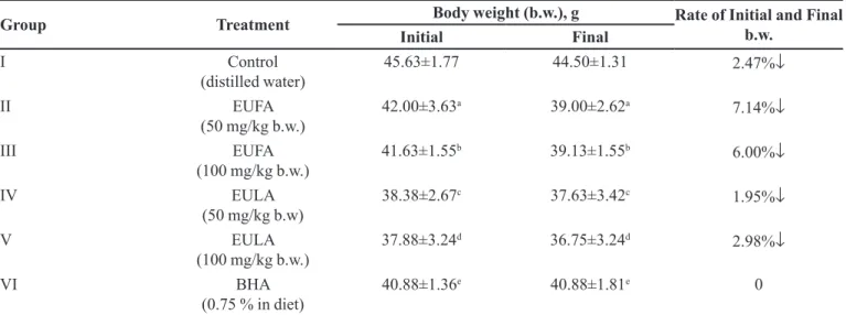
Oral administrations of 50 and 100 mg/kg EUFA and
EULA extracts exhibited a signiicant decrease (p < 0.05)
in body weight (b.w.) when compared to untreated control group. In Table I, the changes in body weights between day
0 and day 14 of the experimental animals of each group were calculated, and also the change rates were expressed
as %. There were signiicant changes in the level of relative weight of tissues (Table II).
No efects were observed on the destructive level of
the BHA-treared group and regularly applied EULA and EUFA for experimental animals through the post-oral. The
ratio of inal b.w. to tissue weights of the experimental
animals did not show a change in regulatory effect of
normal body metabolism. LDH activity values at the
cellular level indicate that EULA, EUFA (50 and 100 mg/kg
body weight/14 days) and BHA (0.75% in diet) had no efect on cell damage (Borges et al., 2008).
Table III represents the modulatory efects of cyt.
P450 and cyt.b5 contents in the liver microsomes of the
Swiss albino mice. There were signiicant increases in the cyt.P450 content by 49.62%, 51.56%, 87.87% and 40.10%
in case of Group II, III, IV and V as compared with the corresponding animals of Group I (control), respectively. Cyt.b5 content was found an increase by 3.87% and 31.65%
in treated-50 and 100 mg/kg b.w. of EUFA, respectively,
as compared to the Group I (control). The activity of cyt.
P450R extibited a signiicant enhancement (p <0.05) in
Group II and III (low and high dose of EUFA) by 1.59% and 21.20%, respectively (Table IV). The cyt.b5R activity
was found a signiicant enhancement with Group II, III
(low and high dose of EUFA) and IV (low dose of EULA)
by 11.36%, 36.93% and 1.44%, respectively (p < 0.05), as
compared with respective control group (Table IV).
The activities of A4H, EROD and NNDNO in the liver microsomes were compared with their respective
Group I (Table III). Group II, III, IV, V and VI animals
in EROD and NNDNO activities were an increase as
compared to the Group I. There was signiicantly increased
the other groups. A4H activity exhibited a reduction efect
in BHA group (Group III), whereas EROD and NNDNO increased (p < 0.05).
The microsomal cyt.P450 system, a product of the cytochrome superfamily family, is the main electron
transport chain in the endoplasmic reticulum membrane.
In the microsomes containing the cyt.P450 system,
electrons low to diferent isomorphic factors of NADPH
or NADH from cyt.P450 and cyt.b5 via flavoprotein
cyt.b5R or cyt.P450R, respectively and have an efective
role in the detoxiication of many xenobiotic compounds. The primary function of phase I metabolism is the ease of drug absorption and its efect on compound preparation for phase II metabolism. Phase II metabolism is detrimental to xenobiotics and drug detoxification in obtaining water-soluble products (Kiruthiga et al., 2015). In vivo
studies, cyt.b5 and cyt.b5R increased levels of all measured components. Thus, the effects of EULA and EUFA in cyt.b5 content may be effective in the metabolism of carcinogens. The levels of AH, EROD and also NNDNO
TABLE I - Impact of EUFA, EULA and BHA on body weight in mice
Group Treatment Body weight (b.w.), g Rate of Initial and Final b.w.
Initial Final
I Control
(distilled water)
45.63±1.77 44.50±1.31 2.47%↓
II EUFA
(50 mg/kg b.w.) 42.00±3.63
a 39.00±2.62a 7.14%↓
III EUFA
(100 mg/kg b.w.) 41.63±1.55
b 39.13±1.55b 6.00%↓
IV EULA
(50 mg/kg b.w) 38.38±2.67
c 37.63±3.42c
1.95%↓
V EULA
(100 mg/kg b.w.) 37.88±3.24
d 36.75±3.24d
2.98%↓
VI BHA
(0.75 % in diet)
40.88±1.36e 40.88±1.81e 0
Data were expressed as mean ± SEM of triplicate assays. Signiicant diferences between dose groups and control were assayed by the use of ANOVA, (p < 0.05).
TABLE II - Impact of EUFA, EULA and BHA on relative weight of tissue
Relative weight of tissue (g): Tissue weight/ last b.w. x 100
Control
Treatment (fourteen days) EUFA
(50 mg/kg b.w.) (100 mg/kg b.w.)EUFA (50 mg/kg b.w.)EULA (100 mg/kg b.w.)EULA (0.75 % in diet)BHA
Liver 5.86±0.79a 5.88± 0.95a
(0.34%↑)*
5.74± 0.70a (2.00%↓)*
5.01 ± 0.42a (14.51%↓)*
4.84 ±0.43a (17.44%↓)*
6.68±0.49a (13.91%↑)*
Kidney 1.79±0.18a 1.93±0.29ab
(7.82%↑)*
1.91±0.14ab (6.72%↑)*
1.61±0.15b (10.11%↓)*
1.61± 0.14b (10.11%↓)*
2.00± 0.22b (11.73%↑)*
Lung 0.76 ±0.14a 0.92±0.26ab
(21.11%↑)*
0.88±0.17ab (15.76%↑)*
0.96±0.14ab (26.29%↑)*
0.75±0.11ab (1.28↓%)*
0.97±0.11b (27.63↑%)*
Forestomach 0.25±0.04a 0.26± 0.05a
(4.00%↑)*
0.25± 0.05a (0.00%)*
0.22± 0.04a (12.00%↓)*
0.23± 0.06a (8.00%↓)*
0.22± 0.10a (12.13%↓)*
Heart 0.50±0.06a 0.56 ± 0.08ab
(12.12%↑)*
0.50±0.04ab (0.00)*
0.46±0.03ab (8.00%↓)*
0.49± 0.07ab (2.00%↓)*
0.59±0.07b (18.14%↑)*
Brain 0.94± 0.07a 1.06±0.07ab
(12.82%↑)*
1.05±0.07ab (11.74%↑)*
1.14±0.1b (21.33%↑)*
1.22±0.07b (29.46%↑)*
1.11±0.04b (18.12%↑)*
*Data were expressed as mean ± SEM of triplicate assays and signiicant diference from control values. Signiicant diferences
activities were found to be highly compatible (National
Toxicology Program, 2006). According to these results,
EULA and EUFA had a harmonious efect on AH, EROD,
NNDNO, cyt.P450 and cyt.b5 activities and showed that
they are able to very efective in detoxiication mechanism of liver. The possible mechanisms of detoxification against toxicity by EULA and EUFA are due to signiicant
modulation of phase I and II enzymes. The efects of the
extracts on these modulating parameters can increase
carcinogenic detoxiication. These efects may also be due to the presence of phenolics, lavonoids and lycopene. Polyphenols and lycopene have been reported to protect
potential external sources of free radicals against the threats (Kuhad, Sethi, Chopra, 2008; Sharma, 2013).
TABLE III - Impact of oral treatments with EUFA, EULA and BHA on phase I hepatic xenobiotic metabolizing enzymes in mice.
XME (phase I
enzymes) Control
Treatment (fourteen days) EUFA
(50 mg/kg b.w.) (100 mg/kg b.w.)EUFA (50 mg/kg b.w.)EULA (100 mg/kg b.w.)EULA (0.75 % in diet)BHA Cyt.P450
(pmol/min/mg)
0.99±0.26 1.48±0.44a
(49.62%↑)*
1.50±0.53b (51.56%↑)*
1.85±0.50c (87.87%↑)*
1.38±0.33d (40.10%↑)*
0.90±0.08e (9.24%↓)*
Cyt. b5
(pmol/min/mg)
1.24±0.16 1.29±0.23a
(3.87%↑)*
1.64±0.54b (31.65%↑)*
0.96±0.42c (23.02%↓)*
0.76±0.24d (39.24%↓)*
0.76±0.15e (38.59%↓)*
Cyt.P450R
(pmoles/min/mg)
0.56±0.06 0.57±0.11a
(1.59%↑)*
0.68±0.04b (21.20%↑)*
0.56±0.13c (0.32%↓)*
0.37±0.05d (34.01%↓)*
0.44±0.07e (21.23%↓)*
Cyt.b5R
(pmoles /min/mg)
7.98±1.08 8.88±1.48a
(11.36%↑)*
10.92±2.08b (36.93%↑)*
8.09±1.42c (1.44%↑)*
3.94±1.24d (50.57%↓)*
6.56±1.31e (17.83%↓)*
A4H
(nmole/min/mg)
12.04±1.50 11.92±1.98a
(0.97%↓)*
14.13±2.18b (17.37%↑)*
9.89±1.89c (17.87%↓)*
8.01±0.76d (33.50%↓)*
8.69±1.10e (27.85%↓)*
EROD
(pmolen/min/mg)
0.292±0.05 0.362±0.08a
(23.84%↑)*
0.42±0.12 b (43.01%↑)*
0.35±0.10c (18.25%↑)*
0.32±0.08d (7.97%↑)*
0.30±0.04e (1.73%↑)*
NNDNO
(pmole/min/mg)
0.30±0.05 0.36±0.08a
(23.84%↑)*
0.42±0.12b (43.01%↑)*
0.35±0.10c (18.25%↑)*
0.32±0.08d (7.97%↑)*
0.30±0.04e (1.73%↑)*
*Data were expressed as mean ± SEM of triplicate assays and signiicant diference from control values. Signiicant diferences
between dose groups and control were assayed by the use of ANOVA, (p < 0.05).
TABLE IV - Impact of EUFA, EULA and BHA on GST activity in mice
XME (phase II) GST (nmole/
min/mg ) Control
Treatment (fourteen days) EUFA
(50 mg/kg b.w.) (100 mg/kg b.w.)EUFA (50 mg/kg b.w.)EULA (100 mg/kg b.w.)EULA (0.75 % in diet)BHA
Liver 9.42±0.42 9.92±1.73a
(5.32%↑)*
7.62±0.99b (19.09%↓)*
8.61±1.38c (8.58%↓)*
9.05±1.48d (3.91%↓)*
9.97±1.56e (5.82%↑)*
Kidney 5.88±0.78 6.01±0.90a
(2.23%↑)*
7.10±0.54b (20.76%↑)*
6.82±1.07c (16.03%↑)*
7.50±0.91d (27.54%↑)*
6.40±0.76e (8.76%↑)*
Lung 9.07±1.18 9.83±4.28a
(8.35%↑)*
8.82±1.47b (2.78%↓)*
10.57±2.10c (16.49%↑)*
10.50±1.51d (15.71%↑)*
9.20±1.78e (1.40%↑)*
Forestomach 11.11±1.42 17.03±3.80a
(53.26%↑)*
13.35±3.59b (20.19%↑)*
12.58±2.66c (13.22%↑)*
14.62±2.71d (31.64%↑)*
16.16±3.91e (45.48%↑)*
Heart 8.84±1.26 10.36±2.07a
(17.19%↑)*
9.32±1.66b (5.51%↑)*
10.36±1.72c (17.18%↑)*
10.86±1.34d (22.92%↑)*
10.14±1.03e (14.78%↑)*
Brain 9.06±1.59 9.29±1.03a
(2.57%↑)*
8.71±0.98b (3.86%↓)*
9.35±1.56c (3.26%↑)*
9.06±1.60d (0.01%↑)*
10.62±1.60e (17.29%↑)*
*Data were expressed as mean ± SEM of triplicate assays and signiicant diference from control values. Signiicant diferences
Table IV was exhibited the changes in GST activities
of experimental groups of mice liver, kidney, lung,
forestomach, heart and brain. GST activities at diferent
doses of EUFA and EULA-treated groups enhanced in
kidney, forestomach, heart, brain and exhibited in a
dose-dependent manner (p <0.05).
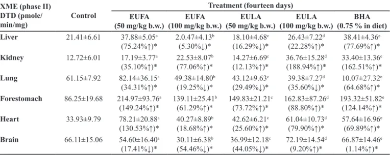
The mice treated with EUFA and EULA at low and high doses were found an increase in the DTD activities in
cytosolic fraction of kidney, forestomach and heart (Table
V) and enhanced in the dose dependent modulation. The activity treated with low of EUFA and EULA extracts was an evident increase in hepatic and extrahepatic,
signiicantly (p<0.05). GST and DTD activities of BHA, treated animals were significantly increased in the all
tissues (Table IV and V).
GST is a detoxiication enzyme that is an important
function in the coagulation of endogenous ligands (reducing
glutathione). GST is efective in protecting against diferent
cytotoxic, mutagenic and carcinogenic chemicals (Lodhi et al., 2014). DTD is the enzyme that is used to determine the
efect of many anticancerogenic substrates. DTD protects against the toxic efect of kinons and metabolites (benzene, aromatic hydrocarbon, hydroquinone, etc.). DTD facilitates the elimination of the semiquinone radical and the bioactive metabolism of kinin. DTD has the property of protecting
the quinone from reactive oxidation intermediates resulting from the oxidation of two electrons (Deepalakshmi, Mirunalini, 2013). The GST and DTD activities in the
kidney, lung heart, brain and forestomach were determined. Under experimental conditions, applications of two diferent
doses of EULA and EUFA were observed to signiicantly
increase GST and DTD activities in the liver. The treatment
of two different doses of EULA and EUFA observed
increases in liver, kidney, lung, forestomach, heart, and forestomach in GST activities and kidney, forestomach, and heart in DTD activities.
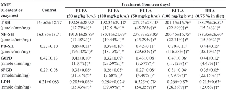
The contents of T-SH, NP-SH and PB-SH were determined in liver homogenate to characterize oxidative status of mice in the hepatic cell and assess endogenous
antioxidant defenses (Table VI). In all extracts-treated and
BHA groups, the amounts of T-SH, NP-SH and PB-SH
were signiicantly higher than the control group (p < 0.05). Furthermore, the T-SH, NP-SH and PB-SH levels of Group II, III, IV and V were found higher than in BHA-treated group.
Cytosolic LDH has been widely used to evaluate cell viability and membrane integrity is active. To further investigate the protective efect of EUFA and EULA, this
study was performed the LDH release assay. As shown in
Table VI, there was a signiicant increase of LDH activity
into medium in Group II, III, IV and V compared to the untreated Group I.
For studying the moderation of the EULA and EUFA, the activities of G6PD and 6PGD activity were
measured in liver (Table VI). G6PD and 6PGD activities
in Group II and V were increased at low and high doses (p < 0.05) and also decreased their activities of Group III,
IV and VI at both of doses, signiicantly (p < 0.05).
Table VII, VIII, IX and X were exhibited the efects
of EUFA and EULA treatment on the levels of SOD,
TABLE V - Impact of EUFA, EULA and BHA on DTD activity in mice
XME (phase II) DTD (pmole/
min/mg) Control
Treatment (fourteen days) EUFA
(50 mg/kg b.w.) (100 mg/kg b.w.)EUFA (50 mg/kg b.w.)EULA (100 mg/kg b.w.)EULA (0.75 % in diet)BHA
Liver 21.41±6.61 37.88±5.05a
(75.24%↑)*
2.0.47±4.13b (5.30%↓)*
18.10±4.68c (16.29%↓)*
26.43±7.22d (22.28%↑)*
38.41±4.36e (77.69%↑)*
Kidney 12.72±6.01 17.19±3.77a
(35.10%↑)*
22.53±8.07b (77.06%↑)*
14.27±6.69c (12.13%↑)*
36.76±15.28d (188.94%↑)*
33.40±13.36e (162.51%↑)*
Lung 61.15±7.92 82.14±36.15a
(34.31%↑)*
49.38±14.80b (19.25%↓)*
43.12±9.63c (29.49%↓)*
39.38±7.27d (35.60%↓)*
10.07±27.32e (64.68%↑)*
Forestomach 86.25±19.68 214.97±93.76a
(149.24%↑)*
139.11±25.41b (61.29%↑)*
149.83±21.21c (73.72%↑)*
162.83±87.26d (88.80%↑)*
193.32±51.82e (124.14%↑)*
Heart 33.93±9.79 78.21±20.88a
(130.53%↑)*
40.27±8.89b (18.68%↑)*
42.62±6.21c (25.60%↑)*
61.04±10.73d (79.90%↑)*
57.64±16.96e (69.89%↑)*
Brain 66.11±15.06 54.60±16.40a
(17.41%↓)*
30.11±6.38b (54.46%↓)*
36.99±12.18c (44.05%↓)*
72.19±14.54d (9.20%↑)*
66.87±14.46e (1.14%↑)*
*Data were expressed as mean ± SEM of triplicate assays and signiicant diference from control values. Signiicant diferences
CAT, GR and GP activities and evaluated in the cytosolic
fraction of hepatic and extrahepatic. These efects were comparable to BHA as a standard antioxidant.
A dose-dependent modulation in speciic activities
of SOD was evident in liver and the results were 40.56% (p < 0.05) in group IV and 0.26% (p < 0.05) in group V. Kidney SOD activities in Group III, IV and V were
increased by 39.92, 1.93 and 20.80%, respectively when
compared with Group I and VI. Kidney and heart SOD
activities in Group II, III, IV and V were improved at low and high doses level (p < 0.05) relative to group I. BHA
in Group VI enhanced the SOD activity signiicantly in forestomach and decreased signiicantly in liver, kidney, lung and brain (Table VII).
Oral application with 50 and 100 mg/kg of EULA and EUFA attenuated increases in the hepatic tissue
and extrahepatic CAT activities. The activity of CATin
forestomach and brain exhibited a signiicant enhancement
TABLE VI - Impact of EUFA, EULA and BHA on T-SH, NP-SH, PB-SH, G6PD and 6PGD activitiesin mice
XME (Content or
enzymes) Control
Treatment (fourteen days) EUFA
(50 mg/kg b.w.) (100 mg/kg b.w.)EUFA (50 mg/kg b.w.)EULA (100 mg/kg b.w.)EULA (0.75 % in diet)BHA T-SH
(µmole/min/mg)
163.68± 18.77 192.80±28.92a
(17.79%↑)*
192.34±39.18b (17.51%↑)*
237.75±23.10c (45.26%↑)*
201.15±16.76d (22.89%↑)*
188.79±26.52e (15.34%↑)*
NP-SH
(µmole /min/mg)
163.35±18.71 191.91±28.83a
(17.48%↑)*
180.41±21.69 b (10.44%↑)*
237.33±23.05c (45.29%↑)*
200.45±16.75d (22.71%↑)*
188.35±26.60e (15.30%↑)*
PB-SH
(µmole/min/mg)
0.32±0.10 0.89±0.13a
(176.10%↑)*
0.38±0.10b (18.15%↑)*
0.42±0.11c (29.63%↑)*
0.70±0.11d (116.53%↑)*
0.44±0.15e (35.10%↑)*
G6PD
(nmole /min/mg)
0.42±0.13 0.45±0.10a
(1.07%↑)*
0.32±0.09b (23.59%↓)*
0.43±0.08c (3.57%↑)*
0.47±0.06d (11.12%↑)*
0.44±0.12e (4.47%↑)*
6PGD
(nmole/min/mg)
0.29±0.08 0.38±0.06a
(31.31%↑)*
0.26±0.08b (7.68%↓)*
0.27±0.08c (4.40%↓)*
0.31±0.04d (7.70%↑)*
0.35±0.05e (22.15%↑)*
LDH
(nmole /min/mg)
0.21±0.083 0.285±0.069a
(35.43%↑)*
0.294±0.074b (39.49%↑)*
0.325±0.78c (54.35%↑)*
0.266±0.87d (26.36%↑)*
0.215±0.67e (2.05%↑)*
*Data were expressed as mean ± SEM of triplicate assays and signiicant diference from control values. Signiicant diferences
between dose groups and control were assayed by the use of ANOVA, (p < 0.05).
TABLE VII - Impact of EUFA, EULA and BHA on SOD activity in mice
Antioxidant enzyme,
SOD (U/mg) Control
Treatment (fourteen days) EUFA
(50 mg/kg b.w.) (100 mg/kg b.w.)EUFA (50 mg/kg b.w.)EULA (100 mg/kg b.w.)EULA (0.75 % in diet)BHA
Liver 11.62±3.80a 7.41±3.35ab
(36.23%↓)*
6.46±2.06b (44.42%↓)*
16.34±2.89ab (40.56%↑)*
11.65±1.46ab (0.26%↑)*
9.03±2.79ab (22.29%↓)*
Kidney 5.18±1.66a 4.81±2.10a
(7.14%↓)*
7.25±3.06a (39.92%↑)*
5.28±1.39a (1.93%↑)*
6.26±2.67a (20.80%↑)*
3.06±1.90α (40.93%↓)*
Lung 7.01±1.9a 5.19±1.65ab
(13.32%↓)*
5.94±1.88ab (10.30%↓)*
5.32±1.6ab (37.82%↓)*
4.44±2.21ab (32.00%↓)*
1.19±0.74b (84.69%↓)*
Forestomach 12.37±1.76a 4.91±1.8b
(60.31%↓)*
4.8±1.92b (61.23%↓)*
9.23±1.85ab (25.38%↓)*
18.98±1.65b (53.44%↑)*
20.97±1.92b (69.52%↑)*
Heart 12.22±1.67a 22.93±2.49b
(87.64%↑)*
13.49±1.67ab (10.39%↑)*
13.11±3ab (7.28%↑)*
13.24±2.96ab (8.35%↑)*
26.10±2.95b (113.75%↑)*
Brain 5.89±2.95a 2.33±1.16ab
(60.44%↓)*
1.35±0.35ab (78.9%↓)*
9.44±5.09ab (60.27%↑)*
3.06±1.61b (48.22%↓)*
2.07±0.46ab (61.97%↓)*
*Data were expressed as mean ± SEM of triplicate assays and signiicant diference from control values. Signiicant diferences
at low and high doses relatived to Group I (Table VIII).
EULA and EUFA extracts enhanced CAT activity in Group IV (liver), III (kidney), IV (kidney), V (lung) and III (heart).
The low and high doses of EULA and EUFA caused
signiicant alterations in liver (Group V), kidney (Group II), heart (Group II and III) and brain (Group II) GP level
when compared to that of the untreated in Group I, p < 0.05
(Table IX).
Table X shows the efects of oral application with
50 and 100 mg/kg of EULA and EUFA on the GR. The
Group II, III and V animals had signiicantly increased in
liver and kidney.
One of the most important markers of hepatic and extrahepatic cell damage is a decrease in the level of SOD enzyme activity (Dorman et al., 2003). SOD is one of the
vital enzymes in the antioxidant defense systems in vivo
and diminishes the toxic O2∙- by converting it into H2O2.
TABLE VIII - Impact of EUFA, EULA and BHA on CAT activity in mice
Antioxidant enzyme,
CAT (U/mg) Control
Treatment (fourteen days) EUFA
(50 mg/kg b.w.) (100 mg/kg b.w.)EUFA (50 mg/kg b.w.)EULA (100 mg/kg b.w.)EULA (0.75 % in diet)BHA
Liver 0.14±0.02a 0.10±0.01b
(37.52%↓)*
(0.13±0.00b (12.54%↓)*
0.17±0.02ab (17.36%↑)*
0.13±0.011ab (12.52%↓)*
0.14±0.048ab (2.14%↓)*
Kidney 0.20±0.04a 0.15±0.017a
(26.50%↓)*
0.26±0.05a (30.00%↑)*
0.24±0.03a (20.00%↑)*
0.12±0.011a (42.00%↓)*
0.19±0.02a (5.00%↓)*
Lung 0.05±0.00a 0.015±0.00b
(70.00%↓)*
0.05±0.00ab (0.00%)*
0.04±0.00ab (28.00%↓)*
0.07±0.02ab (32.00%↑)*
0.02±0.011ab (58.00%↓)*
Forestomach 0.02±0.00a 0.03±0.001a
(72.20%↑)*
0.02±0.00a (11.14%↑)*
0.02±0.001a (16.72%↑)*
0.028±0.01a (55.65%↑)*
0.015±0.025a (16.72%↓)*
Heart 0.09±0.03a 0.031±0.00ab
(53.82%↓)*
0.02±0.001b (4.92%↑)*
0.02±0.00b (36.87%↓)*
0.02±0.00b (30.93%↓)*
0.061±0.003ab (26.41%↓)*
Brain 0.03±0.00a 0.08±0.01ab
(166.00%↑)*
0.06±0.01ab (100.00%↑)*
0.05±0.00b (63.32%↑)*
0.01±0.00ab (80.00%↓)*
0.02±0.00ab (33.32%↓)*
*Data were expressed as mean ± SEM of triplicate assays and signiicant diference from control values. Signiicant diferences
between dose groups and control were assayed by the use of ANOVA, (p < 0.05).
TABLE IX - Impact of of EUFA, EULA and BHA on glutathione peroxidase (GP) activity in mice
Antioxidant enzyme, GP
(nmole/min/mg) Control
Treatment (fourteen days) EUFA
(50 mg/kg b.w.) (100 mg/kg b.w.)EUFA (50 mg/kg b.w.)EULA (100 mg/kg b.w.)EULA (0.75 % in diet)BHA
Liver 11.23±3.02a 5.04±1.85b
(55.12%↓)*
5.44±2.05ab (51.56%↓)*
8.09±2.41ab (27.96%↓)*
14.08±3.74ab (25.38%↑)*
19.77±4.54ab (76.04%↑)*
Kidney 12.84±2.61a 13.05±5.06ab
(1.64%↑)*
5.25±1.6b (59.11%↓)*
5.80±1.89b (54.83%↓)*
5.38±2.74b (58.10%↓)*
12.64±2.34ab (1.56%↓)*
Lung 9.19±2.77a 6.26±2.25b
(31.88%↓)*
3.71±0.78b (59.63%↓)*
3.22±1.06b (64.54%↓)*
4.28±1.81b (53.42%↓)*
6.23±1.92b (32.21%↓)*
Forestomach 3.58±1.44a 3.46 1.13ab
(3.35%↓)*
2.12±1.19ab (34.36%↓)*
1.40±0.72b (60.89%↓)*
1.68±1.32b (53.07%↓)*
3.22±1.16ab (10.06%↓)*
Heart 1.69±0.44a 4.22±1.28b
(149.72%↑)*
2.56±1.40ab (51.48%↑)*
1.63±0.6ab (3.68%↓)*
1.48±0.65ab (12.43%↓)*
2.92±0.63ab (72.78%↑)*
Brain 3.24±1.31a 3.53±3.03a
(8.95%↑)*
2.82±0.97a (12.96%↓)*
2.71±06a (13.36%↓)*
1.18±0.58a (63.59%↓)*
4.13±1.23a (27.47%↑)*
*Data were expressed as mean ± SEM of triplicate assays and signiicant diference from control values. Signiicant diferences
EULA and EUFA caused a signiicant increase in liver,
kidney and heart SOD activity. Thus, the extracts reduce radicals induced oxidative damage and free radicals to
liver, kidney, lung, brain, forestomach and heart. CAT is
an antioxidant enzyme that exists commonly in all living tissues and shows the highest activity in tissues and red
blood cells. CAT protects toxic hydrogen peroxide from
high active hydroxyl radical damage of tissues. For this
reason, the reduction of CAT activity may be due to many deleterious efects due to the removal of hydrogen
peroxide and superoxide radicals. In this study, the standard BHA decreased in the CAT activity levels. The CAT activities of some organs in Groups II, III, IV, and V were reduced or increased. These changes in CAT activity may lead to reduced hepatic and extrahepatic damage. The enhancement in the SOD and CAT activities can
be inluenced the phytochemical contents of EULA and
EUFA (Ozen et al., 2017).
Glutathione (GSH), a non-enzymatic antioxidant,
is the most abundant tripeptide in the liver. GSH protects
from superoxide radicals, ROS, hydrogen peroxide,
and protein thiols of the membrane. EULA and EUFA
significantly affected GST, GR and GP levels in
dose-dependent manner. Polyphenols (phenolics, lavonoids, anthocyanins, lycopene and other antioxidant substances) can be explained by phytochemical evaluation of EULA
and EUFA extracts and supported antioxidant activities in tissues (Ozen et al., 2017).
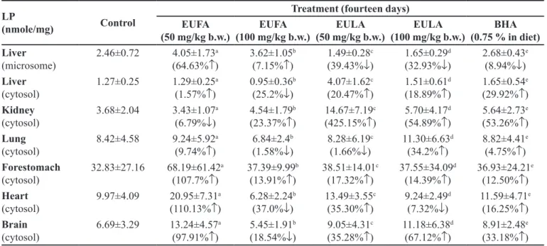
The levels of LP in microsomal and cytosolic
fractions were summarized in Table XI and exhibited
inhibition by Group II, III, IV, V and VI as compared to the control. LP level of liver, lung, heart and brain in high dose of EUFA showed efective as compared control group. The level of MDA, the inal disruption product of
lipid peroxidation in the tissue, was found to enhance the antioxidant defense mechanisms against the oxidative
stress and inhibit lipid peroxidation leading to tissue
damage in EULA and EUFA-treated groups. The increase in MDA levels reveals increased LP leading to excessive free radical damage and is an indicative of the decline of the antioxidant defense mechanism. The LP is the source
of many dangerous diseases such as cancer (Blot et al.,
1993). In this study, LP in the fractions was induced by Fe3+
-ascorbate. The LP inhibition of the EUFA and EULA-treated
groups are apparent. Thus, aquous extracts can regulate
cellular anomalies or chain reactions caused by cellular LP.
The increase in SOD activity accelerates the dismutation
of hydrogen peroxide and superoxide radicals by CAT
(Glasauer, Chandel, 2014). Reduction of LP, coagulation
by CAT and formation of superoxide in the experimental groups may be efective against cell damage of ROS.
In vivo study, it was observed that some enzymes
of the phase I and II, LDH, level of LP and antioxidant
parameters were moderated by oral administration of
EULA and EUFA extracts in experimental animals.
In all of these effects, it can be said that EULA and EUFA is probably efective in increasing carcinogenic detoxiication.
Reducing the effect of reagent that affect the microsomal monooxygenase system can detoxify and
TABLE X - Impact of EUFA, EULA and BHA on glutathione reductase (GR) activity in mice
Antioxidant enzyme, GR
(nmole/min/mg) Control
Treatment (fourteen days) EUFA
(50 mg/kg b.w.) (100 mg/kg b.w.)EUFA (50 mg/kg b.w.)EULA (100 mg/kg b.w.)EULA (0.75 % in diet)BHA
Liver 1.82±0.65a 1.76±0.29a
(4.25%↑)*
2.51±0.45a (37.92%↑)*
1.27±0.56a (30.22%↓)*
1.25±0.22a (31.32%↓)*
1.74±0.20a (4.39%↓)*
Kidney 1.85±0.19a 1.56±0.29a
(15.68%↓)*
2.56±0.39a (38.38%↑)*
1.74±0.37a (5.94%↓)*
2.61±1.37a (41.08%↑)*
2.93±0.56a (58.38%↑)*
Lung 2.09±0.67a 1.15±0.29a
(81.74%↓)*
1.34±0.68a (40.76%↓)*
1.40±0.62a (33.01%↓)*
1.26±0.48a (39.71%↓)*
1.76±0.77a (15.79%↑)*
Forestomach 1.42±0.52a 1.09±0.60a
(23.24%↓)*
1.04±0.34a (26.76%↓)*
1.39±0.25a (2.11%↓)*
1.04±0.34a (26.76%↓)*
1.6±0.48a (12.68%↑)*
Heart 3.77±0.92a 2.31±0.73ab
(38.73%↓)*
2.28±0.74b (39.52%↓)*
1.59±0.67b (57.82%↓)*
1.62±0.49b (57.03%↓)*
2.75±0.63b (27.06%↓)*
Brain 1.84±0.52a 1.61±0.55ab
(12.46%↓)*
0.41±0.23b (76.21%↓)*
1.06±0.65ab (42.39%↓)*
1.22±0.49ab (33.69%↓)*
1.92±0.54ab (4.34%↑)*
*Data were expressed as mean ± SEM of triplicate assays and signiicant diference from control values. Signiicant diferences
activate chemical carcinogens. The changes in LP, LDH, antioxidative parameters, phase I and II enzymes can
accelerate detoxiication reactions. The increase in the
cyt.b5 system is the result of an adequate detoxiication
of the activity of the metabolites by an increase in GST,
DTD, GP, GR, CAT and SOD activities (Guan, He,
2015). Antioxidant enzymes can also be efective in the
detoxification of toxic free radicals produced during
normal cell metabolism as well as abnormal. Superoxide
free radicals having the capacity to affect different
macromolecules can be suiciently detoxiied by SOD
and CAT enzymes.
CONCLUSION
ROS from the metabolic pathways cause
the degradation of living organisms and damage to macromolecules. Peroxidation of lipids, protein
inactivation and DNA mutation are the obvious
consequences of free radicals. Since the reactions are rapid and complex chain reactions take place, only the indications are followed. Cellular defences against ROS
are important detoxification of xenobiotic chemicals, polymerization of cell wall components and biosynthesis
of complex organic molecules.
Thus, there are free and complex systems that eliminate active oxygen in plant cells. Some compounds, such as carotenoids, accelerate the flow of energy in
photosystems and prevent the formation of oxygen.
Some lipid soluble compounds inhibit the formation of lipid peroxidation chain reactions on the cell membrane. Antioxidant compounds such as ascorbate and glutathione eliminate active oxygen by directly detoxifying it. Enzymes
that catalyze the synthesis, degradation and effective mechanism of these antioxidants are important for life.
The impact of EULA and EUFA extracts on mouse hepatic and extrahepatic XMEs, antioxidant enzymes, G6PD, 6PGD, LDH and sulfhydryl groups were
evaluated by assessment of their activities. A signiicant
increase in enzyme activities and structure suggested
for the irst time that EULA and EUFA might efectuate
hepatic and extrahepatic enzymes. As a result, the
diferent components of EULA and EUFA have efective antioxidant and detoxification activities and might be excellent regulatory abilities.
The efective changes indicated that EULA and EUFA extracts have signiicant changes and reliable marker in levels of biotransformation and antioxidative proiles.
ACKNOWLEDGMENTS
We would like to thank the Scientiic and
Technological Research Council of Turkey (TUBITAK)
for inancial support of this work (114Z683) and also the
Samsun Metropolitan Municipality for providing of the EU samples.
TABLE XI - Impact of EUFA, EULA and BHA on LP in mice
LP
(nmole/mg) Control
Treatment (fourteen days) EUFA
(50 mg/kg b.w.) (100 mg/kg b.w.)EUFA (50 mg/kg b.w.)EULA (100 mg/kg b.w.)EULA (0.75 % in diet)BHA Liver
(microsome)
2.46±0.72 4.05±1.73a
(64.63%↑)
3.62±1.05b (7.15%↑)
1.49±0.28c (39.43%↓)
1.65±0.29d (32.93%↓)
2.68±0.43e (8.94%↓)
Liver
(cytosol)
1.27±0.25 1.29±0.25a
(1.57%↑)
0.95±0.36b (25.2%↓)
4.07±1.62c (20.47%↑)
1.51±0.61d (18.89%↑)
1.65±0.54e (29.92%↑)
Kidney
(cytosol)
3.68±2.04 3.43±1.07a
(6.79%↓)
4.54±1.79b (23.37%↑)
14.67±7.19c (425.15%↑)
5.70±4.17d (54.89%↑)
5.64±2.73e (53.26%↑)
Lung
(cytosol)
8.42±4.58 9.24±5.92a
(9.74%↑)
6.84±2.4b (1.58%↓)
8.28±6.19c (1.66%↓)
11.30±6.63d (34.2%↑)
8.82±4.41e (4.75%↑)
Forestomach
(cytosol)
32.83±27.16 68.19±61.42a
(107.7%↑)
37.39±9.99b (13.91%↑)
38.51±14.01c (17.32%↑)
37.55±34.09d (14.39%↑)
36.93±24.21e (12.50%↑)
Heart
(cytosol)
9.97±4.09 20.95±7.31a
(110.13%↑)
6.28±2.24b (37.0%↓)
13.49±3.55c (35.30%↑)
9.24±2.49d (7.32%↓)
11.59±4.71e (16.25%↑)
Brain
(cytosol)
6.69±3.29 13.24±4.57a
(97.91%↑)
5.45±1.91b (18.54%↓)
9.05±4.31c (35.28%↑)
11.18±6.38d (67.12%↑)
8.91±2.48e (33.18%↑)
*Data were expressed as mean ± SEM of triplicate assays and signiicant diference from control values. Signiicant diferences
CONFLICT OF INTEREST
All authors declare no conlict of interest.
REFERENCES
Aebi, H. Catalase in vitro. Meth Enzymol. 1984;105:121-126.
Afrin S, Giampieri F, Gasparrini M, Forbes-Hernandez TY, Varela-López A, Quiles JL, Mezzetti B, Battino M. Chemopreventive and therapeutic efects of edible berries: A
focus on colon cancer prevention and treatment. Molecules.
2016;21(169):1-40.
Beutler E. Glutathione in red blood cell metabolism. In: Beutler E, editor. A manual of biochemical methods. 2th ed. New York: Grune and Stratton; 1975. p 112-114.
Blot WJ, Li JY, Taylor PR, Guo W, Dawsey S, Wang GQ, Yang CS, Zheng SF, Gail M, Li GY, Yu Y, Liu BQ, Tangrea J, Sun YH, Liu F, Fraumeni JF, Zhang YH, Li B. Nutrition intervention trials in Linxian, China: supplementation with speciic vitamin/mineral combinations, cancer incidence, and disease-specific mortality in the general population. J Natl Cancer I. 1993;85(18):1483-1491.
Borges LP, Brandão R, Godoi B, Nogueira CW, Zeni G. Oral administration of diphenyl diselenide protects against cadmium-induced liver damage in rats. Chem-Biol Interact. 2008;171(1):15-25.
Brisswalter J, Louis J. Vitamin supplementation benefits in master athletes. Sports Med. 2014;44(3):311-318.
Deepalakshmi K, Mirunalini S. Modulatory efect of Ganoderma lucidum on expression of xenobiotic enzymes, oxidant-antioxidant and hormonal status in 7, 12-dimethylbenz (a) anthracene-induced mammary carcinoma in rats. Pharmacogn Mag. 2013; 9(34):167-175.
Dorman HD, Koşar M, Kahlos K, Holm Y, Hiltunen R. Antioxidant properties and composition of aqueous extracts from Mentha species, hybrids, varieties, and cultivars. J Agr Food Chem. 2003;51(16):4563-4569.
Emerole G, Thabrew MI. Changes in some rat hepatic microsomal components induced by prolonged administration of chloroquine. Biochem Pharmacol. 1983;32(20):3005-3009.
Flohe L, Otting F. Superoxide dismutase assays. Meth Enzymol. 1984;105:93-104.
Fordham IM, Clevidence BA, Wiley ER, Zimmerman RH. Fruit of autumn olive: a rich source of lycopene. HortScience. 2001;36(6):1136-1137.
George VC, Dellaire G, Rupasinghe HV. Plant flavonoids in cancer chemoprevention: role in genome stability,J Nutr
Biochem. 2017;45:1–14.
Glasauer A, Chandel NS. Targeting antioxidants for cancer therapy. Biochem Pharmacol. 2014;92(1):90-101.
Guan YS, He Q. Plants consumption and liver health. Evid-Based Compl Alt. 2015;2015:ID824185.
Guarisco J, Hall JO, Coulombe R. Butylated hydroxytoluene chemoprevention of aflatoxicosis-effects on aflatoxin B 1 bioavailability, hepatic DNA adduct formation, and biliary excretion. Food Chem Toxicol. 2008;46(12):3727-3731.
Gulcin I. Antioxidant activity of food constituents: an overview. Arch Toxicol. 2012;86(3):345-391.
Guner A, Aslan S, Ekim T, Vural M, Babaç M. Türkiye bitkileri listesi (damarlı bitkiler). Istanbul: Nezahat Gökyiğit, Botanik Bahçesi; 2012. p. 47-83.
Ismail T, Calcabrini C, Diaz AR, Fimognari C, Turrini E, Catanzaro E, Akhtar S, Sestili P. Ellagitannins in cancer chemoprevention and therapy. Toxins. 2016;8(151):1-22.
Ito H, Miki K, Yoshida T. Elaeagnatins AG, C-Glucosidic Ellagitannins from Elaeagnus umbellata. Chem Pharm Bull. 1999;47(4):536-542.
Kaur H, Chauhan S, Sandhir R. Protective efect of lycopene on oxidative stress and cognitive decline in rotenone induced model of Parkinson’s disease. Neurochem Res. 2011;36(8):1435-1443.
Kiruthiga P, Karthikeyan K, Archunan G, Pandian SK, Devi KP. Silymarin prevents benzo (a) pyrene-induced toxicity in Wistar rats by modulating xenobiotic-metabolizing enzymes. Toxicol Ind Health. 2015;31(6):523-541.
K l o t z AV, S t e g e m a n J J , Wa l s h C . A n a l t e r n a t i v e 7-ethoxyresoruin O-deethylase activity assay: a continuous visible spectrophotometric method for measurement of cytochrome P-450 monooxygenase activity. Anal Biochem. 1984;140(1):138-145.
Lodhi P, Tandan N, Singh N, Kumar D, Kumar M. Camellia sinensis (L.) Kuntze extract ameliorates chronic ethanol-induced hepatotoxicity in albino rats. Evid-Based Compl Alt. 2014;2014:ID787153.
Lowry OH, Rosebrough NJ, Farr AL, Randall RJ. Protein measurement with the Folin phenol reagent. J Biol Chem. 1951;193(1):265-275.
Masters B, Williams C, Estabrook HKR, Pullman M. Methods in enzymology. New York: Academic Press; 1967. v. 10, p. 565-573.
Narayan C, Kumar A. Identification and characterization of phenolic compounds in hydro methanolic extract of Achyranthes aspera (HMEA) by UPLC and MALDI-TOF-MS and in vivo antioxidant activity. Orient Pharm Exp Med. 2013;13(1):51-59.
National Toxicology Program. Program NT. NTP technical report on the toxicology and carcinogenesis studies of 2,3,7,8-tetrachlorodibenzo-p-dioxin (TCDD)(CAS No. 1746-01-6) in female Harlan Sprague-Dawley rats (Gavage Studies). Technical Report 521. United States: National Toxicology Program Technical Report Series; 2006. p. 4-232.
Neki N. Oxidative stress and aging. Bangladesh J Med Sci. 2015;14(3):221-227.
Omura T, Sato R. The carbon monoxide-binding pigment of liver microsomes I. Evidence for its hemoprotein nature. J Biol Chem. 1964a;239(7):2370-2378.
Omura T, Sato R. The carbon monoxide-binding pigment of liver microsomes II. Solubilization, puriication, and properties. J Biol Chem. 1964b;239(7):2379-2385.
Ozen T, Korkmaz H. Modulatory effect of Urtica dioica
L. (Urticaceae) leaf extract on biotransformation enzyme systems, antioxidant enzymes, lactate dehydrogenase and lipid peroxidation in mice. Phytomedicine. 2003;10(5):405-415.
Ozen T, Korkmaz H. The efects of Urtica dioica L. leaf extract on aniline 4-hydroxylase in mice. Acta Pol Pharm. 2008; 66(3):305-309.
Ozen T, Yenigun S, Altun M, Demirtas I. Phytochemical constituents, ChEs and urease inhibition, antiproliferative and
antioxidant properties of Elaeagnus umbellata Thunb. Comb
Chem High Throughput Screen. 2017;20(6):559-578.
Patel S. Plant genus elaeagnus: Underutilized lycopene and linoleic acid reserve with permaculture potential. Fruits. 2015;70(4):191-199.
Prochaska HJ. Puriication and crystallization of rat liver NAD (P) H:(quinone-acceptor) oxidoreductase by cibacron blue affinity chromatography: identification of a new and potent inhibitor. Arch Biochem Biophys. 1988;267(2):529-538.
Ray G, Batra S, Shukla NK, Deo S, Raina V, Ashok S, Husain SA. Lipid peroxidation, free radical production and antioxidant status in breast cancer. Breast Cancer Res Tr. 2000;59(2):163-170.
Rudack D, Davie B, Holten D. Regulation of rat liver glucose 6-phosphate dehydrogenase levels by adenosine 3’, 5’-monophosphate. J Biol Chem. 1971;246(24):7823-7824.
Rudack D, Gozukara EM, Chisholm EM, Holten D. The efect of dietary carbohydrate and fat on the synthesis of rat liver 6-phosphogluconate dehydrogenase. BBA-Gen Subj. 1971;252(2):305-313.
Sharma V. Anti-carcinogenic potential of Euphorbia neriifolia leaves and isolated lavonoid against N-Nitrosodiethylamine-induced renal carcinogenesis in mice. Indian J Biochem Bio. 2013;50(6):521-528.
Schlenk D, Buhler D. Role of lavin-containing monooxygenase in the in vitro biotransformation of aldicarb in rainbow trout
(Oncorhynchus mykiss). Xenobiotica. 1991;21(12):1583-1589.
Sedlak J, Lindsay RH. Estimation of total, protein-bound, and nonprotein sulfhydryl groups in tissue with Ellman’s reagent. Anal Biochem. 1968;25:192-205.
Simons PC, Vander Jagt DL. Purification of glutathione S-transferases from human liver by glutathione-affinity chromatography. Anal Biochem. 1977;82(2):334-341.
Varshney R, Kale R. Effects of calmodulin antagonists on radiation-induced lipid peroxidation in microsomes. Int J Radiat Biol. 1990;58(5):733-743.
Wrafter PF, Connelly TM, Khan J, Devane L, Kelly J, Joyce WP. The 100 most inluential manuscripts in colorectal cancer: a bibliometric analysis. Surgeon. 2016;14(6):327-336.