Morphological, biochemical and molecular approaches to
the identification of Meloidogyne incognita, Meloidogyne
javanica and Meloidogyne arenaria in Portugal
Leidy Constanza Mora Rusinque
Dissertation to obtain the Master degree in
Agricultural Engineering
Specialisation in Crop Protection
Tutors: Doctor Maria Filomena de Sousa Nóbrega
Doctor Mariana da Silva Gomes Mota
JURY:
President: Doctor Maria José Antão Pais de Almeida Cerejeira, Associate Professor with Aggregation, Instituto Superior de Agronomia da Universidade de Lisboa.
Committee Members:
Doctor Manuel Galvão de Melo e Mota, Assistant Professor with Aggregation, Universidad de Évora.
Doctor Maria Filomena de Sousa Nóbrega, Senior Researcher, Instituto Nacional de Investigação Agrária e Veterinária,I.P.
Nowadays people know the price of Everything and the value of nothing! “Oscar Wilde”
Cuando la gratitud es tan absoluta las palabras sobran. “Alvaro Mutis”
Quem não sabe a arte, Não a estima. “Luis Vaz Camões”
iii Acknowledgments
This dissertation becomes a reality thanks to the support and help from many people to whom I would like to extend my most sincere gratitude.
To begin with, this work would not have been possible without the help, support, patience and friendship of my dear tutor Doctor Filomena Nóbrega not to mention her continuous advice and encouragement throughout this process; I feel deeply honoured to have worked under your guidance. I also want to extend my thankfulness to Doctor Maria Lurdes Inácio who with her immense enthusiasm, unconditional reassurance and assistance has been a major driving force during the development of this dissertation. I owe you both a huge debt of gratitude. For your guidance, wisdom and camaraderie I am forever grateful.
My deepest thanks and sincere appreciation goes to Professor Doctor Mariana Mota for her kind supervision, for being always available and easily approachable, for her continuous encouragement but mainly for believing in me.
I would also like to acknowledge the staff from the Laboratory of Nematology at INIAV for their support, assistance and willingness to share their knowledge.
I am also extremely grateful to Professor Doctor Isabel Maria de Oliveira Abrantes and Doctor Carla Maria Nobre Maleita from the Functional Ecology Centre, Department of Life Sciences, from Coimbra University for finding out the time to receive us at their Laboratory and for passing down their knowledge on Meloidogyne.
Thank you to Engineer João Heitor for his very useful tips in the identification of Meloidogyne and other nematodes, for his thoughtfulness and for finding the time to collect some vegetal material as well as some laboratorial stuff I was in need of.
A very special thanks goes to my dear friend Maria João Camacho who introduced me not only to my tutors but also to this academic path that I never thought I would take. Her useful comments, her corrections on my Portuguese, her constant support and company in the ups and downs during this process are things I am deeply thankful.
I thank my friends in London, Bogota and many other parts of the world, who in spite of being physically far from here their thoughts and good vibes were always surrounding me.
To my aunt Nelly, my uncle Mario and my cousin Jessica I want to express my profound gratitude for their understanding, love and support especially since I arrived in Lisbon. I am hugely indebted since thanks to you I started and I am finishing this master degree, there are no words that can express how thankful and happy I am of being here.
And last but not least to my beloved parents Betty and Remigio and to my brother Felipe, you have been so supreme, you have nurtured my learning and always supported my dreams. You made me into who I am. Thank you!
Love always Leidy
v Resumo
Os nemátodes-das-galhas-radiculares, Meloidogyne spp., causam anualmente prejuízos de milhões de euros, não só pela redução da quantidade mas também da qualidade dos produtos agrícolas. Assim, o principal objetivo deste trabalho consistiu em contribuir para o desenvolvimento de uma metodologia robusta de diagnóstico e enquadrada nos protocolos da EPPO para identificação das três principais espécies do nemátode das galhas (M. incognita, M. arenaria e M. javanica) nos Laboratorios de Genetica Molecular e Nematologia no INIAV Portugal. Para a verificação do material de referência, foram realizadas observações e respetivas medições das estruturas morfológicas dos juvenis de segundo estádio, assim como estudos moleculares. Não foi possível efetuar estudos bioquímicos com estas amostras, uma vez que não havia disponibilidade de fêmeas adultas. Os resultados obtidos nos estudos morfológicos e biométricos não foram conclusivos. Relativamente ao estudo molecular, também não foi possível identificar corretamente as três espécies.
Com o objetivo de identificar as espécies de Meloidogyne presentes em amostras da região Oeste de Portugal, foram efetuados estudos morfológicos, bioquímicos e moleculares. A morfologia do padrão perineal apontou a presença de duas espécies, M. incognita e M. arenaria, mas devido à variabilidade dos padrões perineais esta caracterização não foi considerada conclusiva. O mesmo aconteceu com as análises enzimáticas e moleculares. Os mesmos estudos foram conduzidos em material originário do Norte de Portugal. Os resultados morfológicos sugeriram a presença de M. javanica, resultado que foi confirmado bioquimicamente e por biologia molecular. Foram ainda realizadas amplificações por PCR usando primers universais para nematodes das amostras de referência e do Oeste de Portugal, com o objetivo de sequenciar os produtos PCR que apresentassem uma banda única do tamanho expectável. As sequências obtidas foram alinhadas e analisadas resultando uma homologia entre as três espécies em estudo e com outras que não pertencem ao género Meloidogyne. Embora não se tenha conseguido definir uma ferramenta de diagnóstico rápida e precisa para a identificação das três principais espécies do género Meloidogyne, a realização deste estudo permitiu determinar que a adequada identificação destes nemátodes só é possível mediante a conjugação dos três métodos de diagnóstico - morfológico, bioquímico e molecular.
Palavras-chave: Nemátodes-das-galhas-radiculares, Meloidogyne, padrões perineais, métodos bioquímicos, PCR.
Abstract
Plant parasitic nematodes are highly damaging pests in many crops of great economic importance. A substantial part of this damage is caused by infestations of root-knot nematodes (RKN), due to their wide geographical distribution as well as a vast range of host plants. The most important and widely distributed root-knot nematode species are Meloidogyne incognita, M. arenaria and M. javanica accounting for almost 95% of the occurrences, and so, an accurate and reliable identification it is primary to establish effective, sustainable and environmentally safe control measures.
The main goals of this study were to characterise morphologically, biochemically and molecularly Portuguese isolates and reference material from Netherlands Food and Consumer Product Safety Authority (NVWA), Wageningen, The Netherlands, in order to find the most suitable tools that can be used in the Laboratory of Molecular Genetics and Nematology at the INIAV in Portugal to assist in the identification of the three most common species of Meloidogyne. Morphological studies incorporated the examination of female’s perineal pattern and measurements of second stage juveniles’ features, which presented enough variation to confirm the unreliability and difficulty of morphological identification alone. Biochemical assays with isozymes such as esterases (EST) were performed on females and the PAGE enzymatic patterns obtained match those already described for M. javanica. Molecular analysis included PCRs using universal primers that target the 28S gene, ITS and IGS ribosomal DNA regions (rDNA) and the region between the cytochrome oxidase (COII) of the mitochondrial DNA (mtDNA) and 16S rDNA genes together with species-specific SCAR primers and sequencing and cloning. The sequences obtained from sequencing of PCR fragments and from cloned fragments were aligned and compared to those found in GenBank database through BLAST analysis with inconclusive results.
Although it was no possible to uncover an efficient tool for Meloidogyne identification this study pinpointed that the three approaches for root-knot nematode identification need to be used if we are to identify this species accurately and effectively. What’s more this research can be used as platform for further studies regarding this species at the Nematology of INIAV Portugal.
Key words: Root-knot nematode, Meloidogyne spp., perineal patterns, esterase phenotype, PCR.
vii Resumo alargado
Os nemátodes parasitas de plantas são conhecidos pelos efeitos nocivos causados em muitas culturas economicamente importantes. Estes fitoparasitas podem ser ectoparasitas, vivendo fora do hospedeiro e alimentando-se a partir do exterior da raiz, ou endoparasitas, vivendo parte do seu ciclo de vida dentro de órgãos da planta hospedeira.
Os nemátodes-das-galhas-radiculares, Meloidogyne spp., são nemátodes endoparasitas sedentários de grande importância económica, parasitando praticamente as raízes de todas as plantas vasculares, e estando amplamente distribuídos tanto em regiões de clima quente e tropical como em regiões de clima temperado. Estes nemátodes causam anualmente prejuízos de milhões de euros, não só pela redução da quantidade mas também da qualidade dos produtos agrícolas.
Entre as espécies do género Meloidogyne as mais importantes são Meloidogyne incognita, Meloidogyne arenaria e Meloidogyne javanica, as quais se encontram amplamente distribuídas nas várias zonas geográficas, sobretudo nas regiões tropicais, representando 95% do total das ocorrências deste género. Por isso, a identificação correta destas espécies é essencial para a implementação de novas e efetivas estratégias de controlo.
Inicialmente, a identificação do género Meloidogyne era apenas baseada nas características morfológicas e biométricas. Contudo, além destas espécies serem morfologicamente semelhantes, apresentam uma grande variabilidade das características para diagnóstico dos juvenis de segundo estádio e dos padrões perineais das fêmeas, tornando a identificação morfológica difícil e laboriosa, mesmo para os especialistas. Por conseguinte, para ultrapassar estas as dificuldades, o padrão de atividade isoenzimática, nomeadamente a análise do polimorfismo das esterases de fêmeas, tem sido considerado um parâmetro bastante fiável. No entanto, esta metodologia só pode ser aplicada a fêmeas adultas. Havendo a necessidade de identificar as espécies de Meloidogyne em todas as fases do seu ciclo, têm sido desenvolvidas metodologias moleculares como alternativa.
Assim, o principal objetivo deste trabalho consistiu em contribuir para o desenvolvimento de uma metodologia robusta de diagnóstico e enquadrada nos protocolos da EPPO para identificação das três principais espécies do nemátode das galhas (M. incognita, M. arenaria e M. javanica) nos Laboratorios de Genetica Molecular e Nematologia no INIAV Portugal.
Visando a produção de material biológico para realização dos estudos morfológicos, biométricos, bioquímicos e moleculares, foram inoculadas plantas de tomate Lycopersicum solanum cv. Rio Grande com juvenis de segundo estádio e raízes infetadas provenientes do NVWA - Netherlands Food and Consumer Product Safety Authority, Wageningen. As plantas foram mantidas na estufa de quarentena durante quatro meses, após os quais as raízes e o solo foram avaliados, confirmando-se a ausência de galhas nas raízes e formas móveis no solo.
Para a verificação do material de referência proveniente de Wageningen, foram realizadas observações e respetivas medições das estruturas morfológicas dos juvenis de segundo estádio, assim como estudos moleculares. Não foi possível efetuar estudos bioquímicos com estas amostras, uma vez que não havia disponibilidade de fêmeas adultas. Os resultados obtidos nos estudos morfológicos e biométricos não foram conclusivos, dado que os valores das medições se sobrepõem e a similaridade das características morfológicas das três espécies é muito elevada. Relativamente ao estudo molecular, também não foi possível identificar corretamente as três espécies.
Com o objetivo de identificar as espécies de Meloidogyne causadoras de galhas nas raízes de tomateiros provenientes da região Oeste de Portugal, foram efetuados estudos morfológicos, bioquímicos e moleculares utilizando as fêmeas que foram retiradas das raízes de cinco plantas diferentes. A morfologia do padrão perineal apontou a presença de duas espécies, M. incognita e M. arenaria, mas devido à variabilidade dos padrões perineais esta caracterização não foi considerada conclusiva. O mesmo aconteceu com as análises enzimáticas e moleculares. Por isso, não foi possível identificar a ou as espécies presentes nestas amostras. Os mesmos estudos foram conduzidos em material originário do Norte de Portugal. Os resultados morfológicos sugeriram a presença de M. javanica, resultado que foi confirmado bioquimicamente através da análise dos padrões da isoenzima esterase e por biologia molecular através da PCR com primers específicos para M. javanica.
Foram ainda realizadas amplificações por PCR usando primers universais para nemátodes das amostras de referência e do Oeste de Portugal, com o objetivo de sequenciar os produtos PCR que apresentassem uma banda única do tamanho expectável. As sequências obtidas foram alinhadas e analisadas resultando uma homologia entre as três espécies em estudo e com outras que não pertencem ao género Meloidogyne.
ix Adicionalmente, os produtos PCR obtidos com os primers TW81/AB28 foram clonados e posteriormente sequenciados, tendo-se obtido resultados semelhantes aos anteriormente referidos.
Embora não se tenha conseguido definir uma ferramenta de diagnóstico rápida e precisa para a identificação das três principais espécies do género Meloidogyne, a realização deste estudo permitiu determinar que a adequada identificação destes nemátodes só é possível mediante a conjugação dos três métodos de diagnóstico - morfológico, bioquímico e molecular. De salientar a importância da manutenção das populações de Meloidogyne spp em estufa e a adequada colheita e preparação das amostras para as análises bioquímica e molecular. A experiência adquirida ao longo deste trabalho servirá como conhecimento de base para futuros estudos na Nematologia do INIAV envolvendo os nemátodes-das-galhas-radiculares.
Palavras-chave: Nemátodes-das-galhas-radiculares, Meloidogyne, padrões perineais, fenótipo de esterase, PCR.
Table of Contents
Acknowledgments ... iii
Resumo ... v
Abstract ... vi
Resumo alargado ... vii
General Introduction ...15
The Genus Meloidogyne ...17
Life cycle and biology...18
Symptoms ...20
Meloidogyne incognita ...20
Meloidogyne arenaria ...21
Meloidogyne javanica ...21
Management and control ...22
CHAPTER ONE: MORPHOLOGYCAL STUDIES ...23
1. INTRODUCTION ...24
1.1. Characteristics for diagnosis ...25
2. MATERIALS AND METHODS ...32
2.1. Nematode Isolates ...32
2.2 Inoculation of plant material ...33
2.3 Morphological and morphometric studies...34
3. RESULTS ...36
3.1 Inoculation ...36
3.2 Reference material ...37
3.3 West Portugal samples ...41
3.4 North Portugal samples ...43
4. DISCUSSION ...44
CHAPTER 2: BIOCHEMICAL STUDIES...46
1. INTRODUCTION ...47
1.1 Isozymes ...49
2. MATERIALS AND METHODS ...51
2.1 Sample preparation ...51
2.2 Polyacrylamide gel electrophoresis...51
xi
CHAPTER 3: MOLECULAR STUDIES ...54
1. INTRODUCTION ...55
1.1 Ribosomal DNA ...56
1.2 Mitochondrial DNA ...56
1.3 Sequence Characterized Amplified Regions (SCARs) ...57
2. MATERIALS AND METHODS ...58
2.1 DNA Extraction ...58
2.2 Polymerase Chain Reaction ...58
2.3 Sequencing ...59
2.4 Cloning and sequencing ...59
3. RESULTS ...61
4. DISCUSSION ...68
General Discussion and Conclusion ...70
References ...73
APPENDIX 1 ... 1
APPENDIX 2 ... 0
APPENDIX 3 ... 0
List of figures
Figure 1 - Life cycle of root-knot nematodes, Meloidogyne spp... ...19
Figure 2 - Root galls formed by Meloidogyne. ...20
Figure 3 - Diagram of perineal pattern of a Meloidogyne female . ...27
Figure 4 - Diagrams of variability of perineal patterns of Meloidogyne arenaria. ...27
Figure 5 - Diagrams of variability of perineal patterns of Meloidogyne incognita. ...28
Figure 6 - Diagrams of variability of perineal patterns of Meloidogyne javanica. ...29
Figure 7 - Reference material of Meloidogyne arenaria, Meloidogyne incognita and Meloidogyne javanica. ...32
Figure 8 - Germinated tomato seeds (Solanum lycopersicum cv. Rio Grande) and transplanted tomato seedlings. ...33
Figure 9 - Perineal pattern area cuts.. ...35
Figure 10 - Roots from inoculated tomato plants (cv. Rio Grande). ...36
Figure 11 - Graphic representation of overlapped morphometric measures of Meloidogyne incognita, Meloidogyne arenaria and Meloidogyne javanica. ...38
Figure 12 - Micrographs of second stage juveniles’ anterior regions with stylets (400X). ...39
Figure 13 - Micrographs of second stage juveniles’ tails (400X). ...40
Figure 14 - Binocular and light microscope photographs of Meloidogyne females from West Portugal samples.. ...41
Figure 15 - Perineal pattern of Meloidogyne incognita (400X). ...42
Figure 16 - Perineal pattern of Meloidogyne arenaria (400X). ...42
Figure 17 - Perineal patterns of Meloidogyne javanica (400X). ...43
Figure 18 - Esterase (EST) phenotypes of the major species of Meloidogyne.. ...50
Figure 19 - Esterase phenotype of Meloidogyne spp. from North Portugal. ...52
Figure 20 - Schematic diagram of the multigenic family of ribosomal DNA (rDNA). ...56
Figure 21 - Meloidogyne mitochondrial genome structure.. ...57
Figure 22 - Amplification products obtained from ten nematode isolates using ITS1 and Vrain2R primers. ...61
Figure 23 - DNA amplification products obtained from seven isolates of Meloidogyne spp. .62 Figure 24 - DNA amplification products obtained from seven isolates of Meloidogyne spp. .63 Figure 25 - DNA amplification products obtained from three isolates of Meloidogyne spp.. .63
Figure 26 - DNA amplification products obtained from three isolates of Meloidogyne javanica.. ...64
xiii List of tables
Table 1 - Reported morphometric values (in µm) for Meloidogyne incognita, Meloidogyne
arenaria and Meloidogyne javanica. ...31
Table 2 - Meloidogyne isolates, host, geographic origin and studies performed ...32 Table 3 - Morphometric values (in µm) of reference samples of Meloidogyne incognita,
Meloidogyne arenaria and Meloidogyne javanica ...37
Table 4 - Morphometric values (in mm) performed on Meloidogyne spp. samples from North Portugal ...43 Table 5 - DNA extraction, dates and material extracted ...58 Table 6 - Alignment results from direct sequencing and cloning ...65
List of abbreviations
bp: Base pairs
DNA: Desoxyribonucleic Acid rDNA Ribosomal DNA
dNTP: Deoxynucleotide triphosphate
EPPO: European and Mediterranean Plant Protection Organization INIAV: Instituto Nacional de Investigação Agrária e Veterinária, I.P. IPM: Integrated pest management strategies
J2: Second Stage Juvenile
NVWA: Netherlands Food and Consumer Product Safety Authority, Wageningen PAGE: Polyacrylamide Gel Electrophoresis
PCR: Polymerase Chain Reaction
RFLP: Restriction Fragment Length Polymorphism RNA: Ribonucleic Acid
RKN: Root-Knot Nematodes
15 General Introduction
The phylum Nematoda comprises a large number of described species, many of which are parasites of animals or plants as well as many free-living species (Cobb, 1915). Nematodes are a major eukaryotic group on earth, due to their ability to adapt to hostile and changing environmental conditions (Abad & Williamson, 2010).
The plant-parasitic nematodes feed on roots or parasitize the aerial portions of the plant. According to their mode of infection, the root parasites are classified as ectoparasites (feed from outside the host root) or as endoparasites (boring into and feeding from inside the host root) and, at the same time, they are classified as mobile (migrating while feeding) or sedentary (stationary while feeding). By far the most economically important group of plant parasitic nematodes is the sedentary endoparasites (Sasser & Freckman 1987; Koenning et al., 1999; Chitwood, 2003). This group is composed of two taxonomic groups, commonly referred as the cyst nematodes (Heterodera and Globodera spp.) and root-knot nematodes (Meloidogyne spp.).
The root-knot nematodes (RKN) are members of the genus Meloidogyne, their name is of Greek origin and means ‘apple-shaped female’. They are a group of highly adapted obligate plant parasites and their control presents a major global challenge (Karsen & Moens, 2006). Their presence was first noted on plants in the early 1800’s when the Englishman M.J Berkeley in 1855 correlated the galls on cucumber roots with nematodes (Hunt & Handoo, 2009). Long after, in the early period of nematode taxonomy (1879-1948), RKN were mostly placed in the same genus as cyst nematodes, until 1949 when Chitwood separated RKN from cyst nematodes. He reassigned these species to the genus Meloidogyne, which was first named by Göeldi in a paper, published in 1887 and reprinted in 1892 (Hirschmann, 1985; Karssen, 2002).
Amongst the many genera of nematodes having some economic impact, Meloidogyne spp. are responsible for a large part of the annual multibillion losses attributed to nematode damage (Sasser et al., 1987). Economic losses due to Meloidogyne spp. are not only confined to yield reductions but also to an increase on production costs for farmers. Apart from these direct losses, many indirect losses such as waste of irrigation, water and fertilizers can occur. Moreover, during the last few years in Europe, chemical treatments have been restricted, limiting the management options or obliging farmers to apply more expensive control measures (Wesemael et al., 2011).
Aim of this study
The identification of Meloidogyne species has become increasingly important for the design of effective nematode management practices such as crop rotation and plant resistance which require precise species identification as well as for quarantine purposes (Hussey, 1990; Zijlstra 2000; Zijlstra & Van Hoof, 2006).
According to Karssen & Moens (2006), the best approach for a reliable identification is to integrate morphology, morphometrics, isozyme and DNA data, together with information on mode of reproduction, host plants and distribution. Therefore, the main purpose of this study was to develop efficient and robust tools in the Laboratory of Molecular Genetics and Nematology at INIAV (National Reference Laboratory of Plant Health) to assist in the identification of the most common and economically important species of Meloidogyne in Portugal. This development plays a crucial role since it helps to provide valid results to the requested analyses of samples of Meloidogyne species present in Portugal fields.
Based on the above, the overall objectives of the present work were:
Morphological characterisation of second stage juveniles and females from reference material and North and West Portugal samples.
Biochemical identification using analysis of the esterase phenotype of specimens from North and West Portugal as well as reference samples.
Molecular identification of reference material and North and West Portugal samples using PCR with universal and species specific primers.
17 The Genus Meloidogyne
In an 1887 paper (reprinted in 1892) Göeldi described different diseases affecting coffee plants in the Rio de Janeiro province including the root-knot nematodes (RKN). He named the nematode Meloidogyne exigua and described and illustrated it briefly. His work is within the first findings on this nematode, highlighting its significance as causing serious problems in crops of high economic importance (Lordello & Lordello, 1983; Karssen 2002). From this description, Chitwood obtained the name we currently use for the RKN nematodes (Mitkowski, & Abawi, 2003). Root-knot nematodes are one of the oldest known parasitic nematodes of plants and have been of interest to nematologists worldwide probably due to their widespread distribution and success as parasites of economically important crops (Dong et al., 2001; Trudgill & Blok, 2001).
This genus comprises more than 80 species (Karssen, 2002) and on a worldwide basis includes the plant parasitic nematodes most economically damaging to crop protection. Meloidogyne incognita, Meloidogyne javanica, Meloidogyne arenaria, Meloidogyne chitwoodi, Meloidogyne fallax and Meloidogyne hapla account for more than 95% of the occurrences of this genus and are the most widely distributed species. Their wide host ranges enhance the impact of these species; the most common species are estimated to be able to infect more than 5500 plant species (Trudgill & Blok, 2001).
Meloidogyne incognita, M. javanica and M. arenaria are highly abundant in tropical climates but also in greenhouses of temperate regions; M. chitwoodi, M. fallax and M. hapla are major species in temperate climate. Furthermore, in respect of changing global trade pattern and crop production system, M. minor and M. enterolobii species are becoming emerging threats (Wesemael et al., 2011) for the temperate and tropical region respectively. As a result, the European and Mediterranean Plant Protection Organization (EPPO) has reported Meloidogyne chitwoodi, M. fallax and M. enterolobii as quarantine pests (EPPO, 2016).
In Portugal, nematology studies started in 1881 when Moraes observed for the first time the formation of galls in vine roots while working on a survey in Phylloxera (Reis, 1970). The description made by Moraes (1882) corresponds to the Meloidogyne genus and he also referred its occurrence to different parts of the country and to be always associated to Vitis vinifera L.
Many other references regarding this nematode appeared, until 1962 when Lima identified the first species in Portugal as being Meloidogyne incognita and M. javanica in peach
orchards. According to Lima (1962) the RKN were the most important plant parasitic nematodes due to its widespread distribution in the country.
Some other species of this genus have been found alone or in mixed populations in different regions of the centre and south, associated with several and important cultivated plants (Abrantes et al., 2008; Maleita et al., 2011). Also, the presence of M. chitwoodi a species of quarantine was detected during a survey for potato cyst nematodes (PCN) Globodera spp. on two samples, one from Porto and one from the island of Madeira (Da conceição et al., 2009).
Life cycle and biology
Nematode growth and reproduction depend on the establishment of specialized feeding sites within the root. Consequently, these nematodes do not kill the host cells from which they feed. Instead, they induce a differentiation process that leads to the formation of giant cells. Each RKN triggers the development of five to seven giant cells (Christie, 1936; Jones, 1981; Abad et al., 2003; Jones & Goto, 2011).
Most Meloidogyne species have a similar life cycle (Figure 1) and their reproduction mode vary considerably, with a few Meloidogyne species producing cross-fertilised eggs after copulation (amphimictic), others reproducing by cross-fertilisation or meiotic parthenogenesis (automictic) and the last and most successful using obligatory mitotic parthenogenesis (apomictic) which is the case of the species under study (Triantaphyllou, 1985; Castagnone-Sereno, 2006; Jones & Goto, 2011). In this last form of reproduction there is neither reduction nor fusion of nuclei, and the egg directly develops into an embryo. When males are present, they can inseminate females, but the sperm nucleus degenerates and does not participate in fertilization (Triantaphyllou, 1962, 1963, 1981). Apomictic RKN species are diploid, triploid and sometimes tetraploid exhibiting a high response capacity to environmental selection (Castagnone-Sereno, 2002).
19 Figure 1 - Life cycle of root-knot nematodes, Meloidogyne spp.. N: Giant cells. (Adapted from Abad et
al., 2008).
Their life cycle begins when they hatch from eggs that are deposited in gelatinous masses on the soil surrounding a plant root. The mobile second-stage (J2s) hatches from the eggs searching for a host plant (Jones & Payne, 1978; Hussey, 1985; Jones & Goto, 2011; Saucet et al, 2016). Generally, they enter into the host tissue near the root tip by physical means (stylet) as well as cell wall degrading enzymes (Wyss et al., 1992; Wieczorek et al., 2014). They move intercellularly in the cell wall compartment towards root tip and turn around when they reach the apical meristem cells and move further to reach the differentiating vascular zone (Bird, 1961; Wyss et al., 1992; Goto et al., 2013; Saucet et al., 2016). There, they look for cells to induce them as multinucleate giant cells (Bird, 1961; Jones & Payne, 1978; Jones, 1981; Jones & Goto, 2011; Bartlem et al., 2014). For their growth and development, they take nutrients from these cells and follow three successive moults to be adults, the third and fourth stages do not feed and are short in duration. Females remain sedentary and are pear to globose in shape and occasionally vermiform males develop and migrate out of the roots (Abad et al., 2008).
Temperature is known to be influential on life cycle and biology of these nematode species’; activities such growth and development, mobility, infection capability and hatching are affected by its surrounding temperature. For instance, sex is determined by environmental conditions, with the frequency of males increasing in conditions of crowding or poor nutrition (Triantaphyllou, 1985; Tzortzakakis & Trudgill, 2005).
Symptoms
The primary symptom of RKN infection is the formation of typical galls on the roots of susceptible host plants (Figure 2). Nutrient and water uptake are substantially reduced because of the damaged root system, resulting in weak and poor-yielding plants (Abad et al., 2003).
Figure 2 - Root galls formed by Meloidogyne.
During parasitism, RKN establish and maintain an intimate relationship with their host. Severe infections result in reduced yields on numerous crops and can also affect consumer acceptance of many plants, including vegetables. The degree of root galling generally depends on three factors: nematode population density, Meloidogyne species and "races," and host plant species and even cultivar (Abad et al., 2003).
While most of root-knot nematode damages occur below ground, numerous symptoms can also be observed above ground. Severely affected plants will often wilt readily and may exhibit nutrient deficiency symptoms because galled roots have only limited ability to absorb and transport water and nutrients to the rest of the plant. Stunting is frequently observed as well as yellowing on host crops grown in root-knot nematode-infested fields, and crop yields are reduced (Starr et al., 2002).
Meloidogyne incognita
Meloidogyne incognita has a wide host range encompassing several hundreds of wild and cultivated plants; its pathogenicity mechanisms are believed to be conserved across plant
21 This species is commonly found in hot regions and restricted to greenhouses in temperate climates (Karssen & Moens, 2006). According to the differentiating hosts from North Carolina there are four physiological races and although considered a species with little variability from the molecular point of view, there are four isozymatic variant phenotypes (Hartman & Sasser, 1985).
Meloidogyne incognita is morphologically similar to other species of Meloidogyne so confusion within this group is common, since the characters used are likely to be variable.
Meloidogyne arenaria
Meloidogyne arenaria is considered one of the most important species of this genus due to the economic impact it has worldwide. It is commonly found in hot regions and rarely in cold places (Carneiro et al., 2008). This species presents the highest morphological, isozymatic and molecular variation within this genus (Eisenback & Triantaphyllou, 1991). The host range of M. arenaria is extremely large and includes members from many plant families including monocotyledons, dicotyledons, and herbaceous and woody plants.
Two host races of M. arenaria have been recognized: race 1 infects and reproduces on groundnut, whereas populations of race 2 do not; the most common populations belong to race 1 and are triploid (3n = 51-56), race 2 populations are less common and are diploid (2n = 34-37) (Hartman & Sasser, 1985).
Meloidogyne javanica
Meloidogyne javanica is widely distributed in warm and tropical climates. The geographic range includes Africa, Australia, South America, Asia, the USA and greenhouses in Europe. According to differential host tests in North Carolina four races of M. javanica were found, where race 1 parasites tobacco, watermelon and tomato; race 2 parasites pepper besides the ones mentioned and race 3 parasites additionally to all the named hosts groundnut (Rammah & Hirschmann, 1990), and finally race 4 infects tobacco, watermelon, tomato, pepper and groundnut being cotton the only one immune (Rammah & Hirschmann, 1990; Carneiro et al., 2003).
Management and control
The eradication of nematodes from soil is a very challenging task. Management strategies have as main objective to increase crop yield by reducing the nematode population on soil and, consequently, limiting the damage to a level economically acceptable (Coyne et al., 2009).
Over the past century, to minimize crop losses caused by RKN, nematicides were widely used but, due to the adverse impacts on the environment and human health their use have been reduced resulting on the elimination of methyl bromide and others compounds from the market (Maleita, 2011).
Nowadays, the most successful approach to nematode control relies on integrated pest management strategies (IPM). IPM combines management options to maintain nematode densities below economic threshold levels. IPM techniques can still be difficult to implement against pathogens as aggressive and resilient as root-knot nematodes. Nevertheless, a combination of management tactics/tools, including cultural practices (rotations with non-host crops and cover crops that favour the build-up of nematode antagonists), resistant cultivars, and chemical soil treatments such as emamectin, dazomet, fenamiphos, metam-sodium and oxamyl, if necessary, generally provide acceptable control of root-knot nematodes. The extent of this success, however, is dependent upon having accurate Meloidogyne spp. identification, definition of damage threshold densities and available and readily acceptable resistant cultivar.
CHAPTER ONE:
1. INTRODUCTION
After their first discovery on the roots of cucumber in an English glasshouse, the RKN were soon recognized as important pathogens on numerous host plants all around the world. Many researches worked to contribute to the morphology of Meloidogyne on its different stages of life but it was Chitwood who found out that the RKN comprised several different species (Eisenback & Hunt, 2009).
Chitwood (1949) used morphological characterization as a first method for identification of RKN species. He studied the morphology of different kinds of nematodes and mentioned that the genus Meloidogyne was extremely adaptable and that their morphological characters showed considerable variations. The species concept introduced by Chitwood was based on female’s perineal pattern morphology added with stylet knob shape and dorsal oesophageal gland orifice (DGO) length differences.
For many years, morphological identification based on the form of the perineal pattern of the mature female and various morphometric and morphological features of the J2 were relied upon in species determination since they are rapid and practical. Nevertheless, the presence of some variability between individuals (Eisenback, 1985; Hirschmann, 1985, Hussey, 1985; Karssen & van Aelst, 2001) and the increase in the number of species (Karssen, 2002) has limited the reliability of this type of identification. For instance, when using morphological characterization, mixed populations are not easily detected, as large numbers of specimens need to be examined for an accurate identification making necessary the development of new techniques for identification of these species (Hunt & Handoo, 2009).
Due to the morphological and morphometric similarity between species of Meloidogyne, the best approach to an optimal identification is to consider a combination of differential characteristics of females, males and second stage juveniles (Carneiro & Cofcewicz, 2008).
25 1.1. Characteristics for diagnosis
1.1.1 Male characteristics. Males are vermiform, annulated non-sedentary, 700-2000 µm long (Hunt & Handoo, 2009). Unlike the J2 and female, the male does not feed, all of the energy required for the development of its reproductive system being obtained while it was a J2. In parthenogenetic species males are very rare and unnecessary for reproduction (Triantaphyllou, 1979).
Head region: The differences in head morphology with respect to shape and size of the head cap, presence or absence of annulation in the head region, and the way the head region fuses with the body are the main characters to be used to distinguish species and populations within species (Eisenback & Hirschmann, 1981).
Stylet: Differences occur in size and shape of the cone, shaft and knobs. In the male the opening of the stylet lumen is located one-fourth the length of the cone from the stylet tip and the oesophageal lumen is smaller in diameter (Eisenback & Hirschmann, 1981).
Dorsal pharyngeal gland orifice (DGO): This is the point at which the dorsal gland empties into the lumen of the oesophagus. This measurement presents variation in general; although some species can be distinguished on the basis of DGO distance (Eisenback & Hunt, 2009)
Excretory pore: Oval shape opening located ventrally use to remove toxic waste from the nematode’s body. The position of the excretory pore in males’ shows large intraspecific variation so, its value as a differential characteristic is limited (Eisenback, 1985).
Hemizonoid: A lens-like structure situated between the cuticle and hypodermal layer on the ventral side of the body just anterior to the excretory pore; generally believed to be associated with the nervous system (Hirschmann, 1985).
Tail: The tail is small, rounded and without bursa, with the phasmids (sensory structures) located near the level of the cloacal opening (Eisenback, 1985).
Spicules: paired, sclerotized structures which are the male copulatory organs. There seem to be slight differences in structure of the spicules between some of the species but no detailed comparative data is available (Hirschmann, 1985). The gubernaculum is the organ that guides the spicule during copulation.
1.1.2 Female characteristics. Round to pear-shaped with short projecting neck, white and sedentary. Vulva and anus located close together, terminal perineum with a fingerprint-like cuticular pattern, usually flattened, rarely elevated (Eisenback & Hunt, 2009). Stylet is slender, generally 12–15 µm long, with small basal knobs. Six large rectal glands secreting gelatinous material in which eggs are deposited. Eggs are not retained in the body (Eisenback, 1985).
Stylet: The conical part of the stylet is curved dorsally, and the stylet lumen opens out ventrally near the stylet tip. The irregular margin of the cone overlaps the cylindrical shaft posteriorly, and the three basal knobs of the stylet may gradually fuse with the shaft or they may be distinctly set off (Eisenback et al. 1980)
Dorsal pharyngeal gland orifice (DGO): The dorsal pharyngeal gland orifice is located behind the stylet knobs (Karssen & Moens, 2006). The range of the distance between the stylet knob base and dorsal pharyngeal gland orifice (DGO) is broad and seems variable within populations (Jepson, 1987).
Excretory pore: The position of the excretory pore may be helpful in differentiating some species, although this character is quite variable within populations and species (Hirschmann, 1985).
Reproductive system: In the pyriform female, it develops into two very long and convoluted ovaries. The shape of the female allows for this increase in the length of the ovaries, a feature that greatly enhances reproductive capacity (Hirschmann, 1985).
Perineal pattern: The morphology of perineal patterns still remains the most important morphological character used for tentative species identification (Karssen, 2002). However, the presence of some variability in the perineal patterns between individuals of the same species (Eisenback, 1985; Hirschmann, 1985; Karssen & van Aelst, 2001) and the varied expertise of the people describing perineal patterns (Karssen, 2002) limit the accuracy of species identification based only on perineal patterns. Furthermore, species identifications based on morphological and morphometric characters requires a lot of skill and is time consuming (Hooper et al., 2005).
27 The perineal pattern is a unique and complex area located at the female posterior body region, comprises the vulva-anus zone (perineum), tail terminus, phasmids, lateral lines and surrounding cuticular striae (Figure 3); the striae are typical for each species (Eisenback, 1985).
Figure 3 - Diagram of perineal pattern of a Meloidogyne female (Einsenback, 1985).
Meloidogyne arenaria: Perineal pattern variable, rounded to ovoid with fine to coarse striae (Eisenback, 1985). Dorsal arch low, flattened with striae smooth or slightly wavy, continuous or broken, slightly bent towards tail tip at lateral line; generally forming shoulders on lateral portion of arch. Dorsal and ventral striae often meeting at an angle at lateral lines (Figure 4). (Hunt & Handoo, 2009).
Figure 4 - Diagrams of variability of perineal patterns of Meloidogyne arenaria (after Chitwood, 1949;
Meloidogyne incognita: Perineal pattern typically oval to rounded with high, squared, dorsal arch; striae usually wavy, lateral field absent or weakly demarcated by forked striae (Figure 5) (Hunt & Handoo, 2009).
Figure 5 - Diagrams of variability of perineal patterns of Meloidogyne incognita (after Chitwood, 1949;
29 Meloidogyne javanica: The perineal patterns of M. javanica are unique because they contain lateral ridges that divide the dorsal and ventral striae (Eisenback, 1985). Generally, the ridges run the entire width of the pattern, but gradually disappear near the tail terminus. The dorsal arch is low and rounded to high and squarish and often contains a whorl in the tail terminal area. The striae are smooth to slightly wavy, and some striae may bend toward the vulval edges (Figure 6) (Hunt & Handoo, 2009).
Figure 6 - Diagrams of variability of perineal patterns of Meloidogyne javanica (after Chitwood, 1949;
1.1.3 Juvenile characteristics. The first stage has a blunt tail tip and moults within egg, the second and third moults occurring within cuticle of second stage. Second stage vermiform, migratory, ranging in size between 250 and 600 µm, (Karssen & Moens, 2006). Due to the small size of J2 it is difficult to identify all the cephalic structures. Second-stage juveniles have similar characteristics to males that can only be observed with an electron microscopy (Hunt & Handoo, 2009).
Body length: There is considerable variation among species with respect to body length. The range of the genus is narrow and the measurements obviously overlap in many species, although the extreme values can be different, and differences in body length may occur also between populations of the same species (Hirschmann, 1985).
Stylet: Stylet morphology is quite distinctive. Differences between species and populations occur especially in size and shape of the stylet knobs and in the way the knobs join the stylet shaft (Eisenback & Triantaphyllou, 1991). Juveniles have a delicate stylet with a size in media of 8 to18 µm long (Eisenback, 1985).
Dorsal pharyngeal gland orifice (DGO): The distance of the DGO to the stylet base is an important distinguishing character, and groups of species may be differentiated by this measurement (Karssen & Moens, 2006). The distance from DGO to the stylet base is situated between 2 to 8 µm (Hunt & Handoo, 2009).
Reproductive system: The genital primordium is very small in the pre-parasitic juveniles, but rapidly increases in size as soon as feeding commences (Hirschmann, 1985).
Tail and hyaline tail terminus: Tail length is one of the most useful features because it varies considerably among species and has little intraspecific variation. Some species are clearly distinct from each other in overall range (Karssen & Moens, 2006). Length of the hyaline tail terminus is often very variable, although in some species it is clearly short and in others always long (Eisenback, 1985).
The hemizonid location in regard to the excretory pore is another character that is also helpful in identification of Meloidogyne (Hunt & Handoo, 2009).
31
Table 1 - Reported morphometric values (in µm) for Meloidogyne incognita, Meloidogyne arenaria and
Meloidogyne javanica. (Eisenback et al. 1981; Eisenback, 1985: Karssen & Moens, 2006; EPPO,
2016;)
()- Numbers in brackets represent the mean value
Characteristic M. incognita M. arenaria M. javanica
Body Length 360-400 (380) 400-490 (470) 400-560 (417)
Tail Length 43-65 (55) 45-70 (57.5) 51-63 (57)
Hyaline Terminus 6-11 (9) 6-15 (10.5) 10-19 (14.5)
DGO 2-3 (2.5) 3-4 (3.5) 3-4 (7.5)
2. MATERIALS AND METHODS
2.1. Nematode Isolates
Isolates of M. incognita, M. arenaria and M. javanica (Figure 7) extracted from soil samples received as reference material from the Netherlands Food and Consumer Product Safety Authority, Wageningen (NVWA) and Portuguese isolates extracted from infected roots and soil samples collected from North and West Portugal, provided by the Laboratory of Nematology (INIAV) were used in this study (Table 2).
Figure 7 - Reference material of Meloidogyne arenaria, Meloidogyne incognita and Meloidogyne
javanica provided by Netherlands Food and Consumer Product Safety Authority, Wageningen (NL)
(NVWA).
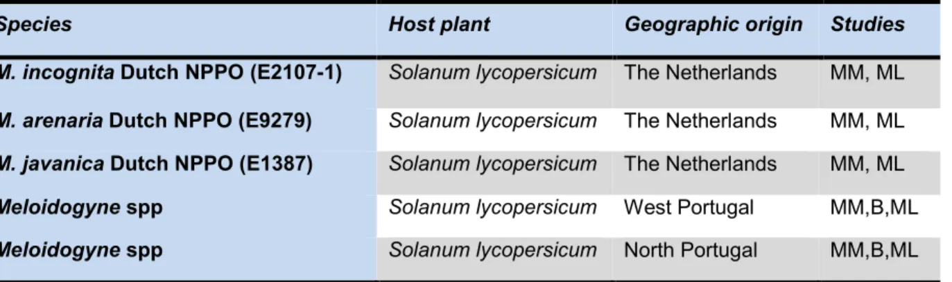
Table 2 - Meloidogyne isolates, host, geographic origin and studies performed in this work
Species Host plant Geographic origin Studies
M. incognita Dutch NPPO(E2107-1) Solanum lycopersicum The Netherlands MM, ML
M. arenaria Dutch NPPO (E9279) Solanum lycopersicum The Netherlands MM, ML
M. javanica Dutch NPPO (E1387) Solanum lycopersicum The Netherlands MM, ML
Meloidogyne spp Solanum lycopersicum West Portugal MM,B,ML
Meloidogyne spp Solanum lycopersicum North Portugal MM,B,ML
33 2.2 Inoculation of plant material
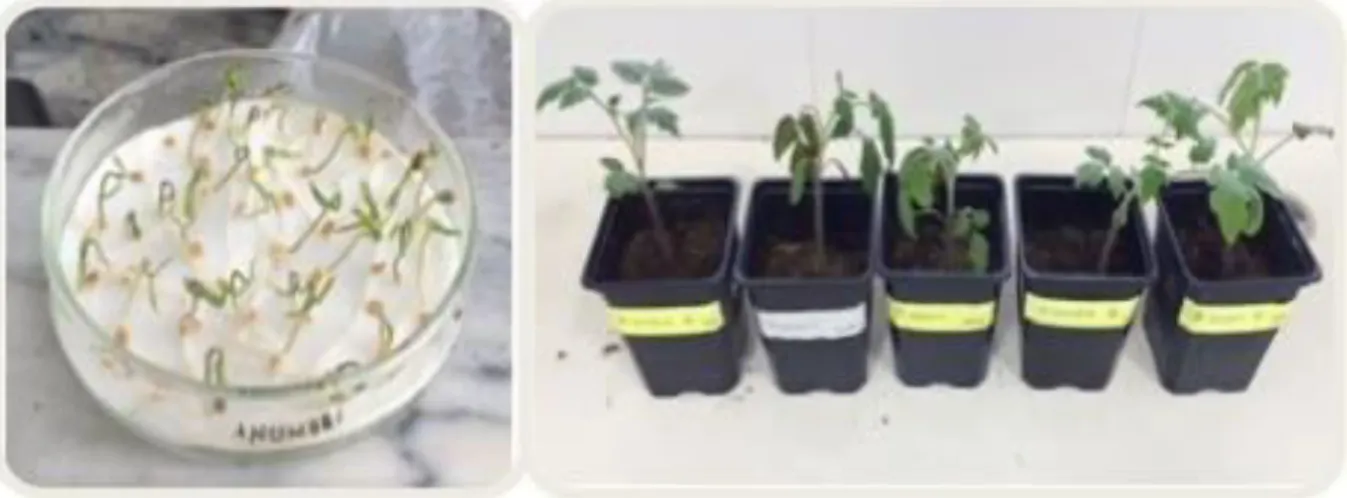
The nematode isolates from NVWA were propagated on tomato plants (Solanum lycopersicum cv. Rio Grande, considered susceptible to species of Meloidogyne) in a greenhouse in order to increase populations and to obtain inoculum to carry out the different studies proposed in this research. Fifteen pots were filled with a sterilised mixture of soil, sand and substrate (1:1:1). Seeds of tomato were previously germinated on moist filter paper at 23°C in Petri dishes (Figure 9). After three days, germinated seeds (those with emerging radicles) were transferred to a tray and two weeks later the seedlings were transplanted into the pots (Figure 8). Five pots with two-week-old tomato seedlings were artificially inoculated placing juveniles (J2) obtained from each of the species under study (M. incognita, M. arenaria, M. javanica) as well as chopped infected roots inside the pots.
The plants were transferred and maintained under greenhouse conditions at 18-27 ºC for 16 weeks.
Figure 8 - Germinated tomato seeds (Solanum lycopersicum cv. Rio Grande) and transplanted tomato
seedlings.
Four months after inoculation, the roots and soil were analysed for the presence or absence of galls and juveniles respectively.
2.3 Morphological and morphometric studies
Morphological and morphometric studies were conducted on second-stage juveniles (J2) obtained from soil extraction and handpicked females from infected roots.
Juvenile preparation
Juveniles were recovered from soil using flotation and sieving together with the centrifugal sugar flotation method (Laboratory of Nematology, INIAV Oeiras) which consist on: mixing 300 gr of soil in approximately 1 Lt of water and let the soil particles settle before pouring the water into the sieves (180 µm, 90 µm and 44 µm); the objective behind using three different sizes of sieves is to retain as much unwanted debris as possible. Once all the water has been poured, the residues caught in the finest sieve are washed into a centrifuge tube to be centrifuged for 4 minutes at 1750 rpm. After this centrifugation nematodes and any other heavy materials are forced to the bottom of the tube so the water is carefully discarded. Next, sugar is added and one more centrifugation takes place for 10 s at 1750 rpm; the nematodes will float in the sugar solution and the heaviest particles will go down to the bottom. At last, the sugar solution containing the nematodes is poured into the finest sieve and rinsed thoroughly with water to avoid damage and to be poured into the beakers.
The recovered J2 were individually transferred to a slide with a drop of water, to be gently heat killed using an alcohol burner and to be later observed at a light microscope.
The measurements made were achieved through drawing lines crossing approximately the middle of the specimen’s body. The study took into account some of the most relevant measurements of second stage juveniles according to EPPO, 2016 as follows:
Body length Stylet length Tail length
Hyalines terminus length DGO
It is important to highlight that usually the influence of the morphological characteristics is higher than the morphometric on the diagnosis since as mentioned by Hunt & Handoo, (2009) the environment may have an effect on the nematode’s morphometrics making them
35 Female preparation
Tomato roots from Portuguese samples were washed to remove soil and any other particles; immediately after, female extraction from vegetal tissues took place with the help of a disposable ophthalmic knife and a dissecting microscope. The females were then placed in glass blocks with lactic acid 45% for two days. Later, one by one were transferred to a slide with a drop of lactic acid 45% and cut with the ophthalmic knife to get the perineal patterns (Figure 9) for its identification at a light microscope.
Figure 9 - Perineal pattern area cuts. A - Hold female’s neck and body; B - excised female with neck
region removed and body contents gently expelled; C - posterior body with perineal pattern removed; D - trimming surplus cuticle around perineal pattern; E - trimmed perineal pattern ready for mounting (Hartman & Sasser, 1985).
Patterns of M. incognita, M. javanica and M. arenaria were compared with diagrams published from previous studies (Chitwood, 1949; Orton Williams, 1972,1973,1975; Eisenback et al., 1981).
3. RESULTS
3.1 Inoculation
Tomato seedlings inoculated with reference material from NVWA Wageningen of the three species of Meloidogyne did not show any symptoms above ground that could indicate nematode infection. Moreover, once the plants were uprooted not Meloidogyne gall formation was observed even though a susceptible cultivar (Rio Grande) was used (Figure 10)
37 3.2 Reference material
As morphological characters of the species under study often vary considerably within populations, the identification must be based on a combination of morphological and morphometric characters. Morphometric measurements performed on second stage juveniles from reference material are reported in Table 3.
Table 3 - Morphometric values (in µm) of reference samples of Meloidogyne incognita, Meloidogyne
arenaria and Meloidogyne javanica
Characteristic M. incognita (n=5) M. arenaria (n=5) M. javanica (n=5) Reported values Eisenback et al. 1981; Eisenback, 1985; Karsen & Moen, 2006; EPPO, 2016. Body length 371.2 – 394.4 ± 9.65 (377.6) 377.7 – 482.6 ± 44.38(421.3) 396.3 – 412.6 ± 5.39 (403.5) M. incognita: 360-400 M. arenaria: 400-490 M. javanica: 400-560 Stylet length 16.2 – 18.2 ± 0.72 (17.2) 13.4 – 15.3 0.79 (14.5) 16.1 – 19.9 ± 070 (18.1) M. incognita: 14-16 (15) M. arenaria: 14-16 (15) M. javanica: 14-18 (16) DGO 2.5 - 3 ± 0.17 (2.7) 3 – 3.5 ± 0.20 (3.3) 2.4 – 2.7 ±0.17 (2.6) M. inc:ognita: 2-3 (2.5) M. arenaria: 3-4 (3.5) M. javanica: 3-4 (3.5) Tail length 47.2 – 55.5 ± 3.71 (50.1) 40.9 – 57.1 7.39 (48.1) 58.2 – 68.5 ± 3.67 (63.2) M. incognita: 43-65 (54) M. arenaria: 45-70 (57.5) M. javanica: 51-63 (57) Hyaline terminus 11.5 – 13.5 ± 0.52 (12.6) 12.8 – 13.8 0.36 (13.1) 10.2 – 13.5± 1.27 (12.3) M. incognita: 6-11 (9) M. arenaria: 6-15 (10.5) M. javanica: 10-19 (14.5)
The mean values are given in brackets.
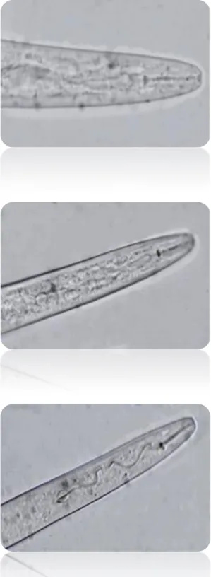
The observed juveniles were vermiform and moderately long, their head not offset from body. The stylet was slender with a sharp pointed stylet cone, cylindrical stylet shaft and rounded stylet knobs set off from the shaft (Figure 12). The tail region was narrow and the hyaline tail terminus was very distinct (Figure 13). Females were not available.
Second-stage juveniles of M. incognita, M. arenaria and M. javanica averaged 377.6 µm, 421.3 µm and 403.5 µm in length respectively and were between the typical ranges for M.
incognita, M. arenaria and M. javanica. The average stylet length of M. incognita and M. javanica and their range was greater in the populations under study. Tail morphometrics were not typical of M. arenaria, M. javanica or M. incognita (Table 3). Tail morphology was consistent with descriptions in the literature. M. javanica’s tail tip was slightly curved which is a typical character of the species (Figure 13).The mean length of the hyaline tail terminus was 12.6 µm, 13.1 µm and 12.3 μm for each species respectively which is not in agreement with previous descriptions. Due to the scarcity of material, measurements were performed on 5 individuals, which may be one of the causes for the observed overlapping since for this kind of studies at least 10 specimens are required.
Morphological and morphometric data required a great deal of work. The data collected was inconclusive and the range of values for each of these characteristics overlapped just as has been observed by many other authors and illustrated in Figure 11. Whereby, these features were not sufficient to distinguish closely related Meloidogyne species.
39
Figure 12 - Micrographs of second stage juveniles’ anterior regions with stylets (400X). A - Meloidogyne arenaria B - Meloidogyne incognita C - Meloidogyne javanica.
A
B
Figure 13 - Micrographs of second stage juveniles’ tails (400X). A - Meloidogyne arenaria B - Meloidogyne incognita C - Meloidogyne javanica.
A
B
41 3.3 West Portugal samples
Infected roots from samples from West Portugal were analysed. J2 were not available so the identification of the species was achieved through the study of the female perineal pattern. This pattern is a valuable morphological feature. However, over the years there has been found significant variability, which undermined the value of this character for comparing Meloidogyne species.
Females obtained from infected roots were completely enclosed by galled tissue containing more than one female. The female’s body was pearly white, pear shaped with anterior body portion commonly off-centre from a median plane and with almost terminal vulva. Their neck was short (Figure 14).
Figure 14 - Stereomicroscope and light microscope photographs of Meloidogyne females from West
Portugal samples. A - whole specimen; B - whole specimen (1000X oil immersion) C - stylet (1000X oil immersion).
Microscopic examination of the perineal pattern morphology of adult females found in the infected roots indicates the presence of two different species, M. incognita and M. arenaria. However, the results are not conclusive since some characteristics are not completely specific and can be found in the three species under study.
Meloidogyne incognita perineal pattern was roughly oval, dorsal striae closely spaced, wavy to zigzag, forking to some extent at the lateral lines. The dorsal arch was trapezoid in shape, sometimes with a distinct tail whorl and numerous transverse striae at the side of the ventral part (Figure 15).
B
Figure 15 - Perineal pattern of Meloidogyne incognita (400X).
Meloidogyne arenaria’s overall shape was rounded. Females showed low dorsal arch and the lines in the post-anal region are wavy and broken. Some striae bend towards the vulva. Phasmids were not visible and the lateral lines were weakly demarcated by forked striae (Figure 16).
43 3.4 North Portugal samples
Morphometrics performed on material from North Portugal are presented on Table 4. There were no major variations when compared to values previously reported in Table 3. However, those values overlap with other Meloidogyne species.
Table 4 - Morphometric values (in mm) performed on Meloidogyne spp. samples from North Portugal
The mean values are given in brackets.
The perineal pattern was typical for M. javanica with a rounded pattern, striae interrupted laterally by a pair of conspicuous incisures extending on both sides of the tail terminus, low dorsal arch trapezoid shape and a tail whorl (Figure 17).
Figure 17 - Perineal patterns of Meloidogyne javanica (400X).
Characteristic M. javanica (n=5) Reported values
Body length 390 - 540 (465) 400 - 560
Stylet length 14.5 - 18 (16.2) 14 -18 (16)
DGO 3 - 4 (3.5) 3 – 4 (3.5)
Tail length 49.9 - 60.8 (55.3) 51 - 63 (57)
4. DISCUSSION
For the onset of this study, it was essential to have isolates of reference well identified and available for a wide range of methodologies to be used. For that reason, this work started with the experimental inoculation of seedlings of tomato (cv Rio Grande) with juveniles (J2) of Meloidogyne obtained from reference material from the Netherlands Food and Consumer Product Safety Authority, Wageningen (NVWA), to obtain and maintain the isolates collection. However, the expected infection of tomato seedlings did not happen probably because the greenhouse conditions, namely the temperature was cooler than expected for April and May and, as it is known these species prefer warmer climates. Also, it can be inferred that the material used may not have been biologically in conditions of producing infection, this due to a contamination as well as the inadequate storage during the delivery from the Netherlands with peaks of high and low temperature that could have affected the sample’s quality.
Morphological features have been valuable tools for RKN identification due to its low cost and ease to learn the skills, with its accuracy depending on the number of characteristics to be evaluated and the number of specimens. On this study were examined the most relevant morphological and morphometric characteristics of Meloidogyne (females, and second-stage juveniles) according to Hirschman (1985) and EPPO (2016). The identification was made using a light microscope and proved to be a difficult task since sometimes it was not possible to see some of the characters or not great detail could be achieved so, for better image quality it is advisable to use differential interference contrast microscopy.
The measurements made on material from the Netherlands such as the juvenile body length, tail length, stylet length and lengths of hyaline tail terminus did not help on verifying the species identity. The obtained morphometric values for most of the characters overlapped and are within the expected range making them fall into more than one species description. For instance, M. incognita had range value between 371 µm to 394.4 µm but measures within this range can be easily misplaced into M. arenaria range value of 377.7 µm to 482.6 µm. For this reason, morphometrics on its own cannot be used to draw a conclusion on the identification of root-knot nematodes as already described by many authors.
Identification of Meloidogyne species was also attempted using morphology of perineal patterns since it is considered important in differentiating species of these nematodes. Nevertheless, on this study it was found that many of them presented a lot of similarities that
45 only spotted on M. javanica but also on M. arenaria’s perineal pattern from West Portugal samples; this kind of incidents can cause confusion and lead to inadequate identifications of Meloidogyne. Same confusion has been already reported by Rammah & Hirschmann, (1990).
Additionally, for a reliable identification based on morphology, individuals from all stages should be examined which is not always possible. For instance, on this research individual samples did not have the three stages available; moreover, due to the parthenogenetic reproduction of the species under study males are not necessary, making them very rare specimens.
Finally, although morphological and morphometric data can be very helpful for tentative identification, it may not be enough to differentiate RKNs and their physiological/cytological races as they are closely related (Zijlstra, 2000).
CHAPTER 2: BIOCHEMICAL
STUDIES
47 1. INTRODUCTION
The taxonomy of the genus Meloidogyne years ago was generally based on morphological and morphometric characters. However, variability of the perineal patterns and diagnostic morphological characters, life stages in different habitats, wide host ranges, indistinct species boundaries or species complexes, sexual dimorphism, species with a potential hybrid origin, polyploidy, and over a century of human-aided dispersal are up to today limitations that have led researchers to find another means of identification. Therefore, the integration of classical methods of identification with techniques such as the use of enzymatic and molecular markers is necessary to a more accurate and reliable species identification.
Extensive enzymatic studies have demonstrated that the major species of Meloidogyne can be differentiated by species-specific enzyme phenotypes, which can be revealed by polyacrylamide gel electrophoresis (PAGE) (Esbenshade & Triantaphyllou, 1990). Furthermore, recent progress in electrophoretic procedures have made possible, and also practical, the detection of the phenotype of one, two, and even more enzymes of a single Meloidogyne female (Carneiro et al., 2000).
Dickson verified that there were differences on the electrophoretic patterns of many enzymes developing the first demonstration that some enzymes may be species-specific and could be used in the identification of Meloidogyne species (Dickson et al., 1971). Later on, in the mid-1980’s Esbenshade and Triantaphyllou developed a biochemical-based diagnostic technique, reliant on isozyme profiles. They reported esterase patterns from 16 Meloidogyne species, with the most common phenotypes being A2 and A3 (M. arenaria), I1 and I2 (M. incognita) and J3 (M. javanica). Variations in esterase and malate dehydrogenase isozyme profiles proved to be extremely informative in differentiating most known Meloidogyne species (Esbenshade & Triantaphyllou, 1990). The main drawback to this method, however, is that the technique is only applicable to young adult females since they all are in a single developmental stage and are associated with the expression of a given gene product. Nonetheless, the adult stage is not readily isolated from the soil as it generally resides in the host. The ineffective second stage juveniles are usually in large numbers overshadowing the adult female stage.
Despite this shortcoming, isozyme electrophoresis remains one of the most reliable and widely used differentiation method (Janseen et al., 2016). Since the female stage is often unavailable in soil samples, the isozyme method requires the time and space to establish and maintain populations in culture from single egg masses or single J2s in order to obtain this stage (Blok &Powers, 2009).